Comparative Study
. 2016 Apr;27(4):1006-14.
doi: 10.1681/ASN.2015040421. Epub 2015 Aug 11.

Comparison of Glomerular and Podocyte mRNA Profiles in Streptozotocin-Induced Diabetes

Affiliations
Comparative Study

Comparison of Glomerular and Podocyte mRNA Profiles in Streptozotocin-Induced Diabetes

Jia Fu et al. J Am Soc Nephrol. 2016 Apr.

Abstract

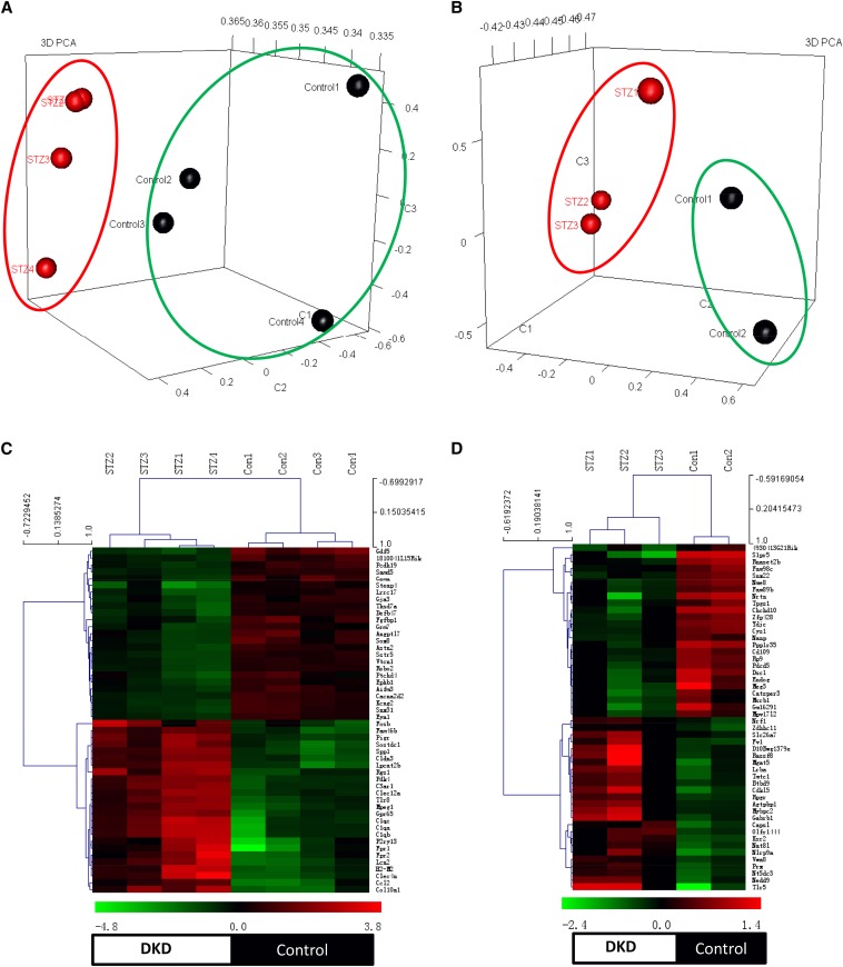
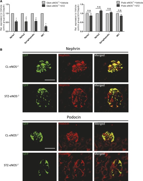
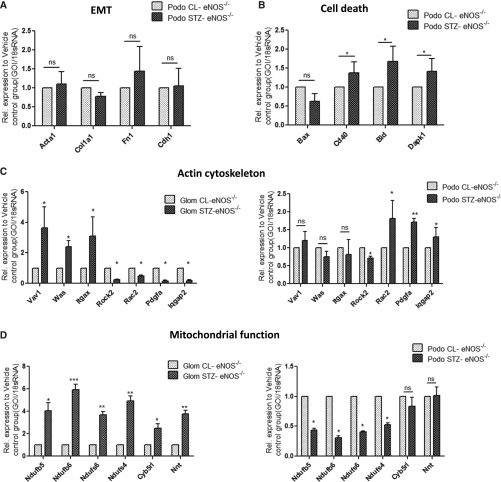
Evaluating the mRNA profile of podocytes in the diabetic kidney may indicate genes involved in the pathogenesis of diabetic nephropathy. To determine if the podocyte-specific gene information contained in mRNA profiles of the whole glomerulus of the diabetic kidney accurately reflects gene expression in the isolated podocytes, we crossed Nos3(-/-) IRG mice with podocin-rtTA and TetON-Cre mice for enhanced green fluorescent protein labeling of podocytes before diabetic injury. Diabetes was induced by streptozotocin, and mRNA profiles of isolated glomeruli and sorted podocytes from diabetic and control mice were examined 10 weeks later. Expression of podocyte-specific markers in glomeruli was downregulated in diabetic mice compared with controls. However, expression of these markers was not altered in sorted podocytes from diabetic mice. When mRNA levels of glomeruli were corrected for podocyte number per glomerulus, the differences in podocyte marker expression disappeared. Analysis of the differentially expressed genes in diabetic mice also revealed distinct upregulated pathways in the glomeruli (mitochondrial function, oxidative stress) and in podocytes (actin organization). In conclusion, our data suggest reduced expression of podocyte markers in glomeruli is a secondary effect of reduced podocyte number, thus podocyte-specific gene expression detected in the whole glomerulus may not represent that in podocytes in the diabetic kidney.

Keywords: diabetic nephropathy; gene transcription; glomerulus; podocyte; signaling.


Figures

Figure 1.
Figure 1.
Heat map and PCA analysis between glomeruli and sorted podocytes from control and diabetic mice. The PCA was performed for transcriptomic data from glomeruli (A) and podocytes (B) between diabetic and non-diabetic mice. Genes with the highest loadings in the first three principal components were plotted in 3D visualization. For both isolated glomeruli and podocytes, PCA revealed that the diabetic and non-diabetic samples form distinct clusters, indicating that samples within each biologic group have more similarity. Each symbol represents each biologic sample (green circles, non-diabetic mice; red circles, diabetic mice). Heatmaps of the top 50 up- or downregulated genes from isolated glomeruli (C) and sorted podocytes (D) between diabetic and control mice (green marks, downregulation; red marks, upregulation). There were four glomerular RNA samples in each group, two podocyte RNA samples from control mice and three samples from diabetic mice. Unsupervised hierarchical clustering was performed for gene expression between samples. Four clusters in each heatmap represented upregulated genes in diabetic mice (top, left cluster), upregulated genes in control mice (top, right cluster), downregulated genes in diabetic mice (bottom, left cluster), and downregulated genes in control mice (bottom right cluster).
Figure 2.
Figure 2.
qPCR analysis and immunostaining of podocyte marker between diabetic and control mice. (A) qPCR was performed for podocyte-specific genes, Neph1, Neph2, synaptopodin, and WT-1, in isolated glomeruli and sorted podocytes from both diabetic and non-diabetic mice. (*P<0.05 compared with citrate-treated control glomeruli, n=3; GOI, gene of interest). (B) Immunostaining of podocyte markers was performed in the kidney of these mice. EGFP was visualized in the podocytes of both STZ-eNOS−/− and CL-eNOS−/−mice. Immunofluorescence staining was performed for nephrin (top panel) and podocin (bottom panel) in the kidney of these mice. The representative images taken from in each group are shown (n=3, original magnification ×400, bar=50 μm).
Figure 3.
Figure 3.
qPCR validation of gene expression related to the major pathways in isolated glomeruli and podocytes between diabetic and control mice. (A) RT-PCR analysis was performed to assess the levels of mRNA for EMT-related genes expression in podocytes sorted from STZ-eNOS−/− and CL-eNOS−/− mice (*P<0.05 compared with those in CL-eNOS−/−, n=3). (B) RT-PCR analysis was performed to assess the levels of mRNA for cell-death–related gene expression in podocytes sorted from STZ-eNOS−/− and CL-eNOS−/− mice (*P<0.05 compared with those in CL-eNOS−/−, n=3). (C) RT-PCR analysis was performed to assess the levels of mRNA for actin cytoskeleton-related gene expression in glomeruli and podocytes isolated from STZ-eNOS−/− and CL-eNOS−/− mice (*P<0.05 compared with those in CL-eNOS−/−, n=3). (D) RT-PCR analysis of mitochondria and oxidative stress-related genes in isolated glomeruli and sorted podocytes from both STZ-eNOS−/− and CL-eNOS−/− mice (n=3, *P<0.05 compared with CL-eNOS−/−). GOI, gene of interest.

References

    1. Steffes MW, Schmidt D, McCrery R, Basgen JM, International Diabetic Nephropathy Study Group : Glomerular cell number in normal subjects and in type 1 diabetic patients. Kidney Int 59: 2104–2113, 2001 - PubMed
    1. Pagtalunan ME, Miller PL, Jumping-Eagle S, Nelson RG, Myers BD, Rennke HG, Coplon NS, Sun L, Meyer TW: Podocyte loss and progressive glomerular injury in type II diabetes. J Clin Invest 99: 342–348, 1997 - PMC - PubMed
    1. Susztak K, Raff AC, Schiffer M, Böttinger EP: Glucose-induced reactive oxygen species cause apoptosis of podocytes and podocyte depletion at the onset of diabetic nephropathy. Diabetes 55: 225–233, 2006 - PubMed
    1. Petermann AT, Pippin J, Krofft R, Blonski M, Griffin S, Durvasula R, Shankland SJ: Viable podocytes detach in experimental diabetic nephropathy: potential mechanism underlying glomerulosclerosis. Nephron, Exp Nephrol 98: e114–e123, 2004 - PubMed
    1. Kang YS, Li Y, Dai C, Kiss LP, Wu C, Liu Y: Inhibition of integrin-linked kinase blocks podocyte epithelial-mesenchymal transition and ameliorates proteinuria. Kidney Int 78: 363–373, 2010 - PMC - PubMed

Publication types