. 2015 Aug 12:6:7556.
doi: 10.1038/ncomms8556.

Injectable cryogel-based whole-cell cancer vaccines

Affiliations

Injectable cryogel-based whole-cell cancer vaccines

Sidi A Bencherif et al. Nat Commun. .

Abstract

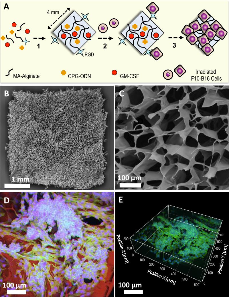
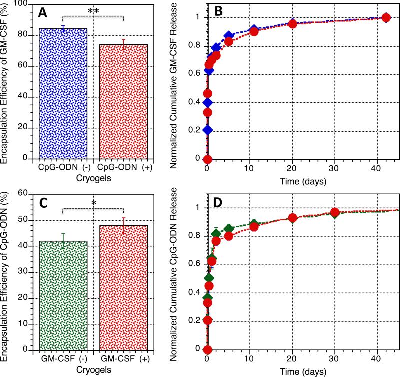
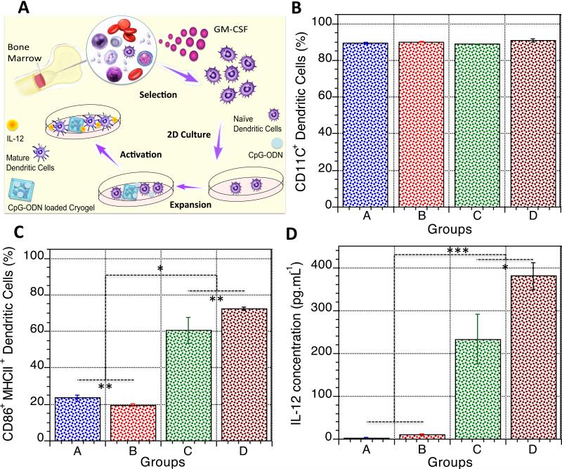
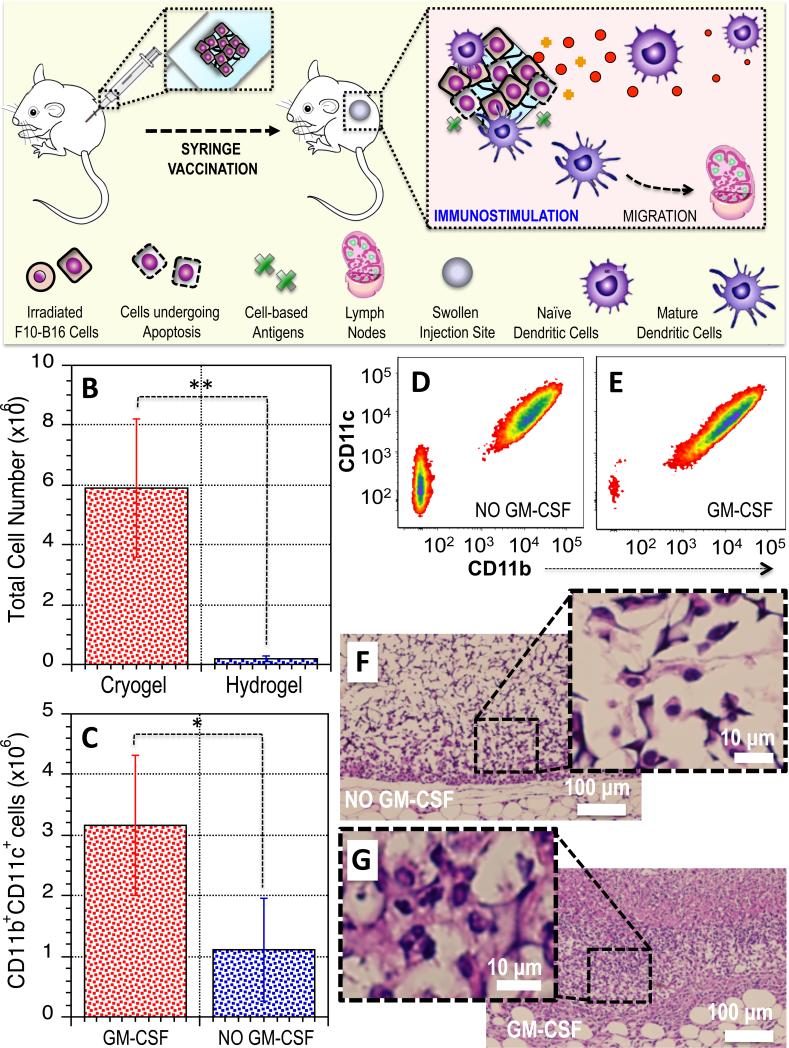
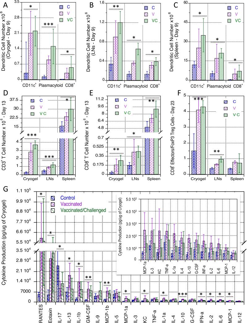
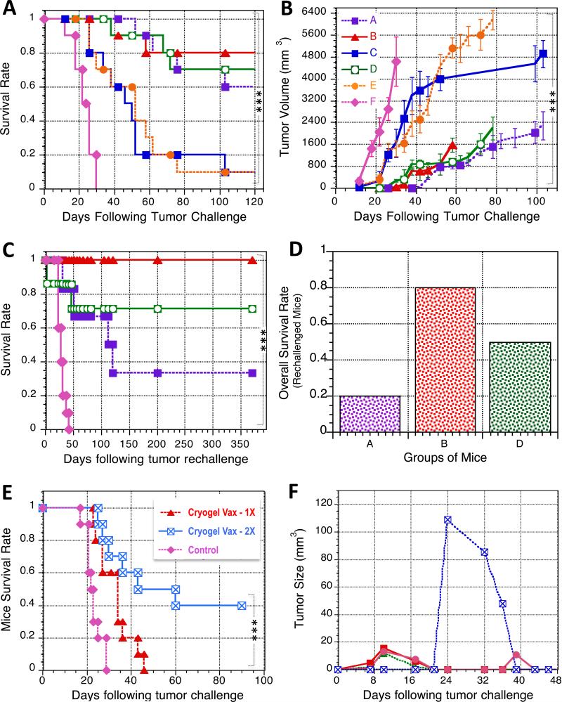
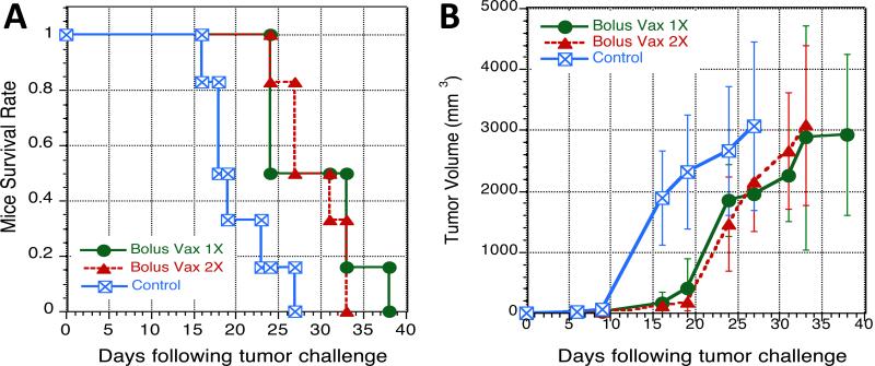
A biomaterial-based vaccination system that uses minimal extracorporeal manipulation could provide in situ enhancement of dendritic cell (DC) numbers, a physical space where DCs interface with transplanted tumour cells, and an immunogenic context. Here we encapsulate GM-CSF, serving as a DC enhancement factor, and CpG ODN, serving as a DC activating factor, into sponge-like macroporous cryogels. These cryogels are injected subcutaneously into mice to localize transplanted tumour cells and deliver immunomodulatory factors in a controlled spatio-temporal manner. These vaccines elicit local infiltrates composed of conventional and plasmacytoid DCs, with the subsequent induction of potent, durable and specific anti-tumour T-cell responses in a melanoma model. These cryogels can be delivered in a minimally invasive manner, bypass the need for genetic modification of transplanted cancer cells and provide sustained release of immunomodulators. Altogether, these findings indicate the potential for cryogels to serve as a platform for cancer cell vaccinations.


Figures

Figure 1
Figure 1. Fabrication and imaging of irradiated tumor cell-loaded cryogel sponge vaccines
A. Preparation of an alginate-derived active vaccine containing viable irradiated B16-F10 cells for the treatment of melanoma in syngeneic C57BL/6 mice. CpG ODN (TLR9-based immune adjuvant) & GM-CSF (cytokine adjuvant) loaded RGD-containing alginate cryogels were prepared by a cryogelation process at subzero temperature. The gels were subsequently seeded with irradiated B16-F10 melanoma cells (depicted as round-shaped cells) and incubated for 6h (depicted as square-shaped spread cells) prior to animal vaccination via subcutaneous injection. B. SEM showing homogeneous macroporous microstructure throughout the square-shaped sponge-like gel construct. C. SEM cross-sectional image of an alginate cryogel showing the interconnected macroporous network. D. 2-D confocal micrograph displaying immobilization of irradiated B16-F10 cells on a typical RGD-containing cryogel after 6h culture. Actin filaments in cells were visualized by staining with Alexa Fluor 488-phalloidin (green), cell nuclei were stained with DAPI (blue), and polymer walls were stained with polylysine-labeled rhodamine (red). E. 3-D reconstructed confocal fluorescence micrograph of irradiated B16-F10 cells in cryogel, depicting cell adhesion, spreading, and elongation after 6h culture.
Figure 2
Figure 2. Controlled release of biologically active immunomodulators from cryogels
A. Encapsulation efficiency of GM-CSF (detected by ELISA) in alginate cryogels following polymerization, washing, and sterilization; GM-CSF was incorporated in the presence (+) or absence (−) of CpG ODN in the cryogel. B. Cumulative release of GM-CSF from alginate cryogels over a period of 6 weeks in vitro; blue: cryogels containing only 1.5 μg GM-CSF, red: cryogels containing 1.5 μg GM-CSF+50 μg CpG ODN. C. Encapsulation efficiency of CpG-ODN (detected by an OliGreen assay) in alginate cryogels post polymerization, washing, and sterilization; CpG ODN was incorporated in the presence (+) or absence (−) of GM-CSF in the cryogel. D. Cumulative release of CpG-ODN from alginate cryogel matrices over a period of 6 weeks; green: cryogels containing only 50 μg CpG ODN, red: cryogels containing 1.5 μg GM-CSF+50 μg CpG ODN. Values represent mean and SD (n= 5). Differences between groups were statistically significant. Data were analyzed using Student's t test, *P < 0.05, **P < 0.01.
Figure 3
Figure 3. In vitro activation of BMDCs in response to CpG ODN-loaded cryogels
A. Cartoon depicting the process of bone marrow cell isolation from murine tibias and femurs; the differentiation, and expansion of BMDCs; and assessment of their activation in response to CpG ODN released from cryogel vaccines. BMDCs were cultured for 24h in the following conditions: medium (A, negative control), blank cryogels/medium (B), CpG ODN loaded cryogels/medium (C), or soluble CpG ODN/medium (D, positive control). B. Fraction of cells (detected by immunostaining in conjunction with flow cytometry) used in each condition that were CD11c+ prior to stimulation. C. Fraction of activated CD86+ MHCII+ BMDCs (detected by immunostaining in conjunction with flow cytometry) obtained in each condition. D. Production of IL-12 (detected by ELISA) in culture media in response to DCs stimulated by exposure to the various conditions. Values represent mean and SD (n= 5). Differences between groups were statistically significant. Data were analyzed using Student's t test and one-way analysis of variance (ANOVA), *P < 0.05, **P < 0.01, ***P < 0.001. Cartoon in A adapted by permission from Macmillan Publishers Ltd: [Nature Reviews Immunology] (Holger Hackstein and Angus W. Thomson, Dendritic cells: emerging pharmacological targets of immunosuppressive drugs, 1:24-34), copyright (2004).
Figure 4
Figure 4. Cryogel vaccines promote cellular infiltration and leukocyte recruitment
A. Schematic representation displaying the subcutaneous injection of cryogel vaccines in mice using a standard hypodermic needle, resulting in local edema and induration at the injection site, and recruitment and activation of DCs. B. Quantification of total cellular infiltration in macroporous cryogel sponges vs. conventional nanoporous hydrogels 4 days post-injection. C. Quantification of the number of CD11b+ CD11c+ cells infiltrating cryogels loaded with GM-CSF or blank (Control, NO GM-CSF) cryogels. D-E. FACS analysis for CD11b+ CD11c+ DCs in blank (Control, D) or GM-CSF releasing cryogels (E). F-G. H&E staining of sectioned cryogel scaffolds 4 days subsequent to subcutaneous injection in the backs of C57BL/6J mice: blank scaffolds (F) and GM-CSF (1.5 μg)–loaded scaffolds (G). Values represent mean and SD (n= 5). Differences between groups were statistically significant. Data were analyzed using Student's t test, *P < 0.05, **P < 0.01.
Figure 5
Figure 5. Cryogel vaccines stimulate the recruitment and activation of innate and adaptive immune cells
Number of CD11c+ DCs, pDCs, and CD8+ DCs at day 9 post-immunization isolated from explanted cryogel vaccines (A), regional LN (B), and spleen (C). Number of CD3+ T cells (D) and CD8+ T cells (E) at day 13 post-immunization isolated from explanted cryogel vaccines, LN, and spleen. F. Ratio of CD8+ T cells versus FoxP3+ Treg cells residing within cryogel vaccines, LN, and spleen at day 23 after immunization. G. In vivo concentrations of the indicated mouse cytokines (detected by a Bio-Plex Pro™ Mouse Cytokines 23-plex Immunoassay) from explanted cryogels at day 13. The inset shows a zoomed-in view of low cytokine levels. C, V, and VC groups correspond to mice injected with blank cryogels at day 0 (C), mice immunized with cryogel vaccines (containing CpG ODN, GM-CSF, and irradiated B16-F10 cells) at day 0 (V), and mice immunized with cryogel vaccines at day 0 + tumor challenged with live B16-F10 cells at day 6 (VC), respectively. Values in A, B, C, D, F, and G represent mean and SD (n = 5). Data were analyzed using one-way analysis of variance (ANOVA), *P < 0.05, **P < 0.01, ***P < 0.001 versus all other experimental conditions unless otherwise noted.
Figure 6
Figure 6. Cryogel vaccines confer long-term prophylactic and therapeutic protection against melanoma
A. A comparison of the survival rate in challenged C57BL/6 mice previously prophylactically vaccinated with (A): Bolus vaccine injection (4 × 10⁵ irradiated B16-F10 cells + soluble 3 μg GM-CSF + soluble 100 μg CpG ODN); (B): Cryogel vaccines (2 × 2 × 105 irradiated B16-F10 cells + 2 × 1.5 μg GM-CSF + 2 × 50 μg CpG-ODN); (C): Cryogel vaccines (2 × 2 × 105 irradiated B16-F10 cells + 2 × 1.5 μg GM-CSF); (D): Cryogel vaccines (2 × 2 × 105 irradiated B16-F10 cells + 2 × 50 μg CpG ODN); (E): Blank cryogel (control; negative control); (F): naïve mice (no immunization). At day 6 following immunization, C57BL/6J mice (10 mice/group) were challenged with 105 B16-F10 tumor cells and monitored for animal survival. B. Tumor growth curve after the 1st tumor challenge (105 cells) on prophylactically vaccinated mice. C. A comparison of the survival rate in re-challenged mice prophylactically treated with (A): Bolus injection (4 × 10⁵ irradiated B16-F10 cells + soluble 3 μg GM-CSF + soluble 100 μg CpG ODN); (B): Cryogel vaccine (2 × 2 × 105 irradiated B16-F10 cells + 2 × 1.5 μg GM-CSF + 2 × 50 μg CpG ODN); (D): Cryogel vaccine (2 × 2 × 105 irradiated B16-F10 cells + 2 × 50 μg CpG ODN); and (F): naïve mice (no immunization). At day 126 following immunization, C57BL/6J mice (10 mice/group) from the first challenge study were challenged a second time with 105 B16-F10 tumor cells and monitored for survival. D. Overall survival rate after two consecutive tumor-challenges in prophylactically immunized mice to evaluate long-term immunological protection in the context of melanoma. Overall survival is defined as the fraction of mice that survive both challenges. E. A comparison of the survival of mice bearing established melanoma tumors (inoculated with 5 × 10⁵ B16-F10 cells and allowed to develop for 3 days, tumor area ≥ 10 mm2) and therapeutically treated with cryogel vaccines (2 × 2 × 105 irradiated B16-F10 cells + 2 × 1.5 μg GM-CSF + 2 × 50 μg CpG ODN) either once at day 3 (Cryogel Vax – 1X) or at both days 3 and 10 (Cryogel Vax – 2X), or naïve mice (Control, no immunization). F. Individual tumor growth curves for each mouse surviving tumor challenge (5 × 10⁵ cells) after a two-time treatment with cryogel vaccines at days 3 and 10. Values represent mean and SD (n = 10 per condition) from at least two independent studies. For simplification, only Fig. 6B shows the legend but it applies also to Fig. 6A, 6C, and 6D. Data were analyzed using chi-squared test, log-rank test (survival curves comparison), or one-way analysis of variance (ANOVA) (**P < 0.01, ***P < 0.001).
Figure 7
Figure 7. Cryogel-free bolus vaccines containing irradiated B16-F10 cells, GM-CSF and CpG ODN confer no therapeutic efficacy
A comparison of the survival (A) and tumor growth (B) of C57BL/6J mice bearing established melanoma tumors (inoculated with 5 × 10⁵ B16-F10 cells and allowed to develop for 3 days, tumor area ≥ 10 mm2, 6 mice/group) and therapeutically treated with a cryogel-free bolus vaccine (Bolux Vax 1X: 4 × 10⁵ irradiated B16-F10 cells + soluble 3 μg GM-CSF + soluble 100 μg CpG ODN) at day 3; or cryogel-free bolus vaccines (Bolux Vax 2X: 4 × 10⁵ irradiated B16-F10 cells + soluble 3 μg GM-CSF + soluble 100 μg CpG ODN) at both days 3 and 10; and (Control, no immunization): naïve mice.

Similar articles

Cited by

References

    1. Mansoor W, Gilham DE, Thistlethwaite FC, Hawkins RE. Engineering T cells for cancer therapy. Br. J. Cancer. 2005;93(10):1085–1091. - PMC - PubMed
    1. Zacharski LR, Sukhatme VP. Coley's toxin revisited: immunotherapy or plasminogen activator therapy of cancer? J Thromb Haemost. 2005;3:424–427. - PubMed
    1. Hoption Cann SA, van Netten JP, van Netten C. Dr William Coley and tumour regression: a place in history or in the future. Postgrad. Med. J. 2003;79:672–680. - PMC - PubMed
    1. Topalian SL, et al. Safety, activity, and immune correlates of anti-PD-1 antibody in cancer. N. Engl. J. Med. 2012;366:2443–2454. - PMC - PubMed
    1. Kantoff PW, et al. Sipuleucel-T immunotherapy for castration-resistant prostate cancer. N. Engl. J. Med. 2010;363:411–422. - PubMed

Publication types