. 2015 Oct 2;290(40):24470-83.
doi: 10.1074/jbc.M115.661439. Epub 2015 Aug 11.

Bacterial rotary export ATPases are allosterically regulated by the nucleotide second messenger cyclic-di-GMP

Affiliations

Bacterial rotary export ATPases are allosterically regulated by the nucleotide second messenger cyclic-di-GMP

Eleftheria Trampari et al. J Biol Chem. .

Abstract

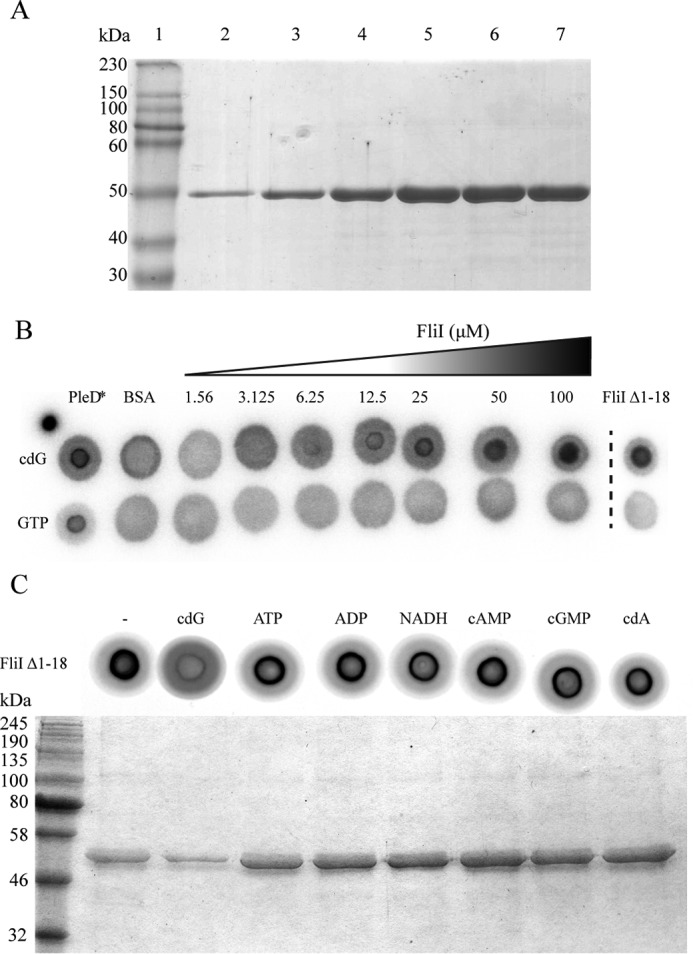
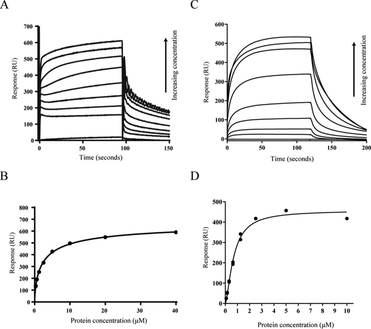
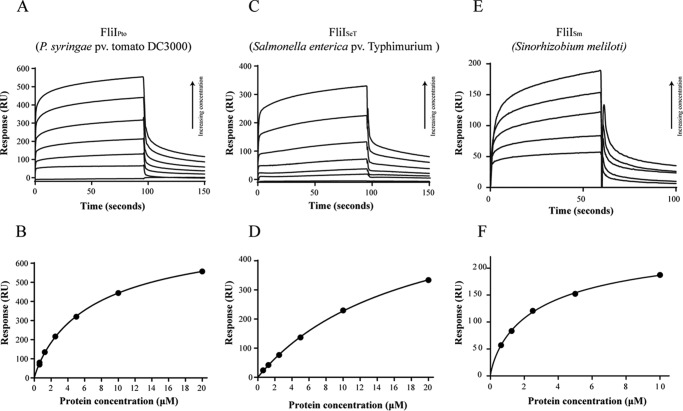
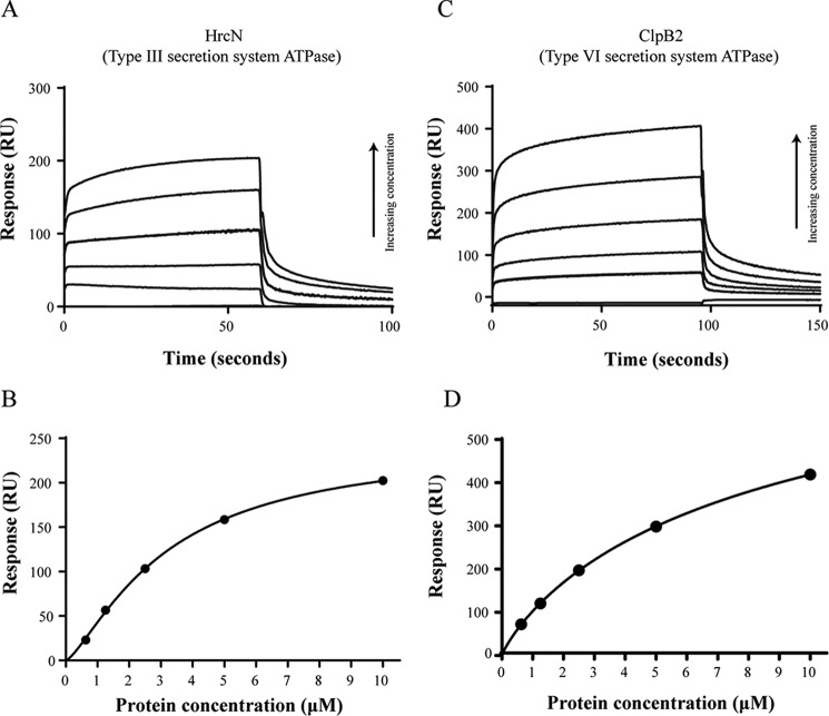
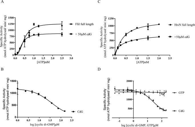
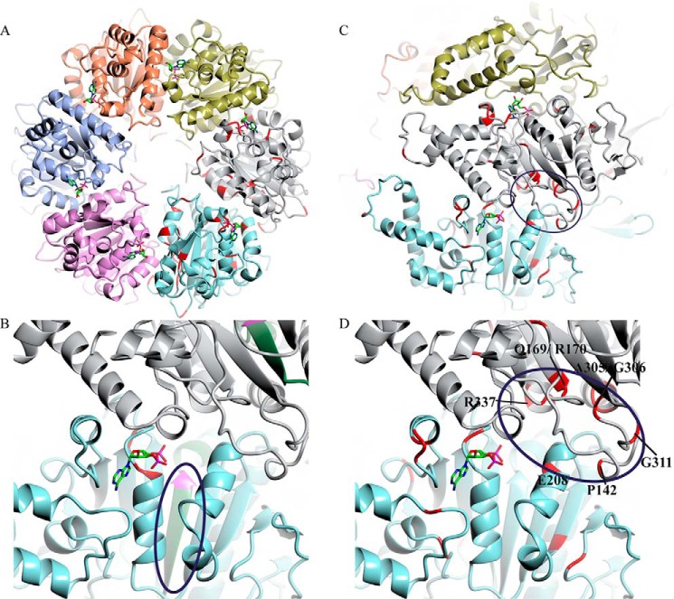
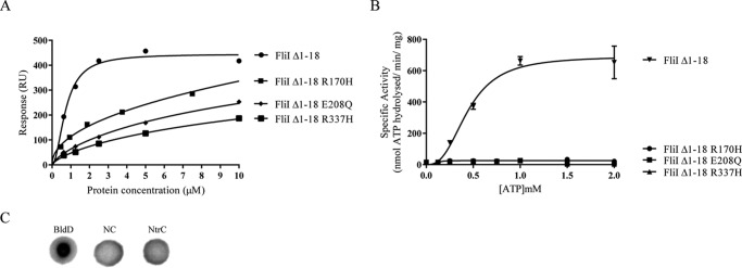
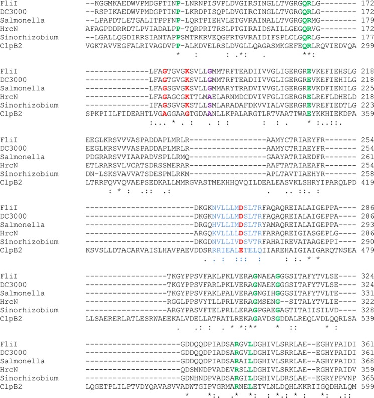
The widespread second messenger molecule cyclic di-GMP (cdG) regulates the transition from motile and virulent lifestyles to sessile, biofilm-forming ones in a wide range of bacteria. Many pathogenic and commensal bacterial-host interactions are known to be controlled by cdG signaling. Although the biochemistry of cyclic dinucleotide metabolism is well understood, much remains to be discovered about the downstream signaling pathways that induce bacterial responses upon cdG binding. As part of our ongoing research into the role of cdG signaling in plant-associated Pseudomonas species, we carried out an affinity capture screen for cdG binding proteins in the model organism Pseudomonas fluorescens SBW25. The flagella export AAA+ ATPase FliI was identified as a result of this screen and subsequently shown to bind specifically to the cdG molecule, with a KD in the low micromolar range. The interaction between FliI and cdG appears to be very widespread. In addition to FliI homologs from diverse bacterial species, high affinity binding was also observed for the type III secretion system homolog HrcN and the type VI ATPase ClpB2. The addition of cdG was shown to inhibit FliI and HrcN ATPase activity in vitro. Finally, a combination of site-specific mutagenesis, mass spectrometry, and in silico analysis was used to predict that cdG binds to FliI in a pocket of highly conserved residues at the interface between two FliI subunits. Our results suggest a novel, fundamental role for cdG in controlling the function of multiple important bacterial export pathways, through direct allosteric control of export ATPase proteins.

Keywords: ATPase; Pseudomonas; bacterial signal transduction; cyclic di-GMP (c-di-GMP); flagellum; second messenger; type III secretion system (T3SS).


Figures

FIGURE 1.
FIGURE 1.
A, Coomassie-stained SDS-PAGE gel showing purified FliIHis fractions eluted with 500 mm imidazole. B, DRaCALA for [32P]cdG and [32P]GTP binding to increasing concentrations of full-length FliI (FliIHis). Positive (10 μm PleD*) and negative (10 μm BSA) binding controls are included, as well as N-terminal truncated FliI (10 μm FliIΔ1–18). C, DRaCALA competition experiment performed on FliIΔ1–18. A variety of nucleotides were included in the reaction to test the specificity of cdG binding. SDS-PAGE gel showing protein bound to the capture compound after preincubation with different nucleotides.
FIGURE 2.
FIGURE 2.
A, SPR sensorgrams showing affinity measurements for FliIHis binding to biotinylated cdG. A range of FliIHis concentrations was used (0.312, 0.625, 1.25, 2.5, 5, 10, 20, and 40 μm), and concentration replicates were included as appropriate together with buffer only and BSA controls. The protein binding and dissociation phases for all sensorgrams are shown. B, affinity fit for FliIHis-cdG binding. The binding response for each concentration was recorded 4 s before the end of the injection, and the KD values for FliIHis binding to cdG (2.4 ± 0.2 μm) were calculated using the BiaEvaluation software and confirmed by GraphPad Prism. C, SPR sensorgrams showing affinity measurements for FliIΔ1–18 binding to biotinylated cdG. A range of protein concentrations was used (0.078, 0.156, 0.3125, 0.625, 1.25, 2.5, 5, and 10 μm), and concentration replicates were included as appropriate together with buffer only and BSA controls. The protein binding and dissociation phases for all sensorgrams are shown. D, affinity fit for FliIΔ1–18-cdG binding. Binding responses were measured 4 s before the end of the injection, and the KD values for FliIΔ1–18 binding to cdG (0.8 ± 0.03 μm) were calculated using the BiaEvaluation software and confirmed by GraphPad Prism.
FIGURE 3.
FIGURE 3.
A and B, SPR sensorgrams and resulting affinity fit for FliIPto binding to biotinylated cdG. C and D, SPR sensorgrams and affinity fit for FliISeT binding to biotinylated cdG. E and F, SPR sensorgrams and affinity fit for FliISm binding to biotinylated cdG. In all three cases, a range of protein concentrations was used (0.625, 1.25, 2.5, 5, 10, and for FliIPto/FliISeT 20 μm), and concentration replicates were included as appropriate together with buffer only and BSA controls. The protein binding and dissociation phases for all sensorgrams are shown. For the affinity fits, binding responses were measured 4 s before the end of the injection, and KD values for each protein were calculated using the BiaEvaluation software and confirmed by GraphPad Prism (Table 3).
FIGURE 4.
FIGURE 4.
A and B, SPR sensorgram and resulting affinity fit for HrcN (type III export ATPase) binding to biotinylated cdG. C and D, SPR sensorgram and resulting affinity fit for ClpB2 (Type VI export ATPase) binding to biotinylated cdG. In both cases, a range of protein concentrations was used (0.625, 1.25, 2.5, 5, and 10 μm), and concentration replicates were included as appropriate together with buffer only and BSA controls. The protein binding and dissociation phases for all sensorgrams are shown. For the affinity fits, binding responses were measured 4 s before the end of the injection, and KD values for each protein were calculated using the BiaEvaluation software and confirmed by GraphPad (Table 3).
FIGURE 5.
FIGURE 5.
A, ATPase activity for FliIHis ± 50 μm cdG. FliIHis specific activity (nmol ATP hydrolyzed/min/mg protein) is shown for increasing ATP concentrations. Addition of cdG causes a decrease of the Vmax of FliIHis ATPase activity. B, IC50 curve for FliIHis ATPase activity upon addition of increasing cdG concentrations. A constant concentration of ATP (1 mm) was included alongside 1 μg of FliIHis protein. C and D, ATPase activity ± 50 μm cdG, and IC50 curve upon addition of increasing cdG concentrations, for HrcN. All parameters remain the same as in A. The IC50 curve also includes results for GTP titration showing no ATPase inhibition.
FIGURE 6.
FIGURE 6.
A, affinity fit for cdG binding to different FliI alleles (FliIHis, Δ1–18, K181A, D265A, and G176A). Sensorgrams obtained using biotinylated cdG (Figs. 1D and 2B) were used to calculate the KD values for FliI binding to cdG (Table 3). At each protein concentration, the responses were recorded 4 s before the end of the injection. B, ATPase activity for different FliI alleles. Protein specific activity in each case (nmol ATP hydrolyzed/min/mg protein) is shown for increasing ATP concentrations.
FIGURE 7.
FIGURE 7.
A, homology model of the predicted hexameric form of SBW25 FliI, based on the crystal structure of FliI from S. typhimurium (Protein Data Bank code 2DPY). Conserved residues between the six cdG-binding proteins tested in this study are marked in red (on the gray and cyan-colored subunits only), and ADP (stick model; taken from template structure) is shown bound at the interfaces between the individual FliI subunits. B, close-up of the interface between two FliI subunits, showing the NVLLLMDSLTR peptide implicated in cdG capture compound binding (circled, in green) and the conserved Walker B aspartate (Asp-265) in pink. C, locations of conserved residues between the six cdG-binding proteins tested in this study (red). D, close-up of the proposed cdG binding pocket (circled). Conserved residues suggested to form the cdG binding site are labeled.
FIGURE 8.
FIGURE 8.
A, affinity fit for cdG binding to different FliI alleles (FliIΔ1–18, FliIΔ1–18 R170H, FliIΔ1–18 E208Q, and FliIΔ1–18 R337H). B, ATPase activity for different FliI alleles. Protein specific activity in each case (nmol ATP hydrolyzed/min/mg protein) is shown for increasing ATP concentrations. C, DRaCALA binding assay for [32P]cdG to 10 μm NtrC (A. vinelandii). Positive (10 μm BldD*) and negative binding controls (NC) were included as appropriate.
FIGURE 9.
FIGURE 9.
Clustal alignment of conserved residues between FliI, HrcN, and ClpB2 proteins in this study. Identities between all six residues are marked with asterisks (*), and similarities across all six with periods (.) or colons (:). The mutated FliI Walker A/B residues (see Fig. 6) are marked in red, the capture compound-binding NVLLLMDSLTR peptide is marked in blue, the position of the conserved cdG binding arginine in FlrA (Arg-176) is marked in purple, and the conserved residues of the proposed cdG binding site are marked in green (see Fig. 7).

References

    1. Naseby D. C., Way J. A., Bainton N. J., Lynch J. M. (2001) Biocontrol of Pythium in the pea rhizosphere by antifungal metabolite producing and non-producing Pseudomonas strains. J. Appl. Microbiol. 90, 421–429 - PubMed
    1. Haas D., Défago G. (2005) Biological control of soil-borne pathogens by fluorescent pseudomonads. Nat. Rev. Microbiol. 3, 307–319 - PubMed
    1. Compant S., Clement C., Sessitsch A. (2010) Plant growth-promoting bacteria in the rhizo- and endosphere of plants: their role, colonization, mechnaisms onvolved and prospects for utilization. Soil Biol. Biochem. 42, 669–678
    1. Xin X. F., He S. Y. (2013) Pseudomonas syringae pv. tomato DC3000: a model pathogen for probing disease susceptibility and hormone signaling in plants. Annu. Rev. Phytopathol. 51, 473–498 - PubMed
    1. Lindeberg M., Cunnac S., Collmer A. (2009) The evolution of Pseudomonas syringae host specificity and type III effector repertoires. Mol. Plant Pathol. 10, 767–775 - PMC - PubMed

Publication types

MeSH terms