. 2015 Oct;201(2):631-49.
doi: 10.1534/genetics.115.180018. Epub 2015 Aug 10.

Efficient Endocytic Uptake and Maturation in Drosophila Oocytes Requires Dynamitin/p50

Affiliations

Efficient Endocytic Uptake and Maturation in Drosophila Oocytes Requires Dynamitin/p50

Guojun Liu et al. Genetics. 2015 Oct.

Abstract

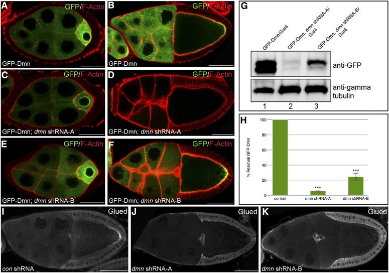
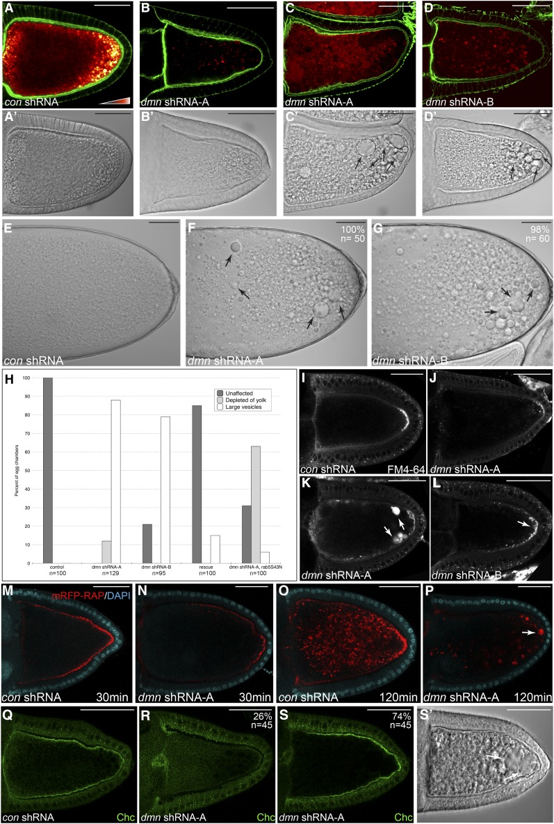
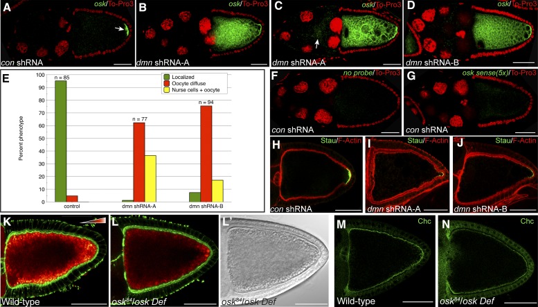
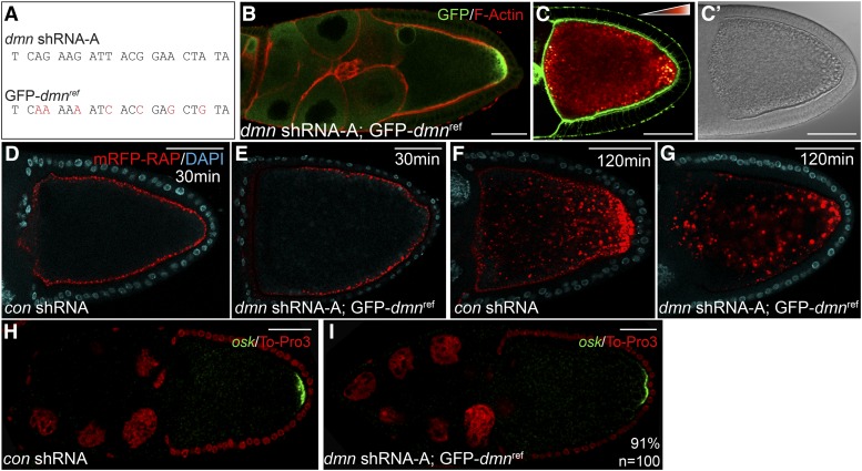
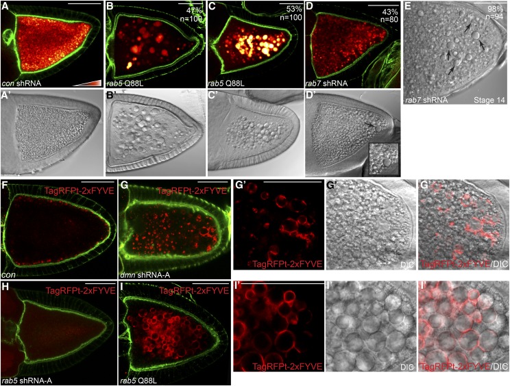
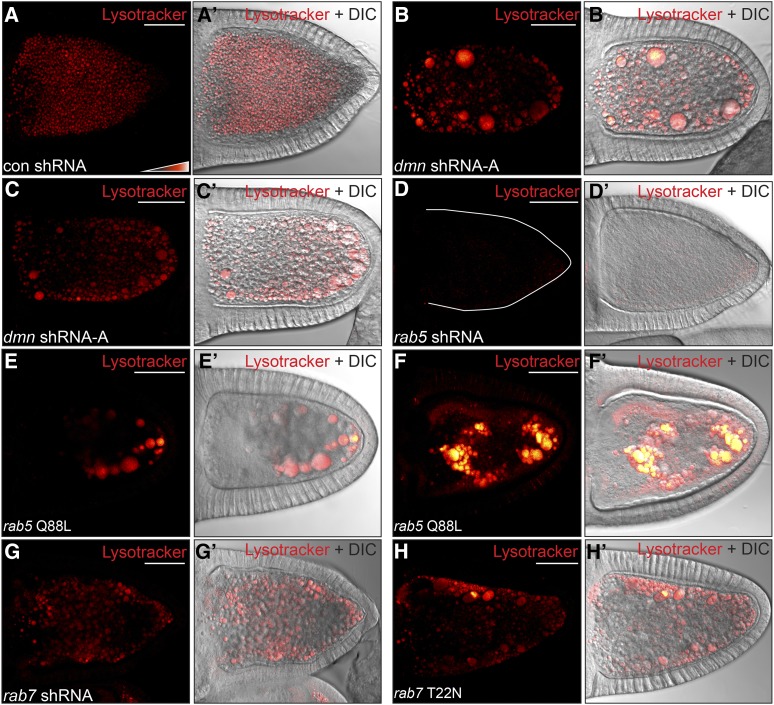
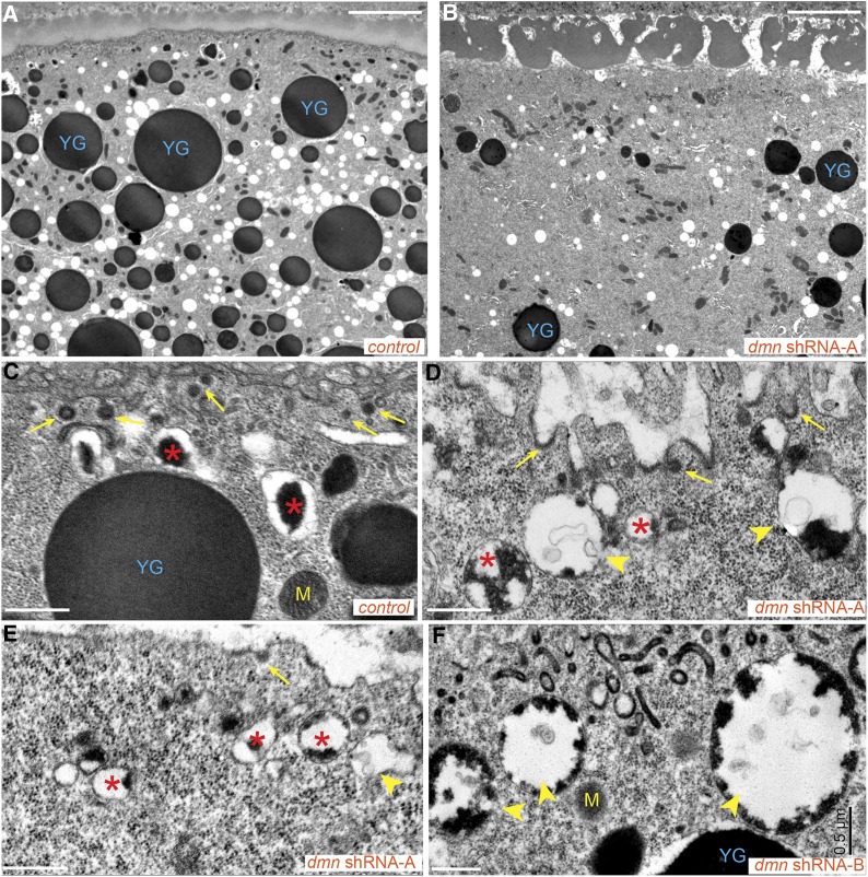
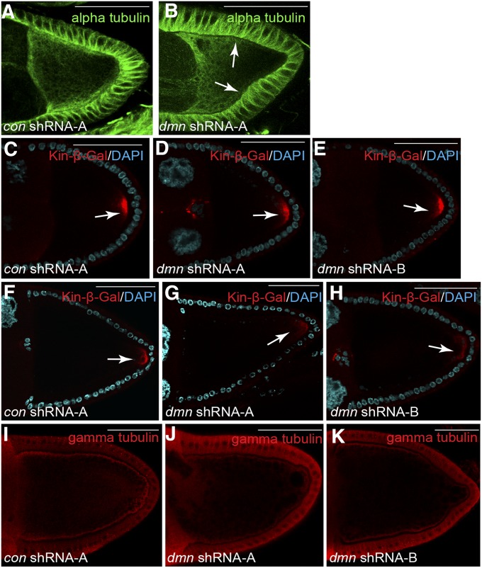
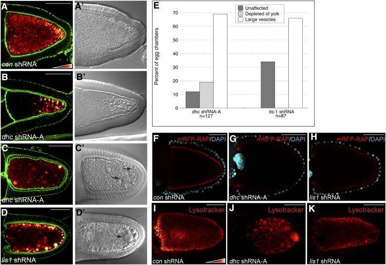
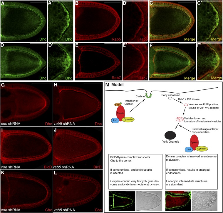
Dynactin is a multi-subunit complex that functions as a regulator of the Dynein motor. A central component of this complex is Dynamitin/p50 (Dmn). Dmn is required for endosome motility in mammalian cell lines. However, the extent to which Dmn participates in the sorting of cargo via the endosomal system is unknown. In this study, we examined the endocytic role of Dmn using the Drosophila melanogaster oocyte as a model. Yolk proteins are internalized into the oocyte via clathrin-mediated endocytosis, trafficked through the endocytic pathway, and stored in condensed yolk granules. Oocytes that were depleted of Dmn contained fewer yolk granules than controls. In addition, these oocytes accumulated numerous endocytic intermediate structures. Particularly prominent were enlarged endosomes that were relatively devoid of Yolk proteins. Ultrastructural and genetic analyses indicate that the endocytic intermediates are produced downstream of Rab5. Similar phenotypes were observed upon depleting Dynein heavy chain (Dhc) or Lis1. Dhc is the motor subunit of the Dynein complex and Lis1 is a regulator of Dynein activity. We therefore propose that Dmn performs its function in endocytosis via the Dynein motor. Consistent with a role for Dynein in endocytosis, the motor colocalized with the endocytic machinery at the oocyte cortex in an endocytosis-dependent manner. Our results suggest a model whereby endocytic activity recruits Dynein to the oocyte cortex. The motor along with its regulators, Dynactin and Lis1, functions to ensure efficient endocytic uptake and maturation.

Keywords: cell polarity; dynactin; endocytosis; kinesin; microtubule motors.


Figures

Figure 1
Figure 1
shRNA-mediated depletion of Dmn. (A and B) Egg chambers expressing GFP-Dmn under the control of a maternal tubulin promoter were fixed and processed for immunofluorescence using an antibody against GFP (green). The egg chambers were also counterstained for F-actin (red). Representative stage 5 (A) and stage 10 (B) egg chambers are shown. (C and D) Egg chambers coexpressing dmn shRNA-A and GFP-Dmn were fixed and processed for immunofluorescence using an antibody against GFP. Representative stage 5 (C) and stage 10 (D) egg chambers are shown. (E and F) Egg chambers coexpressing dmn shRNA-B and GFP-Dmn were fixed and processed for immunofluorescence using an antibody against GFP. Representative stage 5 (E) and stage 10 (F) egg chambers are shown. dmn shRNA-A and dmn shRNA-B were expressed using a maternal α-tubulin-Gal4 driver (see Materials and Methods for details). GFP-Dmn was expressed using a construct in which the maternal tubulin promoter was cloned upstream of the GFP-dmn-coding sequence (Januschke et al. 2002). (G) Ovarian lysates were prepared from strains expressing GFP-Dmn (lane 1), from strains coexpressing dmn shRNA-A and GFP-Dmn (lane 2), or from dmn shRNA-B and GFP-Dmn (lane 3). The lysates were run on an SDS-PAGE gel and analyzed by Western blotting using an antibody against GFP (top). The same blot was then probed using an antibody against γ-tubulin (bottom). The level of γ-tubulin serves as a loading control. The images were captured digitally using a UVP bioimaging system. (H) Western blots from three separate experiments depicted in G were quantified using the VisionWorks software (UVP). The level of GFP-Dmn in strains coexpressing dmn shRNA-A or dmn shRNA-B were compared to the level of GFP-Dmn in the control strain. The error bars indicate standard deviation. ***P = 0.0001. (I–K) Egg chambers expressing a control shRNA against eb1 (H), dmn shRNA-A (I), or dmn shRNA-B (J) were fixed and processed for immunofluorescence using an antibody against Glued. The shRNAs were expressed using a maternal α-tubulin-Gal4 driver. Bar: A, C, and E = 20 μm; B, D, F, I, J, and K = 50 μm.
Figure 2
Figure 2
Dmn depletion phenotypes. (A–D) Egg chambers expressing a control shRNA (A), dmn shRNA-A (B and C), or dmn shRNA-B (D) were fixed and stained to reveal the actin cytoskeleton (green). Autofluorescent yolk particles are displayed using a color-coded range indicator. Black indicates low levels of signal, red indicates moderate signal, and white indicates high levels of signal. DIC images of these egg chambers are shown in A′, B′, C′ and D′. The arrows indicate enlarged vesicular structures. (E–G) DIC images of mature stage 14 egg chambers expressing a control shRNA (E), dmn shRNA-A (F), or dmn shRNA-B (G) are shown. The penetrance of the indicated phenotypes and the number of egg chambers counted are indicated. (H) Quantification of endocytic phenotypes. Egg chambers from the indicated genotypes were scored for the presence of yolk and large vesicular structures. The percentage of each phenotype observed and the number of egg chambers counted for each genotype are indicated. “Rescue” indicates flies that are coexpressing dmn shRNA-A and GFP-dmnref. dmn shRNA-A and rab5S43N were coexpressed using the maternal α-tubulin driver. (I–L) Egg chambers expressing a control shRNA (I), dmn shRNA-A (J and K), or dmn shRNA-B (L) were processed for FM4-64 uptake. The egg chambers were mounted on slides and imaged live. Large vesicles that were positive for FM4-64 are indicated by arrows. (M and N) Egg chambers expressing a control shRNA (M) or dmn shRNA-A (N) were processed for mRFP-RAP endocytosis (red). The egg chambers were incubated with mRFP-RAP for 30 min. They were then fixed and stained with DAPI to reveal nuclei (cyan). (O and P) Egg chambers from these same strains were incubated with mRFP-RAP for 120 min to monitor endocytic maturation (red). They were then fixed and stained with DAPI to reveal nuclei (cyan). The arrow indicates enlarged mRFP-RAP endosomes. (Q–S) Egg chambers expressing a control shRNA (Q) or dmn shRNA-A (R and S) were fixed and processed for immunofluorescence using an antibody against Chc (green). S′ represents the DIC image of the oocyte in S. The white arrows in S′ indicate enlarged endosomes. The percentage of the phenotypes observed and the number of egg chambers counted are indicated. Bar, 50 μm.
Figure 3
Figure 3
oskar mRNA is delocalized in Dmn-depleted egg chambers. (A–D) Egg chambers expressing a control shRNA against eb1 (A), dmn shRNA-A (B and C), or dmn shRNA-B (D) were fixed and processed for in situ hybridization using an anti-sense probe against oskar mRNA (green). The egg chambers were counterstained with ToPro3 to reveal nuclei (red). The egg chambers in B, C, and D were imaged under a higher gain setting in comparison to those in A. This was required to visualize the delocalized oskar mRNA signal. (E) Quantification of oskar mRNA localization phenotypes. The number of egg chambers counted for each genotype and the percentage of each phenotype observed are indicated. The green bars indicate a wild-type localization pattern. The red bars indicate egg chambers in which signal for oskar mRNA is present within the oocyte in a diffuse pattern with residual enrichment at the posterior pole (“Oocyte diffuse”). The yellow bars indicate egg chambers in which oskar mRNA signal could be detected within the nurse cell cytoplasm in addition to the oocyte (“Nurse cells + oocyte”). (F) Egg chambers expressing a control shRNA were fixed and processed for in situ hybridization. For this experiment, no RNA probe was used, but the egg chambers were processed using the mouse anti-DIG antibody and the tyramide amplification step (green). The egg chambers were imaged under the same gain setting used in B, C, and D. The egg chambers were counterstained with ToPro3 to reveal nuclei (red). (G) Egg chambers expressing a control shRNA were fixed and processed for in situ hybridization using a sense probe against oskar mRNA (green). Five times more sense probe was used in this experiment in comparison to the anti-sense probe used in A–D. The egg chambers were imaged using the same gain setting as in B, C, and D. The egg chambers were counterstained with ToPro3 to reveal nuclei (red). (H–J) Egg chambers expressing a control shRNA against eb1 (H), dmn shRNA-A (I), or dmn shRNA-B (J) were fixed and processed for immunofluorescence using an antibody against Staufen (green). The egg chambers were also counterstained to reveal F-actin (red). (K and L) Egg chambers from wild-type flies (K), or oskar protein null flies (L) were fixed and stained to reveal the actin cytoskeleton (green). Autofluorescent yolk particles are displayed using a color-coded range indicator. L′ represents the DIC images of the egg chamber depicted in L. (M and N) Egg chambers from wild-type flies (M) or oskar protein null flies (N) were fixed and processed for immunofluorescence using an antibody against Chc (green). Bar, 50 μm.
Figure 4
Figure 4
Rescue of Dmn depletion phenotypes. (A) The sequence targeted by dmn shRNA-A is indicated. Also indicated are the site-specific mutations made within GFP-dmnref (red). (B) Egg chambers from strains coexpressing dmn shRNA-A and GFP-dmnref were fixed and processed for immunofluorescence using an antibody against GFP (green). The egg chambers were also counterstained for F-actin (red). (C) Egg chambers from strains coexpressing dmn shRNA-A and GFP-dmnref were fixed and stained to reveal the actin cytoskeleton (green). The autofluorescent yolk particles are displayed using a color-coded range indicator. ADIC image is shown in C′. (D and E) Egg chambers expressing a control shRNA (D) or coexpressing dmn shRNA-A and GFP-dmnref (E) were processed for mRFP-RAP endocytosis (red). The egg chambers were incubated with mRFP-RAP for 30 min. They were then fixed and stained with DAPI to reveal nuclei (cyan). (F and G) Egg chambers from these same strains were incubated with mRFP-RAP for 120 min to monitor endocytic maturation (red). They were then fixed and stained with DAPI to reveal nuclei (cyan). (H and I) Egg chambers expressing a control shRNA (H) or coexpressing dmn shRNA-A and GFP-dmnref (I) were fixed and processed for in situ hybridization using an anti-sense probe against oskar mRNA (green). The egg chambers were counterstained with ToPro3 to reveal nuclei (red). Bar, 50 μm.
Figure 5
Figure 5
Blocking endocytic maturation produces enlarged endosomes. (A–E) The following strains were fixed and stained to reveal the actin cytoskeleton: con shRNA (A), rab5 Q88L (B and C), and rab7 shRNA (D and E). The shRNAs and the rab5 Q88L construct were expressed using the maternal α-tubulin-Gal4 driver. The autofluorescent yolk particles are displayed using a color-coded range indicator. DIC images of these egg chambers are in A′, B′, C′, and D′. A stage 14 egg chamber expressing rab7 shRNA is shown in E. The efficacy of Rab5 and Rab7 depletion is shown in Figure S1, A–D. (F and G) Egg chambers from a strain expressing TagRFPt-2xFYVE (F) or from a strain coexpressing dmn shRNA-A and TagRFPt-2xFYVE (G) were fixed and stained to reveal the actin cytoskeleton (green). The inset in D′ shows enlarged endosomes that are present in the Dmn-depleted strain (G′). (H) Egg chambers from a strain coexpressing rab5 shRNA and TagRFPt-2xFYVE were fixed and stained to reveal the actin cytoskeleton (green). (I) Egg chambers from a strain coexpressing rab5 Q88L and TagRFPt-2xFYVE were fixed and stained to reveal the actin cytoskeleton (green). The enlarged RFP-positive vesicles observed upon overexpression of constitutively active Rab5 are indicated (I′). Bar, 50 μm.
Figure 6
Figure 6
The enlarged vesicles are Lysotracker-positive. (A–D) Egg chambers expressing a control shRNA (A), dmn shRNA-A (B and C) or rab5 shRNA (D) were processed live for Lysotracker staining. The egg chambers were then fixed and imaged. Lysotracker signal is displayed using a color-coded range indicator. Black pixels represent no signal, red pixels represent moderate levels of signal, and white pixels represent high signal. (E and F) Egg chambers expressing rab5 Q88L using the maternal α-tubulin-Gal4 driver were processed as in A. Approximately 50% of these egg chambers contained enlarged endosomes that displayed a similar level of Lysotracker staining as control oocytes (E and E′). The remainder contained enlarged endosomes that displayed a very high level of Lysotracker staining (F and F′). (G and H) Egg chambers expressing rab7 shRNA (G) or rab7 T22N (H) using the maternal α-tubulin-Gal4 driver were processed as in A. Depletion of Rab7 (G and G′) or overexpression of dominant negative Rab7 (H and H′) produced Lysotracker-positive enlarged endosomes.
Figure 7
Figure 7
Ultrastructural analysis of Dmn-depleted oocytes. (A and B) Control egg chambers (A) and egg chambers expressing dmn shRNA-A (B) were fixed and processed for electron microscopy. Bar, 5 μm. (C–F) High-magnification views of control egg chambers (C) and egg chambers expressing dmn shRNA-A (D and E) or dmn shRNA-B (F). Bar, 500 nm. “YG” indicates condensed yolk granules. “M” indicates mitochondria. Arrows indicate coated pits and coated vesicles. Arrowheads indicate endocytic intermediate structures containing some yolk and intraluminal vesicles. Asterisks indicate endocytic vesicles with partially condensed yolk proteins. In most of these vesicles, the yolk proteins remained attached to the membrane.
Figure 8
Figure 8
Microtubule polarity in Dmn-depleted oocytes. (A and B) Egg chambers expressing a control shRNA (A) and dmn shRNA-A (B) were fixed and processed for immunofluorescence using an antibody against α-tubluin (green). Arrows indicate reduced α-tubulin staining along the cortex of Dmn-depleted egg chambers. (C–E) Egg chambers expressing a control shRNA (C), dmn shRNA-A (D), or dmn shRNA-B (E) along with the kinesin β-gal reporter were fixed and processed for immunofluorescence using an antibody against β-galactosidase (red). The oocytes were also counterstained with DAPI to reveal nuclei (cyan). Kinesin β-gal is a marker for microtubule plus-ends. Stage 9 egg chambers are depicted. The arrow indicates posterior Kinesin β-gal. (F–H) Stage 10 egg chambers from strains expressing a control shRNA (F), dmn shRNA-A (G), or dmn shRNA-B (H) along with the kinesin β-gal reporter were fixed and processed for immunofluorescence using an antibody against β-gal (red). The oocytes were also counterstained with DAPI to reveal nuclei (cyan). The arrow indicates posterior Kinesin β-gal. (I–K) Egg chambers expressing a control shRNA (I), dmn shRNA-A (J), or dmn shRNA-B (K) were fixed and processed for immunofluorescence using an antibody against γ-tubulin (red). γ-Tubulin is a marker for microtubule minus-ends. Bar, 50 μm.
Figure 9
Figure 9
Dynein and Lis1 are required for endocytosis. (A–D) The following strains were fixed and stained to reveal the actin cytoskeleton: con shRNA (A), dhc shRNA-A (B and C), and lis1 shRNA (D). The shRNAs were expressed using a maternal α-tubulin-Gal4 driver. Autofluorescent yolk particles are displayed using a color-coded range indicator. DIC images of these egg chambers are shown in A′, B′, C′, and D′. The efficacy of Lis1 depletion is shown in Figure S1I. (E) Quantification of endocytic phenotypes. Egg chambers from the indicated genotypes were scored for the presence of yolk and large vesicular structures. The percentage of each phenotype observed and the number of egg chambers counted for each genotype are indicated. (F–H) Egg chambers expressing a control shRNA (F), dhc shRNA-A (G), or lis1 shRNA (H) were processed for mRFP-RAP endocytosis (red). The egg chambers were incubated with mRFP-RAP for 30 min. They were then fixed and stained with DAPI to reveal nuclei (cyan). (I–K) Egg chambers expressing a control shRNA (I), dhc shRNA-A (J), or lis1 shRNA (K) were processed for Lysotracker staining. The Lysotracker signal is shown using a color-coded range indicator. Bar, 50 μm.
Figure 10
Figure 10
Dynein localizes at the oocyte cortex in an endocytosis-dependent manner. (A–C) Wild-type egg chambers were fixed and processed for immunofluorescence using antibodies against Dhc (A and C, green) and Rab5 (B and C, red). A magnified view is shown in A′, B′, and C′. (D–F) Wild-type egg chambers were fixed and processed for immunofluorescence using antibodies against Dhc (D and F, green) and Rab7 (E and F, red). A magnified view is shown in D′, E′, and F′. (G–L) Egg chambers expressing a control shRNA (G, I, and K) or rab5 shRNA (H, J, and L) were fixed and processed for immunofluorescence using antibodies against Dhc (G and H), BicD (I and J), or Chc (K and L). Representative oocytes are shown. (M) A model illustrating the role of Dynein in yolk protein endocytosis. We propose that BicD in association with the Dynein motor functions to localize clathrin heavy chain at the oocyte cortex. If this process is compromised, it results in oocytes with minimal yolk granules. In addition, during later stage of endocytosis, we propose that the Dynein complex functions in the maturation of early endosomes into condensed yolk granules. If this process is defective, it results in accumulation of enlarged endocytic intermediates. The intermediates are positive for the 2xFYVE reporter, are acidic, and contain intraluminal vesicles. Bars in A–M: 50 μm; bars in A′, B′, C′, D′, E′, and F′: 25 μm.

References

    1. Beguinot L., Lyall R. M., Willingham M. C., Pastan I., 1984. Down-regulation of the epidermal growth factor receptor in KB cells is due to receptor internalization and subsequent degradation in lysosomes. Proc. Natl. Acad. Sci. USA 81: 2384–2388. - PMC - PubMed
    1. Behnia R., Munro S., 2005. Organelle identity and the signposts for membrane traffic. Nature 438: 597–604. - PubMed
    1. Burkhardt J. K., Echeverri C. J., Nilsson T., Vallee R. B., 1997. Overexpression of the dynamitin (p50) subunit of the dynactin complex disrupts dynein-dependent maintenance of membrane organelle distribution. J. Cell Biol. 139: 469–484. - PMC - PubMed
    1. Chavrier P., Parton R. G., Hauri H. P., Simons K., Zerial M., 1990. Localization of low molecular weight GTP binding proteins to exocytic and endocytic compartments. Cell 62: 317–329. - PubMed
    1. Chen M. S., Obar R. A., Schroeder C. C., Austin T. W., Poodry C. A., et al. , 1991. Multiple forms of dynamin are encoded by shibire, a Drosophila gene involved in endocytosis. Nature 351: 583–586. - PubMed

Publication types

MeSH terms