. 2015 Aug 27;524(7566):471-5.
doi: 10.1038/nature14899. Epub 2015 Aug 12.

SEC14L2 enables pan-genotype HCV replication in cell culture

Affiliations

SEC14L2 enables pan-genotype HCV replication in cell culture

Mohsan Saeed et al. Nature. .

Abstract

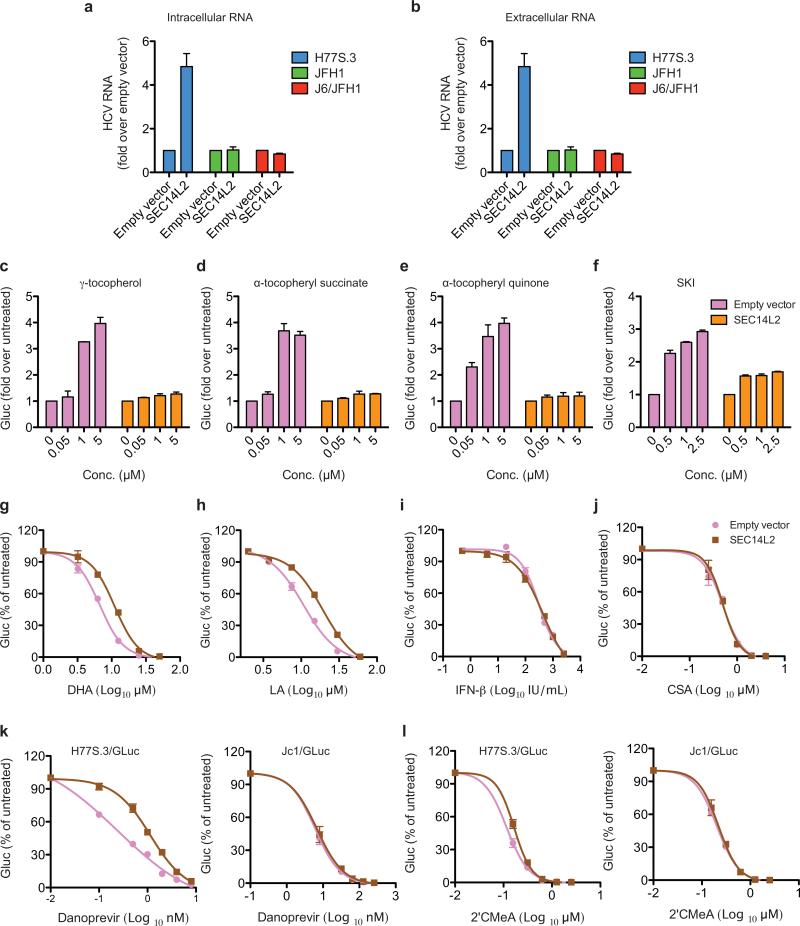
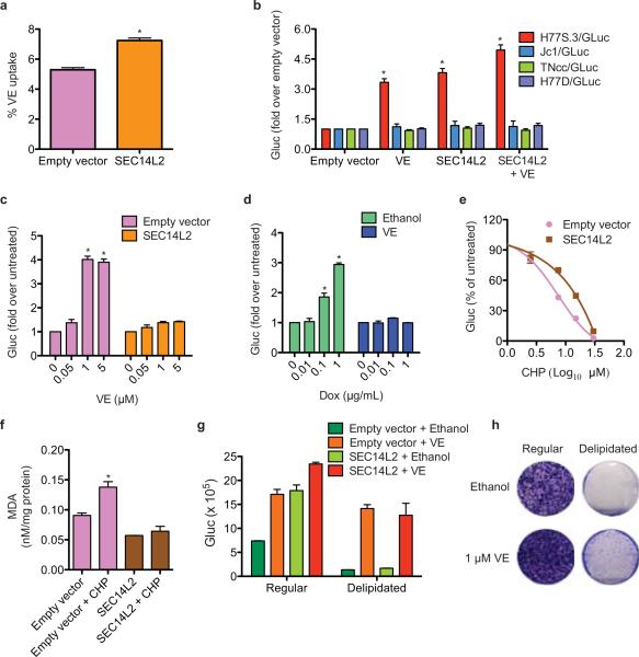
Since its discovery in 1989, efforts to grow clinical isolates of the hepatitis C virus (HCV) in cell culture have met with limited success. Only the JFH-1 isolate has the capacity to replicate efficiently in cultured hepatoma cells without cell culture-adaptive mutations. We hypothesized that cultured cells lack one or more factors required for the replication of clinical isolates. To identify the missing factors, we transduced Huh-7.5 human hepatoma cells with a pooled lentivirus-based human complementary DNA (cDNA) library, transfected the cells with HCV subgenomic replicons lacking adaptive mutations, and selected for stable replicon colonies. This led to the identification of a single cDNA, SEC14L2, that enabled RNA replication of diverse HCV genotypes in several hepatoma cell lines. This effect was dose-dependent, and required the continuous presence of SEC14L2. Full-length HCV genomes also replicated and produced low levels of infectious virus. Remarkably, SEC14L2-expressing Huh-7.5 cells also supported HCV replication following inoculation with patient sera. Mechanistic studies suggest that SEC14L2 promotes HCV infection by enhancing vitamin E-mediated protection against lipid peroxidation. This provides a foundation for development of in vitro replication systems for all HCV isolates, creating a useful platform to dissect the mechanisms by which cell culture-adaptive mutations act.


Figures

Extended Data Figure 1
Extended Data Figure 1. Cured replicon cell clones supported replication of wild-type HCV
Six of the 45 cell colonies obtained from cDNA screen and one control cell clone, S52 (adapted) that carried a cell culture-adapted HCV subgenomic replicon S52/SG-neo(AII), were treated for 15 days with a combination of an HCV NS5A inhibitor (daclatasvir, 1 nM) and an NS5B polymerase inhibitor (2’CMeA, 1 μM) to eliminate the replicating viral genomes. These cells were then transfected with the indicated wild-type HCV replicons and selected with G418 for 3 weeks. The resulting cell colonies were stained with crystal violet. Replication defective replicon, S52 GNN, with a catalytically inactive mutation in the NS5B polymerase served as a negative control.
Extended Data Figure 2
Extended Data Figure 2. The ectopic expression of SEC14L2 confers HCV permissiveness to human hepatoma cells
(a,b) SEC14L2 is not expressed in most cell lines. (a) SEC14L2 mRNA levels were measured in the indicated cells by qPCR, and the values were normalized to those of the housekeeping gene, RPS11. Shown is the fold difference from fetal hepatocytes. The results are plotted as mean±SEM of 2 different cell stocks. (b) SEC14L2 protein expression in the indicated cells was determined by immunoblotting with SEC14L2 rabbit polyclonal antibody. ß-actin is included as a loading control. (c) SEC14L2-expressing Huh-7.5 cells were transfected with the indicated wild-type subgenomic replicons. After 4 weeks of selection, HCV RNA was sequenced in 6 individual colonies from each of the replicons. HCV RNA levels in each of these colonies were determined by qRT-PCR. (d) Hep3B/miR-122 and Huh-7 cells transduced with SEC14L2 or empty vector were transfected with in vitro transcribed RNA from the indicated wild-type replicons and selected for 3 weeks with G418 (500 μg/ml). The resulting cell colonies were stained with crystal violet. S52 GNN with a catalytically inactive mutation in NS5B polymerase was included as a negative control.
Extended Data Figure 3
Extended Data Figure 3. Effect of SEC14L2 expression on the replication of wild-type and cell culture-adapted HCV subgenomic replicons
(a,b) SEC14L2 expression enhances replication of wild-type HCV RNA in a dose-dependent manner. (a) Huh-7.5 cells were transduced with lentiviruses encoding SEC14L2-EGFP fusion protein under a doxycycline-inducible promoter and flow cytometry was performed to obtain single cell clones. Two cell clones selected for downstream analysis were treated with the indicated concentrations of doxycycline for 24 h, followed by flow cytometry to determine the number of EGFP positive cells. Mean fluorescence intensities (MFI) of EGFP are shown at the top of each box. (b) S52/SG-neo colony formation in cells described in a. The results were confirmed by 2 independent transfections. (c) SEC14L2 expression enhances replication of cell culture-adapted HCV replicons to varying extents. Colony formation efficiency of the indicated subgenomic replicons in empty vector- and SEC14L2-expressing Huh-7.5 cells is plotted as CFU/100,000 transfected cells. Results represent mean±SEM from 2 independent transfections.
Extended Data Figure 4
Extended Data Figure 4. SEC14L2 expression enables replication of wild-type full-length HCV genomes and production of low levels of infectious virus particles
(a) HDFR reporter cells transduced with SEC14L2 or empty vector were transfected with in vitro transcribed RNA from H77, Con1, and J6 full-length genomes. Live cell images were captured 6 days after transfection. White arrows indicate the cells with nuclear RFP. (b) The numbers of cells exhibiting nuclear RFP were counted in 3 random microscopic fields at day 6 post-transfection. H77 pol-, with a catalytically inactive mutation in NS5B polymerase, was used as a negative control. (c,d,e) Infectious virus particles are produced from SEC14L2/Huh-7.5 cells harboring selectable full-length HCV genomes. (c) Huh-7.5 cells stably expressing SEC14L2 were incubated at 37°C for 30 min with anti-CD81 antibody or control IgG, and inoculated with the culture medium from SEC14L2/Huh-7.5 cells harboring blasticidin-selectable, full-length (FL-BSD) H77, Con1, and J6 genomes, described in Fig. 3b. After 72 h, selection with blasticidin (2.5 μg/ml) was imposed and the colonies obtained after 3 weeks were stained with crystal violet. (d) The cell colonies obtained in c were pooled and stained with anti-NS5A antibody (9E10 clone). The percentages of positive cells from 2 independent experiments are plotted. As previously described, 9E10 antibody did not detect NS5A from the J6 isolate. (e) The cell colonies obtained in c were pooled and HCV RNA levels were measured by qPCR. The values are plotted as mean±SEM of 2 independent experiments.
Extended Data Figure 5
Extended Data Figure 5. Only full-length SEC14L2 supports replication of wild-type HCV
(a) Huh-7.5 cells stably expressing human or murine SEC14L2 (93% identity) were lysed and protein expression was confirmed by immunoblotting. These cells were then transfected with S52/SG-neo and selected with G418. The resulting cell colonies were stained with crystal violet. (b,c) Only isoform 1 of SEC14L2 supports HCV RNA replication. The SEC14L2 gene is comprised of 12 exons and results in 3 alternatively spliced transcript variants encoding 3 different protein isoforms. Isoform 1 was identified in cDNA screening. (b) Schematic representation of SEC14L2 isoforms. Coding exons are shown as green blocks and the amino acid length of each protein is shown on right. (c) Cell lysates from Huh-7.5 cells stably expressing 3 SEC14L2 isoforms were analyzed by 4-12% SDS-PAGE and immunoblotting was performed with SEC14L2 mouse monoclonal antibody. The bands highlighted with asterisks might be a cleavage product of SEC14L2. UC, untransduced cells. These cells were transfected with S52/SG-neo and selected with G418 for 3 weeks. The resulting cell colonies were stained with crystal violet. (d) SEC14L3 and SEC14L4 (86% and 80% amino acid similarity to SEC14L2, respectively) are not expressed in Huh-7.5 cells. Cell lysates from Huh-7.5 cells stably expressing SEC14L2, SEC14L3, or SEC14L4 were analyzed by 4-12% SDS-PAGE followed by immunoblotting with the indicated antibodies (The signal generated by SEC14L3 antibody in SEC14L2-expressing cells most likely reflects the cross-reactivity of SEC14L3 antibody). These cells were then tested for their ability to support replication of S52/SG-neo. (e,f) Deletion mutants of SEC14L2 do not support HCV RNA replication. (e) Schematic representation of N-terminal EGFP-tagged SEC14L2 deletion mutants. (f) Cell lysates from Huh-7.5 cells stably expressing the full-length SEC14L2 and the deletion mutants were analyzed by 4-12% SDS-PAGE followed by immunoblotting with the indicated antibodies. These cells were then transfected with S52/SG-neo and selected with G418 for 3 weeks. The resulting cell colonies were stained with crystal violet. (g,h) Since N-terminal deletion mutants of SEC14L2 were unstable in Huh-7.5 cells (they formed protein aggregates), we generated chimeric constructs by fusing C-terminal ends of SEC14L2 with the corresponding N-terminal sequences from SEC14L4 and tested their ability to support HCV replication. (g) Schematic representation of C-terminal EGFP-tagged chimeric constructs. (h) Huh-7.5 cells stably expressing the indicated chimeric constructs were lysed and protein expression was confirmed by immunoblotting with anti-GFP antibody. The cells were then transfected with S52/SG-neo and selected with G418 for 3 weeks.
Extended Data Figure 6
Extended Data Figure 6. SEC14L2 does not interact with HCV non-structural proteins under the tested conditions
(a) Yeast two-hybrid assay was performed to examine direct interaction between SEC14L2 and HCV non-structural proteins. SEC14L2-AD (GAL4 activation domain) fusion or control AD vector was co-expressed with DBD (DNA binding domain) fusion of the individual HCV non-structural proteins or control DBD vector and tested for positive yeast two-hybrid interactions under selective nutritional conditions (lacking leucine, tryptophan, histidine, and adenosine). The strong signal obtained for NS5A most likely reflects the intrinsic trans-activating activity of NS5A. (b) Co-immunoprecipitation did not reveal binding of SEC14L2 with HCV NS5A protein. Since two-hybrid assay (panel a) did not yield unambiguous results on interaction between SEC14L2 and NS5A, we employed co-immunoprecipitation assay to probe binding between these proteins. Huh-7.5 cells stably expressing SEC14L2-EGFP and harboring wild-type Con1/SG-neo replicon were lysed and subjected to immunoprecipitation with anti-NS5A antibody, anti-EGFP antibody or control IgGs. The bound proteins were analyzed by immunoblotting. Endogenous MOBKL1B, a previously described binding partner of the NS5A protein, served as a positive control. (c) Subcellular fractionation of SEC14L2-EGFP/Huh-7.5 cells harboring wild-type Con1/SG-neo replicon did not show significant co-fractionation between SEC14L2 and HCV NS5A protein.
Extended Data Figure 7
Extended Data Figure 7. SEC14L2 does not facilitate HCV RNA replication by modulating PI3K/Akt or cholesterol synthesis pathways
(a,b,c,d) Down-regulation of PI3K/Akt pathway does not support HCV replication. (a) Akt phosphorylation in control and SEC14L2-expressing Huh-7.5 cells was analyzed by immunoblotting; ß-actin is included as a loading control. (b) Stable Knockdown of Akt in Huh-7.5 cells with 2 different shRNAs did not facilitate G418 resistant colony formation by S52/SG-neo. (c) Huh-7.5 cells were transduced to stably express PTEN, a negative regulator of PI3K pathway. Despite decreased Akt phosphorylation, these cells did not support colony formation by S52/SG-neo. (d) Suppression of PI3K pathway in Huh-7.5 cells by stable expression of a dominant negative Akt, a dominant negative p85 subunit of PI3K, or a constitutively active FOXO3a did not render them permissive to HCV replication. Dominant negative Akt gets phosphorylated, but since it is kinase-dead, it cannot initiate the downstream signaling. Interestingly, increased Akt phosphorylation was seen in Huh-7.5 cells expressing constitutively active FOXO3a, suggesting a potential feed back mechanism. (e,f) A SEC14L2 mutant (S289A) lacking the cholesterolgenic activity supported HCV replication. (e) Schematic representation of carboxy-terminal EGFP-tagged SEC14L2 point mutants. S288A was generated as a negative control. (f) The colony formation efficiency of S52/SG-neo (plotted as CFU/100,000 transfected cells) and the expression levels of SEC14L2 mutants in Huh-7.5 are shown.
Extended Data Figure 8
Extended Data Figure 8. SEC14L2 expression masks the effects of lipophilic oxidants and anti-oxidants on H77S.3/GLuc replication
(a,b) SEC14L2 enhances transient replication of H77S.3, but not that of JFH-1 or J6/JFH1. Empty vector- and SEC14L2-expressing Huh-7.5 cells were electroporated with the indicated viral RNAs lacking GLuc insertions. (a) Intracellular and (b) extracellular RNA levels were measured 6 days after electroporation. Results are plotted as fold change from empty vector control. (c,d,e,f) The pro-viral effects of various lipophilic antioxidants on H77S.3/GLuc replication were suppressed in the presence of SEC14L2. (c) γ-tocopherol, (d) α-tocopheryl succinate, (e) α-tocopheryl quinone, and (f) sphingosine kinase inhibitor (SKI) were added to Huh-7.5 cells 20 h before transfection with H77S.3/GLuc. Transfections were carried out in the fresh medium lacking these compounds. 6 h after transfection, cells were again fed with each compound and the GLuc expression was measured at 72 h post-transfection. The results are presented as fold change from untreated cells. (g,h,i,j,k,l) SEC14L2 expression suppressed the inhibitory effect of lipophilic oxidants and direct-acting antivirals, but not that of the HCV host factor inhibitors, on H77S.3/GLuc replication. (g) Docosahexaenoic acid (oxidant), (h) Linoleic acid (oxidant), (i) IFN-ß, (j) CSA (cyclosporine A), (k) Danoprevir (NS3 protease inhibitor), and (l) 2’CMeA (NS5B polymerase inhibitor) were added to Huh-7.5 cells 6 h after transfection with H77S.3/GLuc (and Jc1/GLuc in case of Danoprevir and 2’CMeA) and the secreted GLuc activity at 72 h post-transfection was measured. The results are plotted as % inhibition relative to the untreated cells. All results in this figure represent mean±SD of 2 replicate experiments.
Extended Data Figure 9
Extended Data Figure 9. SEC14L2 expression does not enhance replication of lipid peroxidation-resistant RNA viruses
(a-f) Flaviviruses, such as Yellow fever virus (YFV) and dengue virus (DENV) do not respond to SEC14L2 expression. (a,b,d,e) Empty vector- and SEC14L2-expressing Huh-7.5 cells were infected with (a,b) YFV-venus or (d,e) DENV-GFP at a multiplicity of infection (MOI) of 0.01 and 0.1, respectively. The cells were harvested at the indicated times post infection followed by FACS analysis. (a,d) The number of yellow (YFV-venus) and green (DENV-GFP) cells are plotted as % positive cells and (b,e) the mean fluorescence intensities are presented as % relative to empty vector. (c,f) Empty vector- and SEC14L2-expressing Huh-7.5 cells were inoculated with (c) YFV (17D) or (f) DENV (serotype 2 strain 16681) at an MOI of 0.5 and 0.1, respectively. The culture medium was collected at the indicated times post infection and the infectious virus titers were determined by a plaque formation assay on Huh-7.5 cells (YFV-venus) or BHK cells (DENV). The results are presented as plaque-forming units (PFU)/ml. (g-j) Alphaviruses, such as Sindbis virus (SINV) and Ross River virus (RRV) are insensitive to SEC14L2 expression. Empty vector- and SEC14L2-expressing Huh-7.5 cells were infected with (g,h) SINV-GFP or (I,j) RRV-GFP (T48 strain) at an MOI of 0.05. The cells were harvested at the indicated times post infection and FACS analysis was carried out to determine the number of green cells and the mean fluorescence intensities. All results in this figure represent mean±SEM of 2 replicate experiments.
Extended Data Figure 10
Extended Data Figure 10. VE increased SEC14L2-mediated replicon colony formation
(a) VE (α-tocopherol: 1 μM) was added to Huh-7.5 cells 20 h before transfection with the indicated wild-type HCV subgenomic replicons. Transfections were carried out for 6 h in the fresh medium lacking VE, followed by medium change to VE-containing medium. After 48 h, cells were subjected to G418 selection and fed with fresh G418 and VE every 2 days. The resulting cell colonies were stained with crystal violet. Shown are the results of one of the 3 independent experiments. (b) Colony formation efficiency of the indicated subgenomic replicons was measured in SEC14L2- expressing cells in the absence or presence of 1 μM VE. The results are plotted as mean±SEM of CFU/100,000 transfected cells from 3 independent transfections. ** P < 0.005 by two-tailed, paired t-test.
Figure 1
Figure 1. cDNA screening of Huh-7.5 cells identifies SEC14L2 as a critical host factor for HCV RNA replication
(a) Illustration of the shRNA or cDNA screen. (b) Three Huh-7.5 cell populations independently transduced with a pooled lentiviral cDNA library and a population of empty vector-expressing control cells was electroporated with the indicated wild-type HCV replicons or a replication-defective S52 GNN replicon. After 3 weeks of G418 selection, the resulting cell colonies were stained with crystal violet. (c) HCV NS3-NS5B region from 45 cell colonies was amplified by RT-PCR and subjected to direct sequencing. Shown are the % colonies harboring wild-type, mutant, or a mixture of wild-type and mutant sequences. (d) The SEC14L2 cDNA was amplified from 45 colonies (see methods). CR, constant region; L, ladder; UC, untransduced cells; EV, empty vector-transduced cells; NTC, no template control.
Figure 2
Figure 2. SEC14L2 allows replication of wild-type HCV subgenomic replicons
(a) Cell colonies obtained for the indicated replicons in Huh-7.5 cells transduced with empty vector or SEC14L2. (b) HCV RNA levels for wild-type replicons in SEC14L2-expressing cells (black bars) and two cell culture-adapted replicons in control cells (grey bars) are shown. (c) Schematic representation of the doxycycline-inducible SEC14L2-EGFP expression plasmid (top panel). Dose-dependent effect of doxycycline on SEC14L2-EGFP expression (middle panel) and S52/SG-neo replication (bottom panel) is shown in two single cell clones. CFU, colony forming units/100,000 transfected cells. (d) The colonies obtained for 1 μg/ml doxycycline in c were pooled and passaged in G418-free medium in the presence or absence of doxycycline. After 2 weeks, SEC14L2 protein was undetectable in cells passaged without doxycycline (top panel). These cells died when subjected to G418 selection (bottom panel). Bar graphs show means±SD from at least duplicate experiments.
Figure 3
Figure 3. SEC14L2 allows replication of full-length HCV genomes
(a) HCV RNA levels 6 days after electroporation of the indicated full-length HCV genomes in HDFR reporter cells transduced with empty vector or SEC14L2. DMSO or daclatasvir (10 nM) was added 3 days prior to cell harvest. (b) Cell colonies obtained for the indicated blasticidin-selectable, full-length (FL-BSD) HCV genomes in Huh-7.5 cells transduced or not to express SEC14L2. (c) The cell colonies obtained in b were pooled and HCV RNA and protein abundance was determined by qRT-PCR (top panel) and immunoblotting with anti-NS3 antibody (bottom panel). (d) Direct infection of SEC14L2-expressing HDFR reporter cells with 6 serum samples from HCV patients. The top panel shows HCV RNA levels. The bottom panel shows the nuclear localization of RFP in individual cells infected with serum 2 (genotype 1b) and serum 6 (genotype 3a). White arrows indicate cells with nuclear RFP. Bar graphs show means±SD from at least duplicate experiments.
Figure 4
Figure 4. SEC14L2 promotes HCV replication by enhancing the antioxidant effect of VE
(a) Effect of SEC14L2 expression on VE accumulation in Huh-7.5 cells. Results represent the mean±SD from 8 adjacent wells treated with radioactively labeled VE for 4 h. (b) Effect of VE (1 μM) and SEC14L2 on replication of the indicated HCV genomes. (c) Dose-response effects of VE on replication of H77S.3/GLuc in empty vector- and SEC14L2-expressing Huh-7.5 cells. (d) Dose-response effects of SEC14L2 expression on H77S.3/GLuc replication in SEC14L2-EGFP Cl. 1 cells (described in Fig. 2c) in presence or absence of VE. (e) Dose-response effect of cumene hydroperoxide (CHP), a lipophilic oxidant, on replication of H77S.3/GLuc in empty vector- and SEC14L2-expressing Huh-7.5 cells. (f) Intracellular abundance of malondialdehyde (MDA), an end product of lipid peroxidation, in empty vector and SEC14L2-expressing Huh-7.5 cells incubated for 16 h with 15 μM CHP. (g) H77S.3/GLuc replication and (h) S52/SG-neo colony formation in empty vector and SEC14L2-expressing Huh-7.5 cells grown in regular or delipidated medium. Since delipidated serum is suboptimal for hepatoma cell proliferation, the colonies obtained were small and slow growing. Results represent means±SD from 2 (d,f) or 3 (b,c,e,g) biological replicates. * P < 0.01 by one-tailed, Mann-Whitney test (a,b,c,d) or one-tailed, paired t-test (f).

Comment in

Similar articles

Cited by

References

    1. Blight KJ, Kolykhalov AA, Rice CM. Efficient initiation of HCV RNA replication in cell culture. Science. 2000;290:1972–1974. - PubMed
    1. Saeed M, et al. Efficient replication of genotype 3a and 4a hepatitis C virus replicons in human hepatoma cells. Antimicrobial agents and chemotherapy. 2012;56:5365–5373. doi:10.1128/AAC.01256-12. - PMC - PubMed
    1. Saeed M, et al. Replication of hepatitis C virus genotype 3a in cultured cells. Gastroenterology. 2013;144:56–58. e57. doi:10.1053/j.gastro.2012.09.017. - PubMed
    1. Mohd Hanafiah K, Groeger J, Flaxman AD, Wiersma ST. Global epidemiology of hepatitis C virus infection: new estimates of age-specific antibody to HCV seroprevalence. Hepatology. 2013;57:1333–1342. doi:10.1002/hep.26141. - PubMed
    1. Gottwein JM, et al. Novel infectious cDNA clones of hepatitis C virus genotype 3a (strain S52) and 4a (strain ED43): genetic analyses and in vivo pathogenesis studies. Journal of virology. 2010;84:5277–5293. doi:10.1128/JVI.02667-09. - PMC - PubMed

Publication types

MeSH terms