. 2015 Aug 12;10(8):e0135499.
doi: 10.1371/journal.pone.0135499. eCollection 2015.

Alzheimer's Disease-Related Protein Expression in the Retina of Octodon degus

Affiliations

Alzheimer's Disease-Related Protein Expression in the Retina of Octodon degus

Lucia Y Du et al. PLoS One. .

Abstract

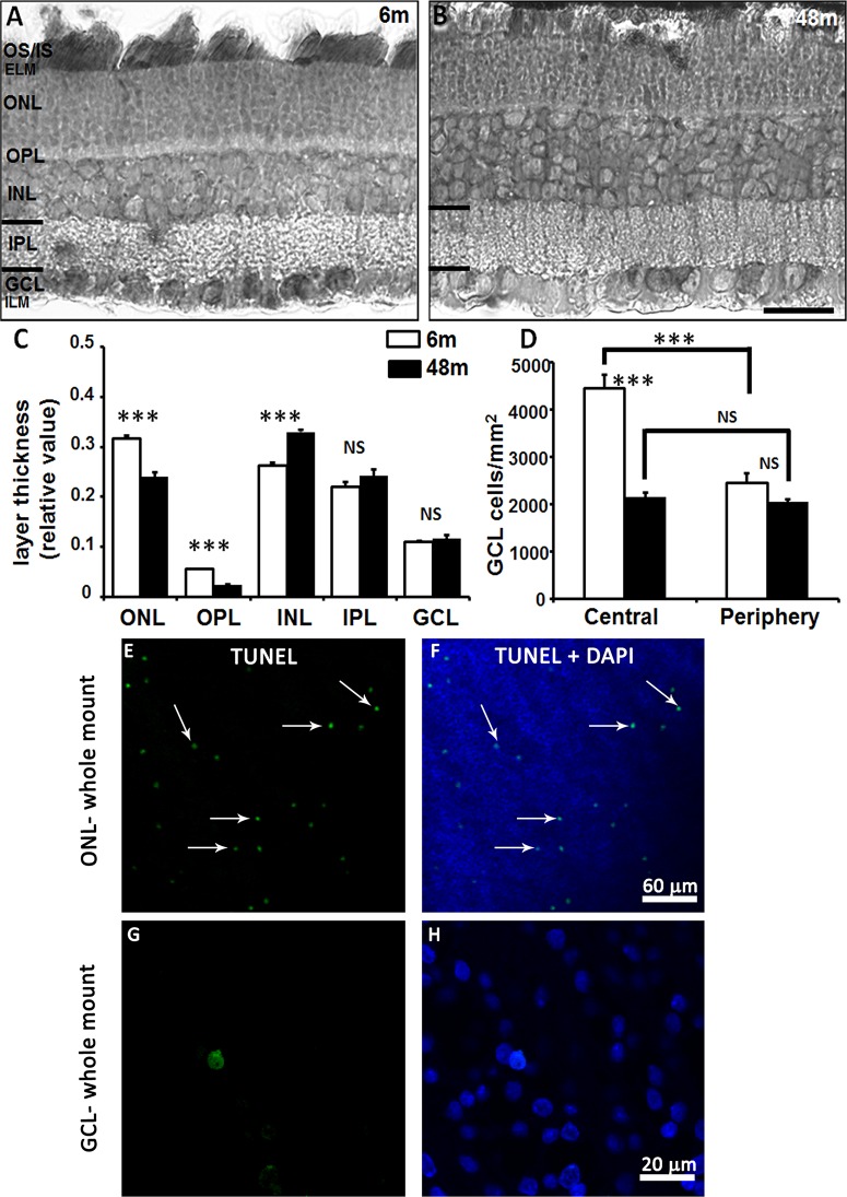
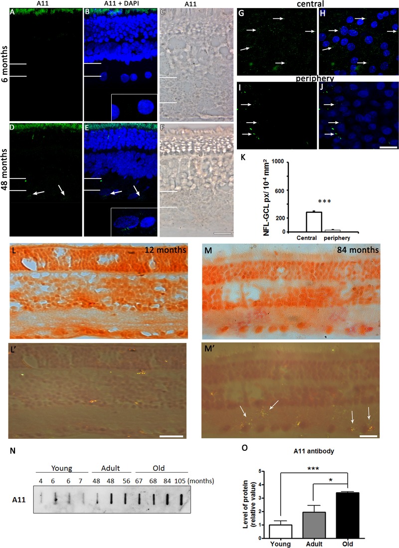
New studies show that the retina also undergoes pathological changes during the development of Alzheimer's disease (AD). While transgenic mouse models used in these previous studies have offered insight into this phenomenon, they do not model human sporadic AD, which is the most common form. Recently, the Octodon degus has been established as a sporadic model of AD. Degus display age-related cognitive impairment associated with Aβ aggregates and phosphorylated tau in the brain. Our aim for this study was to examine the expression of AD-related proteins in young, adult and old degus retina using enzyme-linked or fluorescence immunohistochemistry and to quantify the expression using slot blot and western blot assays. Aβ4G8 and Aβ6E10 detected Aβ peptides in some of the young animals but the expression was higher in the adults. Aβ peptides were observed in the inner and outer segment of the photoreceptors, the nerve fiber layer (NFL) and ganglion cell layer (GCL). Expression was higher in the central retinal region than in the retinal periphery. Using an anti-oligomer antibody we detected Aβ oligomer expression in the young, adult and old retina. Immunohistochemical labeling showed small discrete labeling of oligomers in the GCL that did not resemble plaques. Congo red staining did not result in green birefringence in any of the animals analyzed except for one old (84 months) animal. We also investigated expression of tau and phosphorylated tau. Expression was seen at all ages studied and in adults it was more consistently observed in the NFL-GCL. Hyperphosphorylated tau detected with AT8 antibody was significantly higher in the adult retina and it was localized to the GCL. We confirm for the first time that Aβ peptides and phosphorylated tau are expressed in the retina of degus. This is consistent with the proposal that AD biomarkers are present in the eye.


Conflict of interest statement

Competing Interests: The authors have declared that no competing interests exist.

Figures

Fig 1
Fig 1. Light and confocal fluorescence micrographs illustrating the morphology of the retina in young and adult degus.
Retinal sections from young and adult degus were stained with Toluidine blue (A, B). Quantification of retinal layer thickness (C) and ganglion cell density in young and adult degus (D). TUNEL labeling of the adult retina showed apoptotic cells in the ONL (E, F) but not in the GCL (G, H) in the retinal whole mount. Abbreviations: 6 months old (6m), 48 months old (48m), outer and inner segment of the photoreceptors (OS/IS), external limiting membrane (ELM), outer nuclear layer (ONL), outer plexiform layer (OPL), inner nuclear layer (INL), inner plexiform layer (IPL), ganglion cell layer (GCL), inner limiting membrane (ILM). Scale bar = 20 μm.
Fig 2
Fig 2. APP and Aβ peptide expression.
Fluorescent immunolabeling of APP in 6 months old (A, B) and 48 months old (C, D) retinal sections. DAPI (blue) stains nucleus. GS labeling in adult retina (E). Aβ peptide expression in young (F, G) and adult (H, I) animals. The insert shows a magnification of the NFL-GCL area. Thin arrows indicate labeling in inner retina. Large arrows specify labeling in the GCL area. White lines on the left hand side of the image indicate the extent of the IPL. The number of pixels occupied by the antibody in the NFL-GCL is shown (J). Slot blot assay showing the level of Aβ protein detection with Aβ6E10 for young and adult degus (K). APP protein level was assayed using western blot (L). APP expression as a function of age was quantified in (M). Abbreviations: glutamine synthetase (GS), nerve fiber layer- ganglion cell layer (NFL-GCL), pixels (px). Scale bar = 20 μm.
Fig 3
Fig 3. Expression of Aβ peptides.
Fluorescent and enzyme-linked immunolabeling show Aβ4G8 labeling of Aβ peptides in 6 months old (A-C) and 48 months-old (D-F) retinal sections. DAPI (blue) stains nucleus. White lines on the left hand side of the image indicate the extent of the IPL. Aβ peptides were also labeled using fluorescence immunohistochemistry in the adult central (I, J) and peripheral (K, L) GCL. Arrows indicate the location of Aβ deposits. Negative control immunolabeling without the antibody is (G, H). The number of pixels occupied by the Aβ4G8 antibody in the central and peripheral NFL-GCL is shown (M). Slot blot assay for detection of Aβ peptides as a function of age (N) was quantified in (O). Abbreviations: nerve fiber layer- ganglion cell layer (NFL-GCL), pixels (px). Scale bar = 20 μm.
Fig 4
Fig 4. Expression of Aβ oligomers.
Fluorescent and enzyme-linked immunolabeling show the absence of Aβ oligomers in 6 months old (A-C) and faint labeling in the adult, 48 months old (D-F) retinal sections. Aβ oligomers were also detected in 48 months old central (G, H) and peripheral (I, J) GCL. Arrows indicate the location of Aβ oligomers. The insert shows a magnification of the NFL-GCL. White lines on the left hand side of the image indicate the extent of the IPL. The number of pixels occupied by the A11 antibody in the central and peripheral NFL-GCL is shown (K). Congo red staining of a 12 month retina (L) and polarized light microscopy (L’) did not show presence of amyloid fibrils. Congo red was seen only in an 84 month old retina (M, M’). A11 detection of proteins in a slot blot as a function of development (N) shows the age-related expression of the peptide (O). Scale bar = 20 μm.
Fig 5
Fig 5. Tau and PHF-tau protein expression.
Fluorescent immunolabeling shows tau expression in 6 months old (A, B) and 48 months old (C, D) retinal sections. DAPI (blue) stains nucleus. PHF-tau proteins labeled with AT8 in young (E, F) and adult (G, H) retinal sections. The insert shows a magnification of the NFL-GCL. Large arrows indicate positive labeling in the GCL area and thin arrows highlight the labeling pattern along the vertical axis, which is reminiscent of Müller cells. Thin white lines on the left hand side of the image indicate the extent of the IPL. Whole mount labeling using Tau5A6 (I, J) and AT8 in the adult central GCL (K, L). Arrows indicate the location of tau and phosphorylated tau respectively. The number of pixels occupied by the Tau5A6 and AT8 antibodies in the central NFL-GCL is shown (M). Western blot of phosphorylated tau and PHF levels in young and adult retinae using Ser235 and AT8 antibodies (N). Quantification of the bands shows an age-related increase in expression of phosphorylated tau and PHF detected with Ser235 and AT8 (O). Anti-actin was used as loading control. Scale bar = 20 μm

References

    1. Braidy N, Munoz P, Palacios AG, Castellano-Gonzalez G, Inestrosa NC, Chung RS, et al. (2012) Recent rodent models for Alzheimer's disease: clinical implications and basic research. J Neural Transm 119: 173–195. 10.1007/s00702-011-0731-5 - DOI - PubMed
    1. Danesh-Meyer HV, Birch H, Ku JY, Carroll S, Gamble G (2006) Reduction of optic nerve fibers in patients with Alzheimer disease identified by laser imaging. Neurology 67: 1852–1854. - PubMed
    1. Paquet C, Boissonnot M, Roger F, Dighiero P, Gil R, Hugon J (2007) Abnormal retinal thickness in patients with mild cognitive impairment and Alzheimer's disease. Neurosci Lett 420: 97–99. - PubMed
    1. Chang LY, Lowe J, Ardiles A, Lim J, Grey AC, Robertson K, et al. (2014) Alzheimer's disease in the human eye. Clinical tests that identify ocular and visual information processing deficit as biomarkers. Alzheimers Dement 10: 251–261. 10.1016/j.jalz.2013.06.004 - DOI - PubMed
    1. Koronyo-Hamaoui M, Koronyo Y, Ljubimov AV, Miller CA, Ko MK, Black KL, et al. (2011) Identification of amyloid plaques in retinas from Alzheimer's patients and noninvasive in vivo optical imaging of retinal plaques in a mouse model. Neuroimage 54 Suppl 1: S204–217. 10.1016/j.neuroimage.2010.06.020 - DOI - PMC - PubMed

Publication types