Comparative Study
. 2016 Feb 15;524(3):433-47.
doi: 10.1002/cne.23879. Epub 2015 Sep 8.

Evolutionary origin of Tbr2-expressing precursor cells and the subventricular zone in the developing cortex

Affiliations
Comparative Study

Evolutionary origin of Tbr2-expressing precursor cells and the subventricular zone in the developing cortex

Verónica Martínez-Cerdeño et al. J Comp Neurol. .

Abstract

The subventricular zone (SVZ) is greatly expanded in primates with gyrencephalic cortices and is thought to be absent from vertebrates with three-layered, lissencephalic cortices, such as the turtle. Recent work in rodents has shown that Tbr2-expressing neural precursor cells in the SVZ produce excitatory neurons for each cortical layer in the neocortex. Many excitatory neurons are generated through a two-step process in which Pax6-expressing radial glial cells divide in the VZ to produce Tbr2-expressing intermediate progenitor cells, which divide in the SVZ to produce cortical neurons. We investigated the evolutionary origin of SVZ neural precursor cells in the prenatal cerebral cortex by testing for the presence and distribution of Tbr2-expressing cells in the prenatal cortex of reptilian and avian species. We found that mitotic Tbr2(+) cells are present in the prenatal cortex of lizard, turtle, chicken, and dove. Furthermore, Tbr2(+) cells are organized into a distinct SVZ in the dorsal ventricular ridge (DVR) of turtle forebrain and in the cortices of chicken and dove. Our results are consistent with the concept that Tbr2(+) neural precursor cells were present in the common ancestor of mammals and reptiles. Our data also suggest that the organizing principle guiding the assembly of Tbr2(+) cells into an anatomically distinct SVZ, both developmentally and evolutionarily, may be shared across vertebrates. Finally, our results indicate that Tbr2 expression can be used to test for the presence of a distinct SVZ and to define the boundaries of the SVZ in developing cortices.

Keywords: Pax6; Tbr2; cortical neurons; neural precursor cell types; subventricular zone.


Figures

Figure 1
Figure 1
Tbr2-expressing cells may have been present in the developing forebrain of the common ancestor for mammals and reptiles. Cladogram showing the relationship between mammals, turtles, birds and lizards. Based on recent genetic analysis (Crawford et al., 2014), turtles and birds have been placed in the recently proposed clade archelosauria, while lizards and other reptiles, such as snakes, are in the superorder lepidosauria. Tbr2-expressing cells are found in the developing forebrain of each vertebrate we examined: mammals, turtles, birds and lizards. Mammals and birds possess a distinct SVZ based on the distribution of Tbr2+ cells (red asterisks). Tbr2+ cells are present in both the dorsal cortex and dorsal ventricular ridge (DVR) of developing turtles but only the DVR shows evidence of a distinct SVZ-like structure (red/black asterisk). Tbr2+ cells are also present in the developing lizard forebrain, but there is no evidence of abventricular divisions or an anatomically defined SVZ (black asterisk). The presence of Tbr2+ cells in mammals and both reptilian clades, archelosauria and lepidosauria, supports the concept that the common ancestor for mammals and reptiles possessed Tbr2+ cells and possibly a distinct SVZ.
Figure 2
Figure 2
Nissl-stained sections of the embryonic turtle forebrain from Stage 16 through Stage 27 (Yntema, 1968), showing development of the dorsal cortex and dorsal ventricular ridge (DVR). The DVR becomes prominent after S19. Inset boxes at each stage are shown at higher magnification for the dorsal cortex (A1-D1) and for the DVR (A2-D2). The proliferative ventricular zone (VZ) is thicker in the DVR than in the dorsal cortex. The VZ becomes thinner in the dorsal cortex after S20, but remains relatively thicker in the DVR for a longer span of development. Scale bar in A = 500 μm, and applies to A-D; Scale bar in panel A1 = 50 μm, and applies to panels A1-D1, and A2-D2.
Figure 3
Figure 3
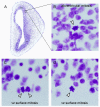
Nissl-stained sections of S20 turtle forebrain (A) showing that mitotic cells undergo division at the surface of the ventricle (arrowheads, C, D), and away from the ventricle (arrowhead, B) as in mammals.
Figure 4
Figure 4
Nissl-stained sections from embryonic lizard forebrain showing development of the dorsal cortex and dorsal ventricular ridge (DVR) over Carnegie stages CS3, CS5, CS9 and CS11. Through CS9 the proliferative ventricular zone (VZ) remains at a relative constant thickness, but becomes noticeably thinner by CS11. (A1-D1 and A2-D2) Inset boxes show higher magnification images of lizard VZ cells. Many mitotic cells were observed undergoing division at the surface of the ventricle (arrowheads), but we did not observe any cells undergoing division away from the ventricle at any stage of development in the dorsal cortex or DVR of the lizard forebrain.
Figure 5
Figure 5
Mitotic cells undergoing division at the surface of the ventricle in the lizard forebrain express the mitotic cell marker phosphorylated vimentin (4A4). The 4A4+ mitotic cells possess a pial process that courses toward the pial surface of the developing brain, and in mammals.
Figure 6
Figure 6
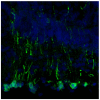
Ventricular zone (VZ) cells in the dorsal cortex of the turtle forebrain express the transcription factor Pax6 (red), as do mammalian VZ cells. Proliferative zone cells that express the intermediate progenitor cell marker Tbr2 (green) are also present in the VZ. A magenta-green version of this figure can be viewed online as Supplementary Figure 6.
Figure 7
Figure 7
A) Tbr2-expressing cells (red) in the ventricular zone (VZ) of the turtle dorsal cortex also express the mitotic cell marker PCNA (green), indicating that they are in the mitotic cell cycle (white arrowheads). B) The Tbr2+ cells are scattered throughout the VZ of turtle dorsal cortex. C) Higher magnification images from the inset in panel A showing that some Tbr2+ cells (white arrowheads) in the dorsal cortex also express the mitotic cell marker PCNA. A magenta-green version of this figure can be viewed online as Supplementary Figure 7.
Figure 8
Figure 8
A) Tbr2-expressing cells (red) in the dorsal ventricular ridge (DVR) of the developing turtle forebrain form a subventricular zone (SVZ) and express the mitotic cell marker PCNA (green). B) Inset from panel A showing a higher magnification image of Tbr2+ cells assembling into a distinct SVZ-like structure in the DVR. A PCNA+ / Tbr2+ cell is indicated with the white arrowhead and shown at higher magnification in panel C. C) Some Tbr2+ cells in the DVR and the dorsal cortex (Cx) also express PCNA, indicating that they are mitotic. A magenta-green version of this figure can be viewed online as Supplementary Figure 8.
Figure 9
Figure 9
Tbr2-expressing cells are present in the developing forebrain of the lizard Anolis carolinensis. The Tbr2+ cells (red/magenta) are located in the thin ventricular zone (VZ) of both the dorsal cortex and dorsal ventricular ridge (DVR), but are not organized into a subventricular band superficial to the VZ. White arrows indicate the lateral ventricle. Dorsal (D) and medial (M) orientation indicated. Blue, DAPI.
Figure 10
Figure 10
Tbr2-expressing cells in two avian species, chick and dove, are organized into a subventricular zone (SVZ) structure. (A, B) Tbr2+ cells (red) are located superficial to Pax6-expressing cells (green) in the ventricular zone (VZ) of the chick hyperpallium (HP) and mesopallium (Meso). Mitotic Pax6-expressing cells were also observed superficial to the SVZ (white arrow, inset in panel B), as described for translocating cells in the mammalian neocortex. (C) Tbr2+ cells (red) are concentrated in a tight band that is located superficial to the VZ in the developing forebrain of the dove Zenaida macroura. The distribution of Tbr2-expressing cells in the developing avian forebrain supports the concept that these species possess a distinct SVZ. A magenta-green version of this figure can be viewed online as Supplementary Figure 10.

References

    1. Angevine JB, Bodian D, Coulombre AJ, Edds MV, Hamburger V, Jacobson M, Lyser KM, Prestige MC, Sidman RL, Varon S, Weiss PA. Embryonic vertebrate central nervous system: revised terminology. Anatomical Record. 1970;166:257–261. - PubMed
    1. Avila A, Vidal PM, Tielens S, Morelli G, Laguesse S, Harvey RJ, Rigo JM, Nguyen L. Glycine receptors control the generation of projection neurons in the developing cerebral cortex. Cell death and differentiation. 2014;21:1696–1708. - PMC - PubMed
    1. Bayer SA, Altman J. Neocortical Development. Raven Press; New York: 1991.
    1. Betizeau M, Cortay V, Patti D, Pfister S, Gautier E, Bellemin-Menard A, Afanassieff M, Huissoud C, Douglas RJ, Kennedy H, Dehay C. Precursor diversity and complexity of lineage relationships in the outer subventricular zone of the primate. Neuron. 2013;80:442–457. - PubMed
    1. Blanton MG, Shen JM, Kriegstein AR. Evidence for the inhibitory neurotransmitter gamma-aminobutyric acid in aspiny and sparsely spiny nonpyramidal neurons of the turtle dorsal cortex. Journal of Comparative Neurology. 1987;259:277–297. - PubMed

Publication types

MeSH terms