Review
. 2015 Sep;16(9):1071-83.
doi: 10.15252/embr.201540891. Epub 2015 Aug 12.

Expanding the ubiquitin code through post-translational modification

Affiliations
Review

Expanding the ubiquitin code through post-translational modification

Lina Herhaus et al. EMBO Rep. 2015 Sep.

Abstract

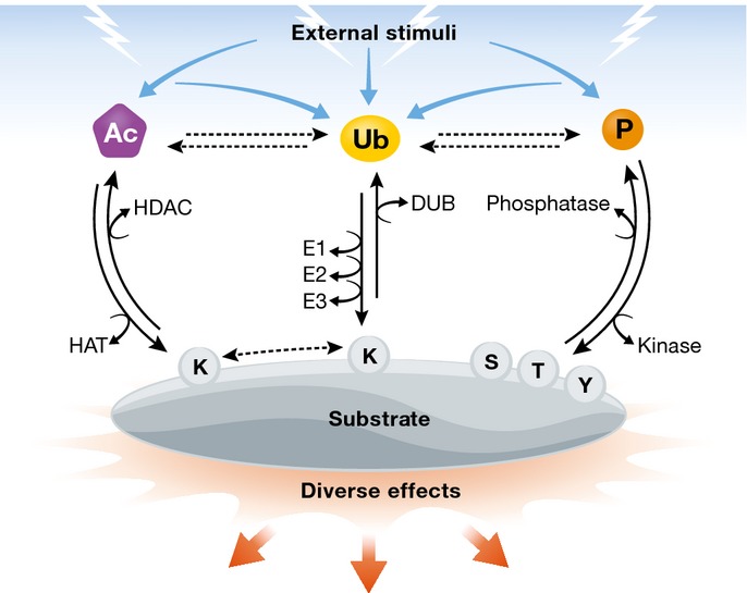
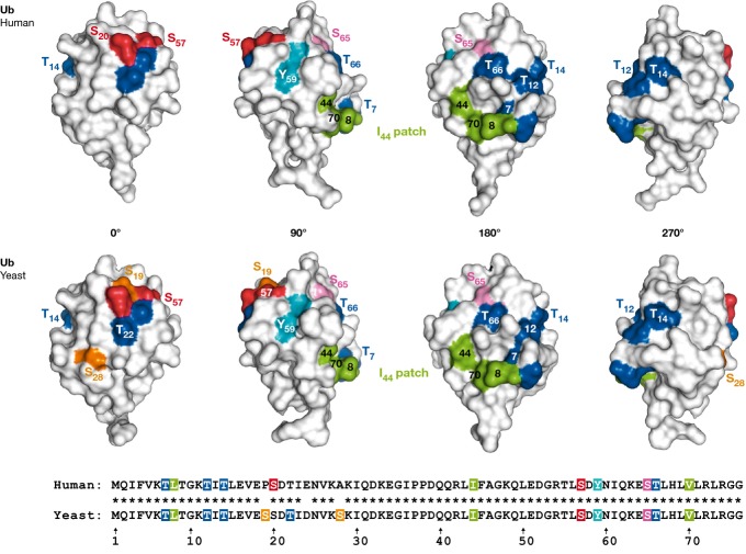
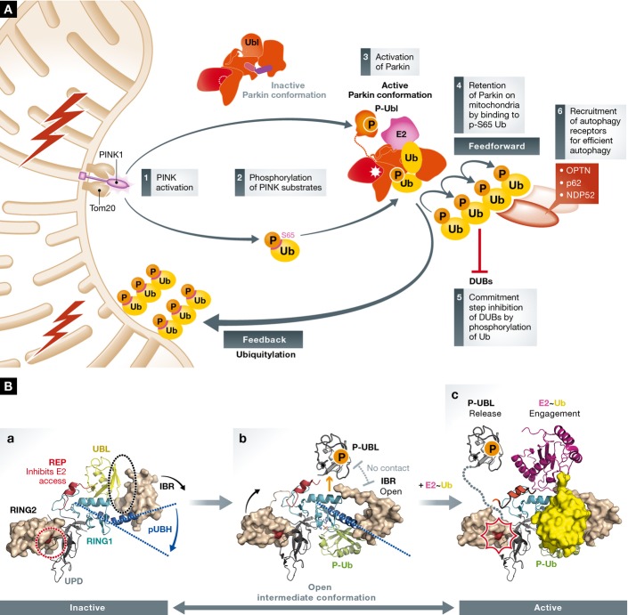
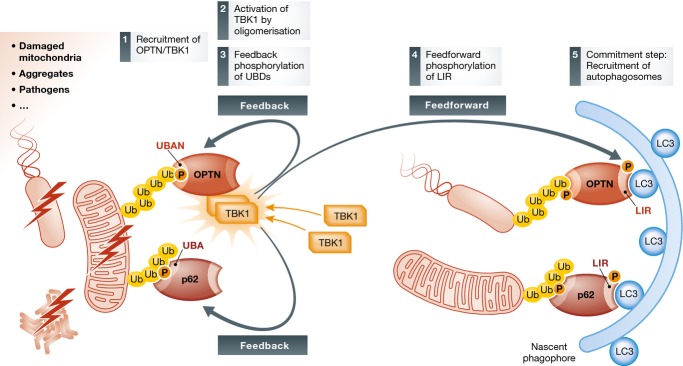
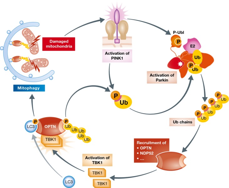
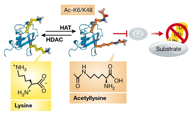
Ubiquitylation is among the most prevalent post-translational modifications (PTMs) and regulates numerous cellular functions. Interestingly, ubiquitin (Ub) can be itself modified by other PTMs, including acetylation and phosphorylation. Acetylation of Ub on K6 and K48 represses the formation and elongation of Ub chains. Phosphorylation of Ub happens on multiple sites, S57 and S65 being the most frequently modified in yeast and mammalian cells, respectively. In mammals, the PINK1 kinase activates ubiquitin ligase Parkin by phosphorylating S65 of Ub and of the Parkin Ubl domain, which in turn promotes the amplification of autophagy signals necessary for the removal of damaged mitochondria. Similarly, TBK1 phosphorylates the autophagy receptors OPTN and p62 to initiate feedback and feedforward programs for Ub-dependent removal of protein aggregates, mitochondria and pathogens (such as Salmonella and Mycobacterium tuberculosis). The impact of PINK1-mediated phosphorylation of Ub and TBK1-dependent phosphorylation of autophagy receptors (OPTN and p62) has been recently linked to the development of Parkinson's disease and amyotrophic lateral sclerosis, respectively. Hence, the post-translational modification of Ub and its receptors can efficiently expand the Ub code and modulate its functions in health and disease.

Keywords: mitophagy; phosphorylation; post‐translational modification; ubiquitin.


Figures

Figure 1
Figure 1. Generation and control of PTMs
External stimuli result in the generation of different PTMs on target proteins. Phosphorylation, acetylation and ubiquitylation can influence each other and are attached to and removed from substrate proteins by different enzymes. Phosphorylation occurs mainly on serine, threonine and tyrosine residues, whereas acetylation and ubiquitylation target lysine residues. The attachment or removal of PTMs determines substrate fate.
Figure 2
Figure 2. Structure of wild-type human and yeast ubiquitin
The phosphorylation sites of human (T7, T12, T14, S20, S57, Y59, S65 and T66) and yeast (T7, T12, T14, S19, T22, S28, S57, Y59, S65 and T66) Ub are indicated, as well as the residues lining the ubiquitin hydrophobic patch (L8, I44, V70).
Figure 3
Figure 3. PINK1-mediated phosphorylation of ubiquitin and Parkin
(A) Upon mitochondrial damage, PINK1 is recruited and activated at the mitochondrial surface resulting in the sequential phosphorylation of both Ub and the Parkin Ubl domain on their respective Ser65 sites. This leads to Parkin activation and ubiquitylation of multiple substrates on the mitochondrial outer membrane (MOM), which in turn become favourable substrates for PINK1. In such a way, PINK1 and Parkin generate high-density p-S65-Ub chains on MOM proteins. This increases the binding and retention time of Parkin on mitochondria, leading to the amplification of Ub signals. Phosphorylation of Ub chains also serves as a commitment step, as p-Ub chains are resistant to deubiquitylation by the majority of DUBs. The multiple ubiquitylation signals then commit mitochondria for degradation by attracting multiple autophagy receptors, such as OPTN and NDP52. (B) Conformational domain rearrangements for Parkin activation. (a) Inactive PARKIN: Closed auto-inhibited structure of full-length PARKIN (PDB: 4k95). The catalytic cysteine 431 of the RING2 domain (in red) is blocked by the UPD. The I44 patch of the Ubl domain interacts with the RING1 helix and the IBR interacts with Ubl, thereby covering the S65 phosphorylation site of the Ubl domain. Additionally, the C-terminal Ubl and REP block the E2 access to the RING1 domain. (b) Dynamic intermediate Parkin (PDB: 5caw; 143–461 a.a.) in complex with p-Ub. The interaction between p-Ub and Parkin straightens the kinked pUBH helix, opens the IBR and releases the Ubl and its phosphorylation site from the Parkin RBR core. (c) Model of the active form of Parkin. The RING domain of a Cbl-UBCH7 crystal structure (PDB: 1fbv; magenta) conjugated to ubiquitin (PDB: 4q5e; yellow) is superposed to PARKIN RING1 (PDB: 5caw). The Ubl and REP domains are released, making the RING1 domain accessible to the E2∼Ub. The interaction of Parkin with the E2∼Ub could induce conformational changes in that active site of the RING2 domain and fully activate Parkin.
Figure 4
Figure 4. TBK1 in control of ubiquitin-dependent autophagy
TBK1 mediates feedback and feedforward regulation of the two autophagic adaptor proteins p62 and OPTN. Phosphorylation on their UBDs promotes binding to ubiquitylated cargo, whereas phosphorylation of the LIR motifs promotes the recruitment of autophagosomal membranes. Thus, TBK1 amplifies cargo recognition and commits it to degradation.
Figure 5
Figure 5. Integrative model for PINK1/Parkin- and TBK1/OPTN-dependent mitophagy
Mitochondrial damage leads to the activation of the kinase PINK1, which phosphorylates Ub and Parkin on S65. The phosphorylation-dependent activation of Parkin results in the generation of phosphorylated polyUb chains on MOM proteins. Autophagic receptors such as OPTN are recruited to the site of mitochondrial damage, together with the kinase TBK1, which becomes activated through dimerization and autophosphorylation. Active TBK1 phosphorylates the Ub-binding regions within OPTN. The p-Ub MOM substrates are subsequently bound by autophagic receptors, which link damaged mitochondria to autophagosomal membranes through binding to LC3s thereby resulting in mitophagy.
None
None

References

    1. Deribe YL, Pawson T, Dikic I. Post-translational modifications in signal integration. Nat Struct Mol Biol. 2010;17:666–672. - PubMed
    1. Varshavsky A. The early history of the ubiquitin field. Protein Sci. 2006;15:647–654. - PMC - PubMed
    1. Hochstrasser M. Origin and function of ubiquitin-like proteins. Nature. 2009;458:422–429. - PMC - PubMed
    1. Hershko A, Ciechanover A. The ubiquitin system. Annu Rev Biochem. 1998;67:425–479. - PubMed
    1. Schulman BA. Twists and turns in ubiquitin-like protein conjugation cascades. Protein Sci. 2011;20:1941–1954. - PMC - PubMed

Publication types

MeSH terms