. 2015 Nov;138(Pt 11):3386-99.
doi: 10.1093/brain/awv235. Epub 2015 Aug 11.

Iatrogenic CJD due to pituitary-derived growth hormone with genetically determined incubation times of up to 40 years

Affiliations

Iatrogenic CJD due to pituitary-derived growth hormone with genetically determined incubation times of up to 40 years

Peter Rudge et al. Brain. 2015 Nov.

Abstract

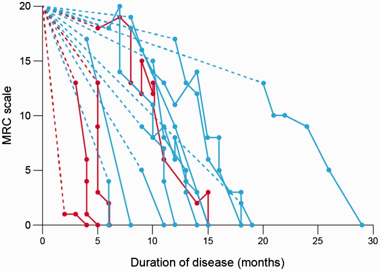
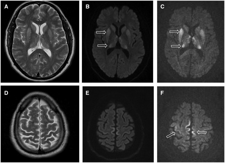
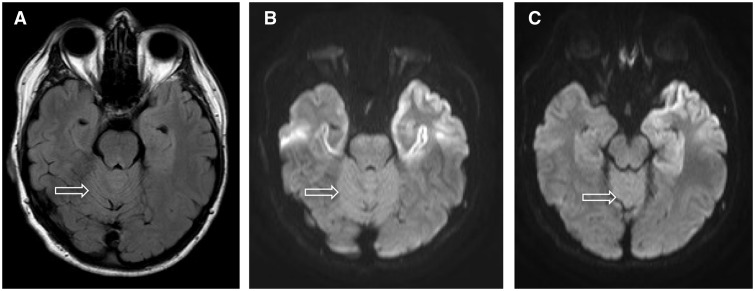
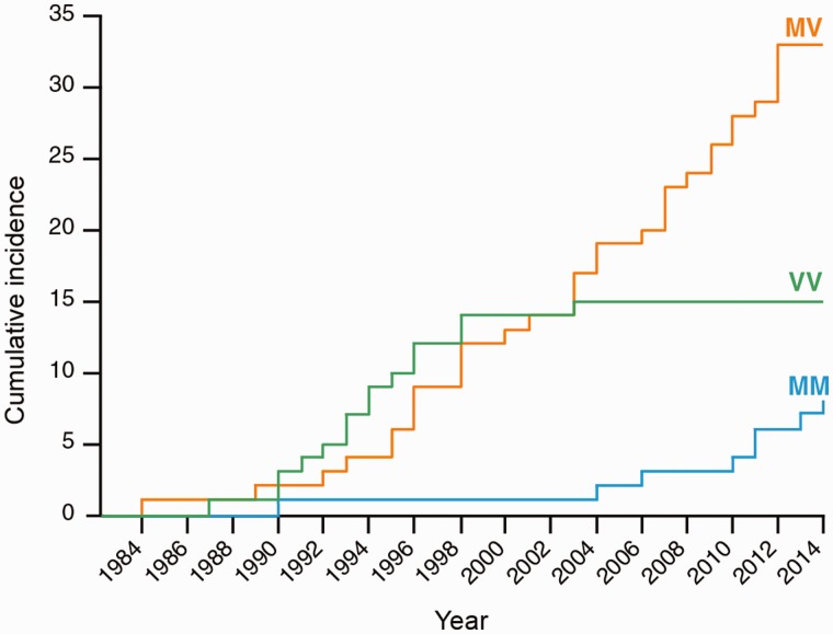
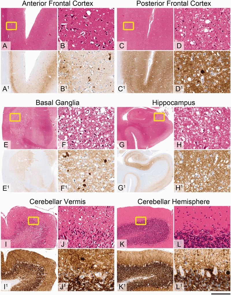
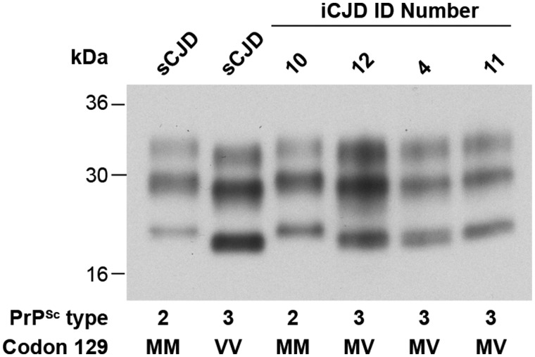
Patients with iatrogenic Creutzfeldt-Jakob disease due to administration of cadaver-sourced growth hormone during childhood are still being seen in the UK 30 years after cessation of this treatment. Of the 77 patients who have developed iatrogenic Creutzfeldt-Jakob disease, 56 have been genotyped. There has been a marked change in genotype profile at polymorphic codon 129 of the prion protein gene (PRNP) from predominantly valine homozygous to a mixed picture of methionine homozygous and methionine-valine heterozygous over time. The incubation period of iatrogenic Creutzfeldt-Jakob disease is significantly different between all three genotypes. This experience is a striking contrast with that in France and the USA, which may relate to contamination of different growth hormone batches with different strains of human prions. We describe the clinical, imaging, molecular and autopsy features in 22 of 24 patients who have developed iatrogenic Creutzfeldt-Jakob disease in the UK since 2003. Mean age at onset of symptoms was 42.7 years. Gait ataxia and lower limb dysaesthesiae were the most frequent presenting symptoms. All had cerebellar signs, and the majority had myoclonus and lower limb pyramidal signs, with relatively preserved cognitive function, when first seen. There was a progressive decline in neurological and cognitive function leading to death after 5-32 (mean 14) months. Despite incubation periods approaching 40 years, the clinical duration in methionine homozygote patients appeared to be shorter than that seen in heterozygote patients. MRI showed restricted diffusion in the basal ganglia, thalamus, hippocampus, frontal and the paracentral motor cortex and cerebellar vermis. The electroencephalogram was abnormal in 15 patients and cerebrospinal fluid 14-3-3 protein was positive in half the patients. Neuropathological examination was conducted in nine patients. All but one showed synaptic prion deposition with numerous kuru type plaques in the basal ganglia, anterior frontal and parietal cortex, thalamus, basal ganglia and cerebellum. The patient with the shortest clinical duration had an atypical synaptic deposition of abnormal prion protein and no kuru plaques. Taken together, these data provide a remarkable example of the interplay between the strain of the pathogen and host prion protein genotype. Based on extensive modelling of human prion transmission barriers in transgenic mice expressing human prion protein on a mouse prion protein null background, the temporal distribution of codon 129 genotypes within the cohort of patients with iatrogenic Creutzfeldt-Jakob disease in the UK suggests that there was a point source of infecting prion contamination of growth hormone derived from a patient with Creutzfeldt-Jakob disease expressing prion protein valine 129.

Keywords: PRNP; RRP9; growth hormone; iatrogenic Creutzfeldt-Jakob disease; prion disease.


Figures

Figure 1
Figure 1
Clinical progression assessed on the MRC Prion Disease Rating Scale. Broken line connects time of first symptom to the first assessment on the MRC scale assuming patient would have scored 20 before disease onset. Continuous lines join consecutive assessments indicated by dots. Last assessment is the point at which the patient first scored 0 on the rating scale; death occurred shortly after this in all cases. Horizontal axis = time in months from onset of disease; vertical axis = MRC Prion Rating Scale score; red lines = codon 129 MM patients; blue = codon 129 MV patients.
Figure 2
Figure 2
MRI findings in a 46-year-old male patient with iatrogenic CJD due to growth hormone. Basal ganglia and thalamic hyperintensity is seen on T2-weighted images (A), DWI at b = 1000s/mm2 (B) and (C) DWI at b = 3000s/mm2. Cortical hyperintensity in the paracentral lobule bilaterally and right precentral gyrus is not visualized on T2-weighted images (D) but is seen on DWI b = 1000s/mm2 (E) and (F) is more conspicuous at DWI at b = 3000s/mm2 (Patient 12).
Figure 3
Figure 3
MRI findings in a 47-year-old male patient with iatrogenic CJD due to growth hormone. Hyperintensity in the superior cerebellar vermis is seen on axial FLAIR images (A), and is more conspicuous at DWI at b = 1000s/mm2 at the same level (B). (C) A more superior axial image at DWI at b = 1000s/mm2 shows the increased vermian signal abnormality compared to the surrounding occipital cortex (Patient 8).
Figure 4
Figure 4
Cumulative incidence of patients stratified by PRNP codon 129 polymorphism. There is a highly significant difference in the distribution of the three polymorphisms at codon 129 over time and incubation period (ANOVA, P < 10−7), with each pairwise comparison of genotypes being statistically significant in post hoc analyses (P < 0.001) as indicated in the text.
Figure 5
Figure 5
Neuropathological findings in a representative iatrogenic CJD case. Sections (A–L) stained with haematoxylin and eosin. Sections (A1L1) immunostained for abnormal prion protein deposition with anti-PrP monoclonal antibody ICSM35. Throughout the grey matter there is variably prominent microvacuolar degeneration and abnormal prion protein deposition in a diffuse synaptic manner and as micro-plaques. In the anterior frontal cortex (A and B, haematoxylin and eosin) there is mild patchy microvacuolar degeneration and abnormal prion protein deposits (A1 and B1, ICSM35) are restricted to the deep cortical layers. In the posterior frontal lobe, including motor cortex (C and D, haematoxylin and eosin) there is more prominent micro-vacuolation and dense pan-cortical deposition of the abnormal prion protein (C1 and D1, ICSM35). In the basal ganglia (E, haematoxylin and eosin; E1 ICSM35) there is very severe vacuolation and diffuse deposition of abnormal prion protein in the neuropil of the putamen (F, haematoxylin and eosin; F1 ICSM35) and caudate nucleus. In the hippocampus (G, haematoxylin and eosin) there is particularly prominent micro-vacuolar degeneration in the Sommer’s sector, subiculum and entorhinal cortex (H, haematoxylin and eosin) with corresponding deposits of abnormal prion protein (G1 and H1, ICSM35). In the cerebellum there is very severe cortical atrophy in vermis with prominent microvacuolation in the molecular layer, loss of Purkinje cells, granular cells and accompanying Bergmann gliosis (I, haematoxylin and eosin). The cortex in the cerebellar hemisphere is much better preserved with mild degree of microvacuolar degeneration in the molecular layer (J, haematoxylin and eosin). In both, cerebellar vermis and hemisphere there are scattered micro-plaques and diffuse, dense synaptic labelling for abnormal prion protein. Scale bars = 4 mm in A, A1, C, C1, E, E1, I, I1, K and K1; 8 mm in G and G1; 200 µm in B, B1, D, D1, F, F1, H, H1, J, J1, L and L1.
Figure 6
Figure 6
PrP immunoblot of patient brain. Proteinase K (PK)-digested 10 % (w/v) brain homogenates from patients with sporadic CJD or iatrogenic CJD were analysed by enhanced chemiluminescence using anti-PrP monoclonal antibody 3F4. The provenance of each brain sample is designated above each lane and the type of PrPSc (London classification; Hill et al., 2003) detected in each sample and the PRNP codon 129 genotype of the patient (M, methionine; V, valine) is designated below. Control samples of human PrPSc types 2 and 3 from patients with sporadic CJD (lanes 1 and 2) are compared with the PrPSc molecular strain types seen in Patients 4, 10,11 and 12 with iatrogenic CJD (lanes 3–6).

References

    1. Asante EA, Linehan J, Desbruslais M, Joiner S, Gowland I, Wood A, et al. BSE prions propagate as either variant CJD-like or sporadic CJD-like prion strains in transgenic mice expressing human prion protein. EMBO J 2002; 21: 6358–66. - PMC - PubMed
    1. Beringue V, Le Dur A, Tixador P, Reine F, Lepourry L, Perret-Liaudet A, et al. Prominent and persistent extraneural infection in human PrP transgenic mice infected with variant CJD. PLoS One 2008; 3: e1419. - PMC - PubMed
    1. Bishop MT, Will RG, Manson JC. Defining sporadic Creutzfeldt-Jakob disease strains and their transmission properties. Proc Natl Acad Sci USA 2010; 107: 12005–10. - PMC - PubMed
    1. Brandel JP, Preece M, Brown P, Croes E, Laplanche JL, Agid Y, et al. Distribution of codon 129 genotype in human growth hormone-treated CJD patients in France and the UK. Lancet 2003; 362: 128–30. - PubMed
    1. Brandner S. Diversity of prion diseases: (no) strains attached? Acta Neuropathol 2011; 121: 1–4. - PubMed

Publication types

MeSH terms