Long-term Surgical Outcomes of Epiretinal Membrane in Patients with Retinitis Pigmentosa
- PMID: 26268934
- PMCID: PMC4535036
- DOI: 10.1038/srep13078
Long-term Surgical Outcomes of Epiretinal Membrane in Patients with Retinitis Pigmentosa
Abstract
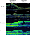
Macular complications such as an epiretinal membrane (ERM), a cystoid macular edema and a macular hole lead to unexpected central vision impairment especially for patients with retinitis pigmentosa (RP). To evaluate the long-term surgical outcomes of pars plana vitrectomy (PPV) for ERM in patients with RP, we retrospectively reviewed the charts of a consecutive series of 10 RP patients who underwent PPV for ERM at Kyushu University Hospital. Visual acuity (VA) testing, a fundus examination, and an optical coherence tomography (OCT) analysis were conducted. The standard PPV using three sclerotomies was performed for ERM. PPV was performed in 12 eyes of 10 patients. One eye was excluded from the outcome assessment due to short period observation (18 months). There was no significantly deleterious change from the baseline to final VA between the operation eyes and the fellow eyes (P = 0.19). Moreover, morphological improvement was obtained in 9 of 11 eyes based on OCT. Our present data suggest that PPV may be tolerable in the management for ERM in RP patients over the long-term. Furthermore, the appearance of the ellipsoid zone was an important factor in the prediction of visual outcome and determination of surgical indication.
Figures


References
-
- Wong P. Apoptosis, retinitis pigmentosa, and degeneration. Biochem Cell Biol 72, 489–98 (1994). - PubMed
-
- Hartong D. T., Berson E. L. & Dryja T. P. Retinitis pigmentosa. Lancet 368, 1795–809 (2006). - PubMed
-
- Berson E. L. Retinitis pigmentosa. The Friedenwald Lecture. Invest Ophthalmol Vis Sci 34, 1659–76 (1993). - PubMed
-
- You Q. S. et al. Prevalence of retinitis pigmentosa in North China: the Beijing Eye Public Health Care Project. Acta Ophthalmol 91, e499–500 (2013). - PubMed
-
- Hirakawa H., Iijima H., Gohdo T. & Tsukahara S. Optical coherence tomography of cystoid macular edema associated with retinitis pigmentosa. Am J Ophthalmol 128, 185–91 (1999). - PubMed
Publication types
MeSH terms
LinkOut - more resources
Full Text Sources
Other Literature Sources
Medical
Miscellaneous

