. 2015 Oct;89(20):10637-47.
doi: 10.1128/JVI.01868-15. Epub 2015 Aug 12.

MicroRNA-Attenuated Clone of Virulent Semliki Forest Virus Overcomes Antiviral Type I Interferon in Resistant Mouse CT-2A Glioma

Affiliations

MicroRNA-Attenuated Clone of Virulent Semliki Forest Virus Overcomes Antiviral Type I Interferon in Resistant Mouse CT-2A Glioma

Miika Martikainen et al. J Virol. 2015 Oct.

Abstract

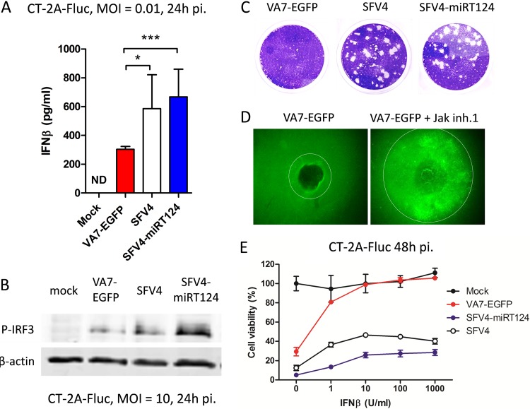
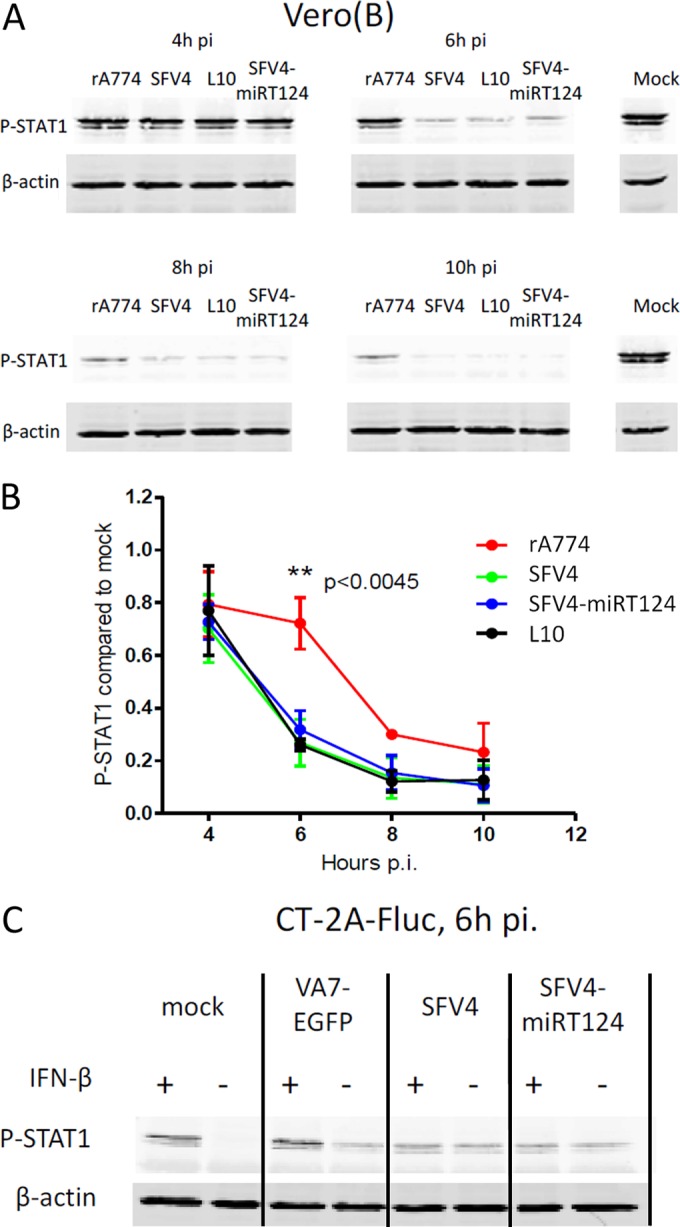
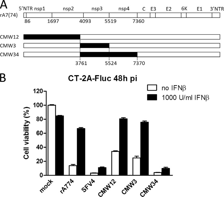
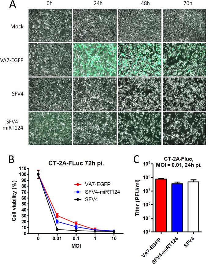
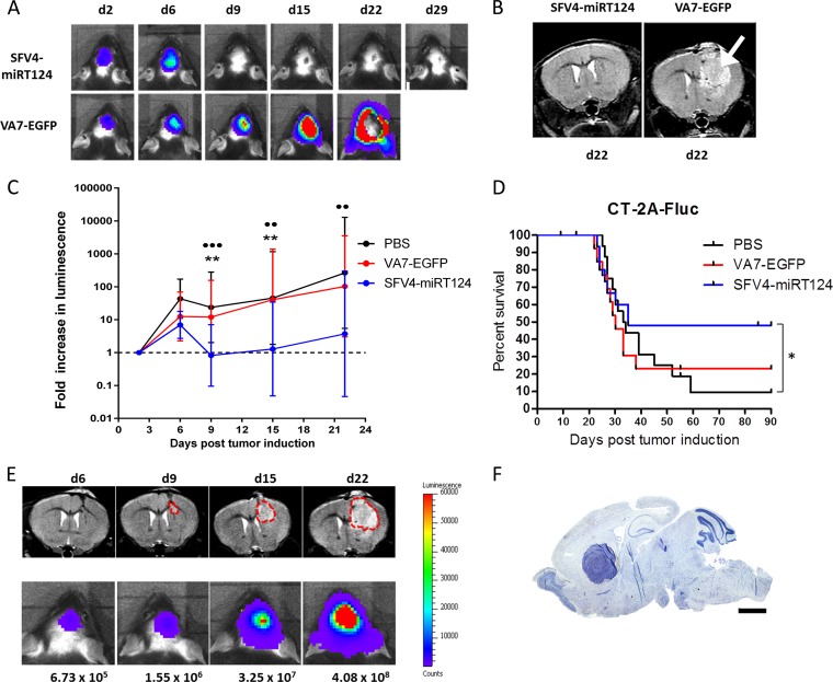
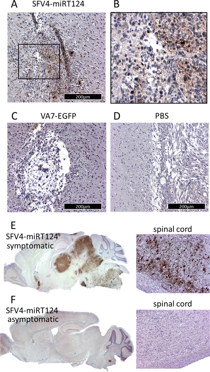
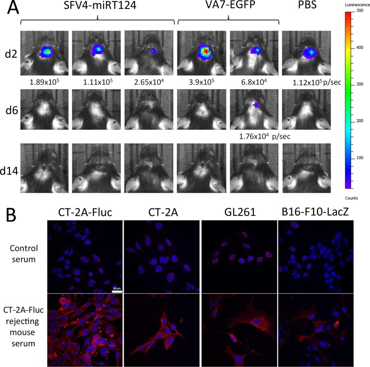
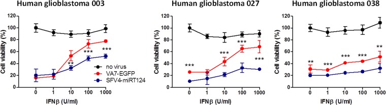
Glioblastoma is a terminal disease with no effective treatment currently available. Among the new therapy candidates are oncolytic viruses capable of selectively replicating in cancer cells, causing tumor lysis and inducing adaptive immune responses against the tumor. However, tumor antiviral responses, primarily mediated by type I interferon (IFN-I), remain a key problem that severely restricts viral replication and oncolysis. We show here that the Semliki Forest virus (SFV) strain SFV4, which causes lethal encephalitis in mice, is able to infect and replicate independent of the IFN-I defense in mouse glioblastoma cells and cell lines originating from primary human glioblastoma patient samples. The ability to tolerate IFN-I was retained in SFV4-miRT124 cells, a derivative cell line of strain SFV4 with a restricted capacity to replicate in neurons due to insertion of target sites for neuronal microRNA 124. The IFN-I tolerance was associated with the viral nsp3-nsp4 gene region and distinct from the genetic loci responsible for SFV neurovirulence. In contrast to the naturally attenuated strain SFV A7(74) and its derivatives, SFV4-miRT124 displayed increased oncolytic potency in CT-2A murine astrocytoma cells and in the human glioblastoma cell lines pretreated with IFN-I. Following a single intraperitoneal injection of SFV4-miRT124 into C57BL/6 mice bearing CT-2A orthotopic gliomas, the virus homed to the brain and was amplified in the tumor, resulting in significant tumor growth inhibition and improved survival.

Importance: Although progress has been made in development of replicative oncolytic viruses, information regarding their overall therapeutic potency in a clinical setting is still lacking. This could be at least partially dependent on the IFN-I sensitivity of the viruses used. Here, we show that the conditionally replicating SFV4-miRT124 virus shares the IFN-I tolerance of the pathogenic wild-type SFV, thereby allowing efficient targeting of a glioma that is refractory to naturally attenuated therapy vector strains sensitive to IFN-I. This is the first evidence of orthotopic syngeneic mouse glioma eradication following peripheral alphavirus administration. Our findings indicate a clear benefit in harnessing the wild-type virus replicative potency in development of next-generation oncolytic alphaviruses.


Figures

FIG 1
FIG 1
SFV4-miRT124 replicates despite the presence of IFN-I. (A) IFN-β measured from cell culture medium of infected CT-2A-Fluc cells. VA7-EGFP-induced IFN-β was significantly reduced compared to that with SFV4. SFV4-miRT124 induced significantly more IFN-β than VA7-EGFP or SFV4 (P < 0.001). Data are means of three replicates ± SD. (B) Ser396-phosphorylated IRF3 detected from infected or uninfected (mock) CT-2A-Fluc cells by Western blotting. (C) Plaque assay of CT-2A-Fluc cells. Both SFV and SFV4-miRT124 formed plaques in CT-2A-Fluc cultures (under agarose cover). (D) Plaque expansion of VA7-EGFP is increased when virus is administered with Jak inhibitor 1 (inh.1). (E) SFV4 and SFV4-miRT124 potently induce a cytopathic effect in mouse IFN-β-treated CT-2A-Fluc cells. Cells were infected with an MOI of 10. Cell viability was measured in an MTT assay. Data are presented as means (from 5 replicates) ± SD.
FIG 2
FIG 2
Virulent SFV inhibits STAT1 activation in infected cells. (A) Tyr701-phosphorylated STAT1 quantified in infected Vero(B) cells after stimulation with 1,000 U/ml human IFN-β. (B) rA774 showed significantly reduced potency for inhibiting STAT1 activation. Band intensities were quantified by using ImageJ. Data presented are means of three replicate analyses ± SD. (C) SFV4 and SFV4-miRT124 but not VA7-EGFP show P-STAT1 inhibition in CT-2A-Fluc cells. Results show phosphorylated STAT1 detected from CT-2A-Fluc cells that were either treated or not with 1,000 U/ml mouse IFN-β 6 h postinfection.
FIG 3
FIG 3
SFV4 nonstructural genes required for IFN-I-resistant replication in CT-2A cells. (A) Schematic of the chimeric viruses we used. Segments marked in black were derived from neurovirulent SFV4. Numbers indicate nucleotide positions. (B) Cell viability was measured in an MTT assay 48 h after infection, with (black bars) or without (white bars) pretreatment with 1,000 units/ml IFN-β. Means ± SD from 4 parallel wells are presented.
FIG 4
FIG 4
VA7-EGFP, SFV4, and SFV4-miRT124 showed similar replication potencies in CT-2A-Fluc cells under normal culture conditions. (A) Phase-contrast and fluorescence microscopy images (overlaid) were taken before and 24, 48, and 70 h after infection (MOI, 0.01). (B) Cell viability was measured in an MTT assay 72 h postinfection with an MOI of 0.01. Means of 4 replicates ± SD are presented. (C) Infectious titers were determined 24 h postinfection from culture medium collected from 12-well plates seeded with 4 × 10⁵ CT-2A-Fluc cells. Titers were measured from a pooled sample of three wells infected in parallel for the plaque assay using Vero(B) cells. Means of 3 replicate measurements ± SD are presented.
FIG 5
FIG 5
Tumor growth and mouse survival. (A) Representative IVIS images of animals treated with SFV4-miRT124 or VA7-EGFP (1 × 10⁶ PFU i.p. at day 3). Signal disappearance can be seen between day 6 and day 9 following SFV4-miRT124 therapy. (B) Representative MRIs of SFV4-miRT124- and VA7-EGFP-treated mice at day 22. A large tumor mass was clearly detectable following VA7-EGFP therapy (arrow). (C) The tumor-emitted luminescence signal was quantified as the average radiance (photons per second per square centimeter per steradian). The increase was measured as the fold change compared to the first measurement, performed at day 2. Data are plotted as geometric means ± SD. Undetectable signals were given a value of 0.1. Statistical analysis was done using an unpaired, two-tailed t test. Comparison results for VA7-EGFP or PBS treatments versus SFV4-miRT124 treatment are marked with stars or circles, respectively: *** or •••, P < 0.001; ** or ••, P < 0.01. (D) Kaplan-Meier survival plot for the mice. Statistical analysis used Fisher's exact test. *. P < 0.05. (E) Comparison of IVIS and MRI signal development in an untreated mouse. (F) Toluidine blue staining of tumor mass in mouse displaying endpoint symptoms.
FIG 6
FIG 6
Immunohistochemical analysis of mouse brains; virus antigens in CT-2A-Fluc glioma tissue were measured from brain samples collected 5 days post-i.p. virus injection. (A) Tumor mass of SFV4-miRT124; (B) magnification of the inset marked in panel A. (C and D) Results in VA7-EGFP-treated mice (C) and PBS-treated mice (D). (E and F) Detection of virus replication in the brain and spinal cord following i.p. injection of SFV4 miRT124. Samples were collected from a mouse suffering from neurological symptoms (E) and an asymptomatic mouse (F).
FIG 7
FIG 7
Mice surviving the initial CT-2A-Fluc inoculation become resistant to tumor rechallenge. (A) Mice imaged with the IVIS system after CT-2A-Fluc rechallenge. (B) C57BL/6 mouse tumor cell-reactive antibodies detected with immunofluorescence from sera of rechallenged mice. Blue, cell nuclei (stained with DAPI); red, tumor cell-reactive antibodies.
FIG 8
FIG 8
SFV4-miRT124 induces increased CPE in human glioblastoma cell lines. Cell viability was measured in an MTT assay 48 h postinfection (MOI, 10). Human recombinant IFN-β was administered simultaneously with virus. Means of 4 replicates ± SD are presented. The statistical analysis was done with Student's t test: ***, P < 0.001; **, P < 0.01.

References

    1. Stupp R, Mason WP, van den Bent MJ, Weller M, Fisher B, Taphoorn MJB, Belanger K, Brandes AA, Marosi C, Bogdahn U, Curschmann J, Janzer RC, Ludwin SK, Gorlia T, Allgeier A, Lacombe D, Cairncross JG, Eisenhauer E, Mirimanoff RO. 2005. Radiotherapy plus concomitant and adjuvant temozolomide for glioblastoma. N Engl J Med 352:987–996. doi:10.1056/NEJMoa043330. - DOI - PubMed
    1. Chiocca EA, Rabkin SD. 2014. Oncolytic viruses and their application to cancer immunotherapy. Cancer Immunol Res 2:295–300. doi:10.1158/2326-6066.CIR-14-0015. - DOI - PMC - PubMed
    1. Markert JM, Liechty PG, Wang W, Gaston S, Braz E, Karrasch M, Nabors LB, Markiewicz M, Lakeman AD, Palmer CA, Parker JN, Whitley RJ, Gillespie GY. 2009. Phase Ib trial of mutant herpes simplex virus G207 inoculated pre- and post-tumor resection for recurrent GBM. Mol Ther 17:199–207. doi:10.1038/mt.2008.228. - DOI - PMC - PubMed
    1. Kicielinski KP, Chiocca EA, Yu JS, Gill GM, Coffey M, Markert JM. 2014. Phase 1 clinical trial of intratumoral reovirus infusion for the treatment of recurrent malignant gliomas in adults. Mol Ther 22:1056–1062. doi:10.1038/mt.2014.21. - DOI - PMC - PubMed
    1. Freeman A, Zakay-Rones Z, GomoriI J, Linetsky E, Rasooly L, Greenbaum E, Rozenman-Yair S, Panet A, Libson E, Irving C, Galun E, Siegal T. 2006. Phase I/II trial of Intravenous NDV-HUJ oncolytic virus in recurrent glioblastoma multiforme. Mol Ther 13:221–228. doi:10.1016/j.ymthe.2005.08.016. - DOI - PubMed

Publication types

MeSH terms