. 2015 Oct 20;132(16):1528-1537.
doi: 10.1161/CIRCULATIONAHA.115.016217. Epub 2015 Aug 12.

Molecular Basis of Hypokalemia-Induced Ventricular Fibrillation

Affiliations

Molecular Basis of Hypokalemia-Induced Ventricular Fibrillation

Arash Pezhouman et al. Circulation. .

Abstract

Background: Hypokalemia is known to promote ventricular arrhythmias, especially in combination with class III antiarrhythmic drugs like dofetilide. Here, we evaluated the underlying molecular mechanisms.

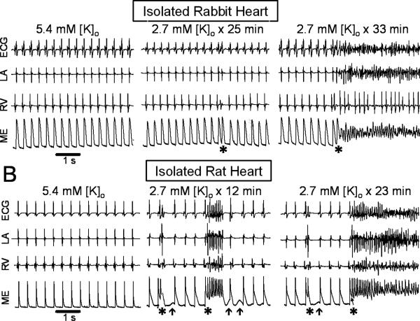
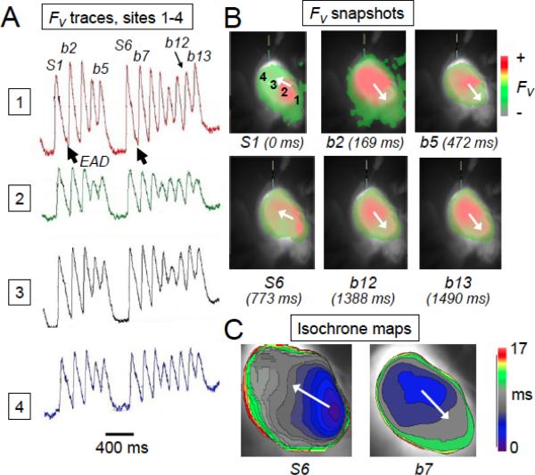
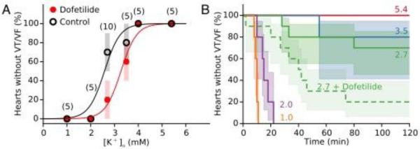
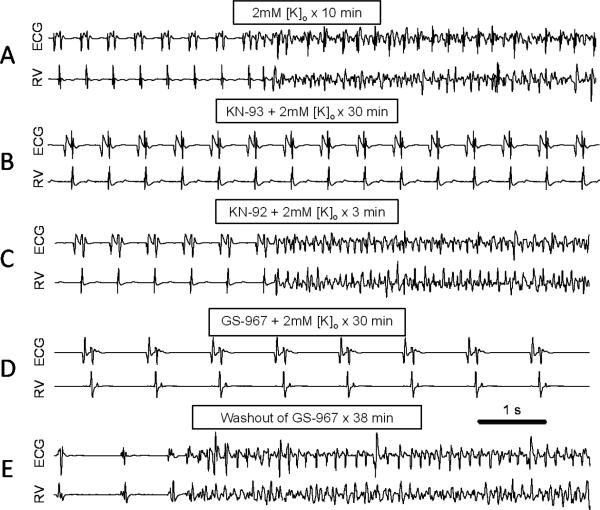
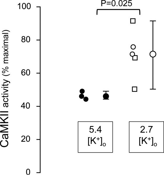
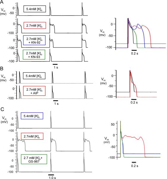
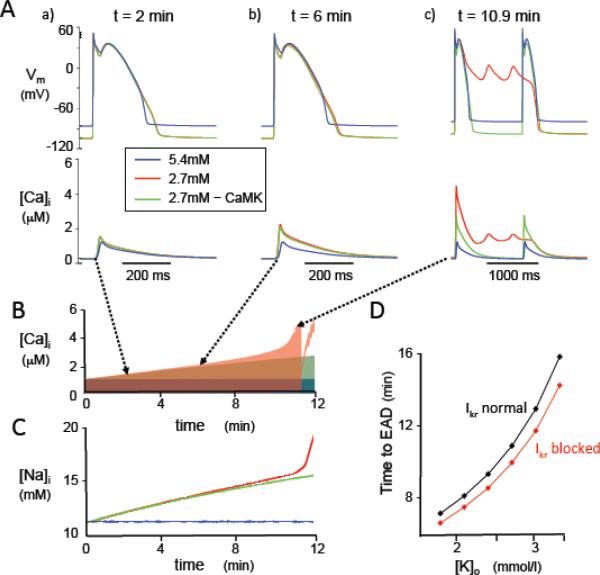
Methods and results: Arrhythmias were recorded in isolated rabbit and rat hearts or patch-clamped ventricular myocytes exposed to hypokalemia (1.0-3.5 mmol/L) in the absence or presence of dofetilide (1 μmol/L). Spontaneous early afterdepolarizations (EADs) and ventricular tachycardia/fibrillation occurred in 50% of hearts at 2.7 mmol/L [K] in the absence of dofetilide and 3.3 mmol/L [K] in its presence. Pretreatment with the Ca-calmodulin kinase II (CaMKII) inhibitor KN-93, but not its inactive analogue KN-92, abolished EADs and hypokalemia-induced ventricular tachycardia/fibrillation, as did the selective late Na current (INa) blocker GS-967. In intact hearts, moderate hypokalemia (2.7 mmol/L) significantly increased tissue CaMKII activity. Computer modeling revealed that EAD generation by hypokalemia (with or without dofetilide) required Na-K pump inhibition to induce intracellular Na and Ca overload with consequent CaMKII activation enhancing late INa and the L-type Ca current. K current suppression by hypokalemia and dofetilide alone in the absence of CaMKII activation were ineffective at causing EADs.

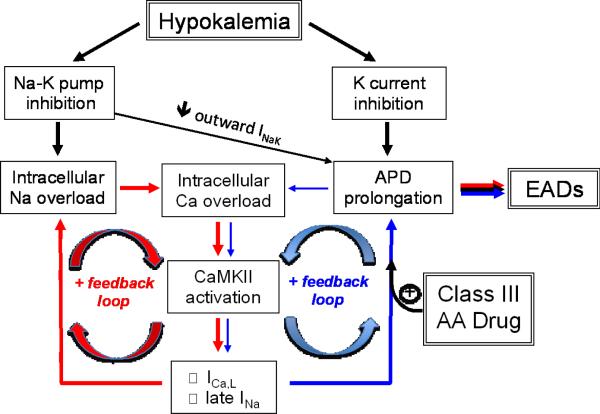
Conclusions: We conclude that Na-K pump inhibition by even moderate hypokalemia plays a critical role in promoting EAD-mediated arrhythmias by inducing a positive feedback cycle activating CaMKII and enhancing late INa. Class III antiarrhythmic drugs like dofetilide sensitize the heart to this positive feedback loop.

Keywords: anti-arrhythmia agents; arrhythmias, cardiac; long QT syndrome; potassium; signal transduction.


Figures

Figure 1
Figure 1
Hypokalemia-induced VF in adult isolated Langendorff rabbit (A) and rat (B) hearts exposed to 2.7 mmol/l [K]o. Left panel: sinus rhythm during normokalemia, as recorded with a pseudo-ECG, bipolar electrograms from the left atrium (LA) and right ventricle (RV) and an epicardial intracellular microelectrode (ME). Middle and right panel: onset of EAD-mediated triggered activity (*) from sinus rhythm during hypokalemia. Arrows in B indicate DADs.
Figure 2
Figure 2
Optical action potential recordings and activation maps during EAD-mediated triggered activity in an isolated-perfused rat heart exposed to hypokalemia (2.7 mmol/l [K]). A. Optical voltage fluorescence (FV) traces from 4 adjacent sites labeled 1-4 in panel B, illustrating two runs of EAD-mediated triggered activity (b1-b13) arising spontaneous from sinus rhythm (beats S1 and S6). B. Optical voltage fluorescence snapshots corresponding to the labeled beats in A. C. Isochronal activation maps of a sinus beat (S6) and the subsequent triggered beat (b7) indicating the different sites of origin.
Figure 3
Figure 3
K dependence of hypokalemia-induced VT/VF in isolated rabbit hearts, in the absence and presence of dofetilide. A. When extracellular K concentration was lowered from the control value of 5.4 mmol/l to 4.0, 3.5, 2.7, 2.0 or 1.0 mmol/l, the incidence of VT/VF within 90 minutes progressively increased to 100% at 2.0 and 1.0 mmol/l. Data show median and 83% CI for the number of hearts indicated in parentheses, in the absence (black circles) or presence (red circles) of 1 μmol/l dofetilide. The significantly increased by dofetilide (P=0.03). Curves are fits to a Hill equation in the absence (black curve) and presence of dofetilide (red curve). B. Kaplan-Meier survival curves comparing time to onset of VT/VF, with 83% CI indicated by shading (solid lines=without dofetilide, dashed line=with dofetilide). Rate of VT/VF onset was significantly faster at 1.0 and 2.0 mmol/l [K] than at higher [K] (P=0.0002-0.002), and significantly faster at 2.7 mmol/l [K] in the presence of dofetilide than in its absence (P=0.02).
Figure 4
Figure 4
Suppression of hypokalemia-induced VF by CaMKII inhibition or late INa blockade.Representative ECG and RV electrograms from isolated rabbit ventricles at the times indicated after reducing [K]o from 5.4 to 2.0 mmol/l. A. No drug, illustrating the spontaneous transition from sinus rhythm to VT/VF after 10 min. B. KN-93 (1 μmol/l), illustrating maintenance of sinus rhythm after 30 min. C. Same heart as in B, 3 min after replacing KN-93 with KN-92 (1 μmol/l), illustrating spontaneous onset of VT/VF. D. GS-967 (1 μmol/l), illustrating maintenance of sinus rhythm after 30 min. E. Same heart as in D, 38 min after washing out GS-967, illustrating onset of spontaneous VT/VF.
Figure 5
Figure 5
Basal (autophosphorylated) tissue CaMKII activity level, expressed as percent of maximal Ca/CaM-stimulated CaMKII activity, in isolated rabbit ventricular tissue arteriallyperfused with 5.4 mmol/l or 2.7 mmol [K]o for 30 mins. Individual data points, median, 95% confidence intervals and p value are indicated. For 2.7 mmol/l data, open circles indicate hearts in which no VT/VF occurred during hypokalemia (2 hearts), and open squares indicate hearts in which transient (1-2 min episodes in 2 hearts) or sustained VT/VF developed (1 heart) during hypokalemia.
Figure 6
Figure 6
Suppression of hypokalemia-induced EADs by CaMKII inhibition and late INa blockade in isolated patch-clamped rabbit ventricular myocytes. Membrane voltage (Vm) traces during pacing at PCL 6 s. Top to bottom traces: A. [K]o = 5.4 mmol/l; 2.7 mmol/l; 2.7 mmol/l + 1 μmol/l KN-92; 2.7 mmol/l + 1 μmol/l KN-92. B. [K]o = 5.4 mmol/l + AIP; 2.7 mmol/l + AIP (dialyzed for 30 min). C. [K]o = 5.4 mmol/l; 2.7 mmol/l; 2.7 mmol/l; 2.7 mmol/l + 1 μmol/l GS-967 (bottom). Right panels show superimposed action potentials under the various conditions for A-C.
Figure 7
Figure 7
Effects of hypokalemia on membrane voltage (Vm), [Ca]i, and [Na]i in the rabbit ventricular AP model paced at a CL of 1s. A. Vm at various times corresponding to panel B, for [K]o = 5.4 mmol/l (blue traces); [K]o = 2.7 mmol/l (red traces); and [K]o = 2.7 mmol/l with CaMKII signaling disabled (green traces). B-C. Corresponding time course of changes in [Ca]i, (B) and [Na]i (C). [Ca]i and [Na]i accelerate rapidly with the onset of EADs, and systolic [Ca]i then transiently decreases when EADs transition to repolarization failure. D. Time to onset of the first EAD versus level of [K]o, with normal IKr (black) or IKr blocked (red) to simulate the effects of dofetilide.
Figure 8
Figure 8
Schema of positive feedback loops (red and blue arrows) promoting intracellular Na and Ca overload, CaMKII activation and EADs during hypokalemia. The potentiation of the blue positive feedback loop by Class III antiarrhythmic (AA) drugs such as dofetilide is also shown. INaK=Na-K pump current.

References

    1. Schulman M, Narins RG. Hypokalemia and cardiovascular disease. Am J Cardiol. 1990;65:4E–9E. discussion 22E-23E. - PubMed
    1. Macdonald JE, Struthers AD. What is the optimal serum potassium level in cardiovascular patients? J Am Coll Cardiol. 2004;43:155–61. - PubMed
    1. Cooper HA, Dries DL, Davis CE, Shen YL, Domanski MJ. Diuretics and risk of arrhythmic death in patients with left ventricular dysfunction. Circulation. 1999;100:1311–5. - PubMed
    1. Siscovick DS, Raghunathan TE, Psaty BM, Koepsell TD, Wicklund KG, Lin X, Cobb L, Rautaharju PM, Copass MK, Wagner EH. Diuretic therapy for hypertension and the risk of primary cardiac arrest. N Engl J Med. 1994;330:1852–7. - PubMed
    1. Sanguinetti MC, Jurkiewicz NK. Role of external Ca and K in gating of cardiac delayed rectifier K currents. Pflugers Arch. 1992;420:180–6. - PubMed

Publication types