. 2015 Oct 2;290(40):24067-78.
doi: 10.1074/jbc.M115.659029. Epub 2015 Aug 12.

Neutrophil elastase promotes interleukin-1β secretion from human coronary endothelium

Affiliations

Neutrophil elastase promotes interleukin-1β secretion from human coronary endothelium

Mabruka Alfaidi et al. J Biol Chem. .

Abstract

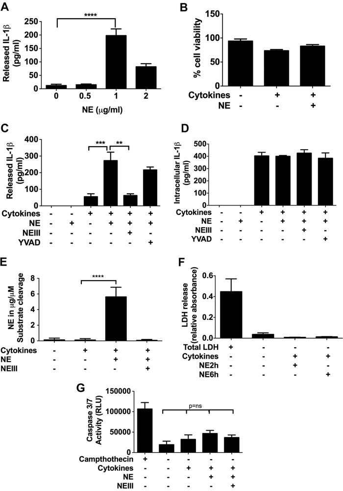
The endothelium is critically involved in the pathogenesis of atherosclerosis by producing pro-inflammatory mediators, including IL-1β. Coronary arteries from patients with ischemic heart disease express large amounts of IL-1β in the endothelium. However, the mechanism by which endothelial cells (ECs) release IL-1β remains to be elucidated. We investigated neutrophil elastase (NE), a potent serine protease detected in vulnerable areas of human carotid plaques, as a potential "trigger" for IL-1β processing and release. This study tested the hypothesis that NE potentiates the processing and release of IL-1β from human coronary endothelium. We found that NE cleaves the pro-isoform of IL-1β in ECs and causes significant secretion of bioactive IL-1β via extracellular vesicles. This release was attenuated significantly by inhibition of neutrophil elastase but not caspase-1. Transient increases in intracellular Ca(2+) levels were observed prior to secretion. Inside ECs, and after NE treatment only, IL-1β was detected within LAMP-1-positive multivesicular bodies. The released vesicles contained bioactive IL-1β. In vivo, in experimental atherosclerosis, NE was detected in mature atherosclerotic plaques, predominantly in the endothelium, alongside IL-1β. This study reveals a novel mechanistic link between NE expression in atherosclerotic plaques and concomitant pro-inflammatory bioactive IL-1β secretion from ECs. This could reveal additional potential anti-IL-1β therapeutic targets and provide further insights into the inflammatory process by which vascular disease develops.

Keywords: IL-1; atherosclerosis; endothelium; extracellular vesicles; inflammation; neutrophil elastase.


Figures

FIGURE 1.
FIGURE 1.
NE enhances IL-1β secretion from HCAECs by a caspase-1-independent mechanism. A, IL-1β is released by HCAECs after 48-h stimulation with cytokines (TNF-α/IL-1α, 10 ng/ml), followed by NE activation (0.5–2 μg/ml) for 2 h as measured by ELISA (n = 3). B, cell viability (measured by trypan blue) is not reduced significantly following exposure to 1 μg/ml NE (n = 3). C, HCAECs, incubated for 48 h with or without cytokines and then incubated further for 6 h in serum-free media with or without NE (1 μg/ml) in the presence or absence of inhibitors (NEII, 500 μm; YVAD, 50 μm; n = 5), show that IL-1β release is increased by NE independent of caspase-1. D, levels of IL-1β in cell lysates are not increased following NE incubation (n = 5). E, increased NE activity in EC lysates treated with NE for 6 h compared with unstimulated cells (n = 3). F, lactate dehydrogenase levels are unchanged following NE treatment, as measured in conditioned media or in cell lysates as a total of lactate dehydrogenase (n = 3). G, caspase-3/7 activity is unchanged in HCAECs following NE treatment. HCAECs in 96-well plates (2 × 10⁴ cells/well), incubated with or without cytokines (TNF-α/IL-1α, 10 ng/ml each) for 48 h, were subjected to NE (1 μg/ml) in serum-free media for 6 h (n = 3). Camptothecin (10 μg/ml) was used to induce apoptosis as a positive control. RLU, relative light units. All data are mean ± S.E. and were analyzed by one-way ANOVA with Tukey's multiple comparison test. *, p < 0.05; **, p < 0.01; ***, p < 0.001; ****, p < 0.0001.
FIGURE 2.
FIGURE 2.
NE selectively cleaves proIL-1β in primed EC lysates without caspase-1/NLRP3 activation. A, Western blot analysis of cell lysates of primed EC with or without NE and assessed for IL-1β (i), Caspase-1 (ii), and NLRP-3 (iii). The blots are representative of three experiments, with α-tubulin levels as a loading control. For IL-1β, recombinant IL-1β (rIL-1β, 20 μg, 17 kDa) was loaded as a positive control and represents the commonly detected mature form, whereas proIL-1β (31 kDa) indicates the inactive pro-form. For caspase-1, activated THP-1 cell lysates were used as a positive control for the p20 isoform. B, densitometric analysis of 20-kDa IL-1β levels (n = 3). C, Western blot illustrating the cleavage of recombinant proIL-1β (rProIL-1β, 20 μg) by NE. Recombinant ProIL-1β was incubated at 37 °C (5% CO2, v/v) alone or in the presence of NE (1 μg/ml) for 30 min, 2 h, and 6 h. D, Western blot for recombinant mature IL-1β (20 μg) in the presence or absence of NE, showing no cleavage (n = 3). E, ELISA for recombinant mature IL-1β, showing no difference in levels following NE treatment. Data are mean ± S.E. and were analyzed by one-way ANOVA and Tukey's post-test. *, p < 0.05; **, p < 0.01.
FIGURE 3.
FIGURE 3.
Neutrophil elastase activates microvesicle shedding from endothelial cells in a time-dependent manner. HCAECs were left untreated or treated with cytokines (IL-1α/TNF-α) for 48 h and then labeled with annexin-V/Alexa Fluor 488. After the addition of 1 μg/ml of NE, cells were visualized in a heated chamber (5% CO2 v/v) using a confocal microscope to scan MV release. Images captured after 10 min, 30 min, 2 h, and 6 h show an early generation of MVs after 10 min of NE stimulation but more prominent at later time points. The arrowhead indicates fluorescent MVs, and the arrow indicates earliest blebbing in EC treated with NE. Scale bars = 50 μm. The representative images are from three independent experiments and were altered digitally to remove background fluorescence.
FIGURE 4.
FIGURE 4.
NE induces secretion of extracellular vesicles containing bioactive IL-1 from HCAECs. A, flow cytometric characterization of MVs released in response to NE. MVs were isolated and stained with annexin V/PE-Cy7 as described under “Experimental Procedures.” Analysis of MVs (red) using Megamex beads (blue) shows that they are within the 0.2- 0.9-μm size limit. B, a significant increase in MVs stained with annexin V is seen in NE-treated cells compared with untreated controls. Analysis was performed by FlowJo software (n = 3). FCS, forward scatter. Data are mean ± S.E., one-way ANOVA followed by Tukey's post- test. *, p < 0.05; **, p < 0.01. C and D, detection of IL-1β in isolated MVs by immunoblotting. Equal amounts of protein (20 μg) were loaded in each lane, with recombinant IL-1β (rIL-1β, 20 μg) used as a positive control (17 kDa). Data are representative of four experiments. E, luciferase assay for the measurement of IL-1β bioactivity in HeLa cells exposed to freshly harvested conditioned media (total supernatants from cytokine-primed cells (TNF-α/IL-1α, 10 ng/ml each with or without NE, 1 μg/ml) or recombinant IL-1β (0.1 nm) for 6 h with or without anti-IL-1β (1 μg/ml). Specificity for IL-1β is shown by a reduction of IL-8 luciferase detection following incubation with IL-1β-neutralizing antibody. Data are expressed as mean ± S.E. (n = 3) and were analyzed by one-way ANOVA followed by Tukey's test. ****, p < 0.0001. F, immunoelectron microscopic analysis of IL-1β in ECs ± NE treatment. Anti-IL-1β-conjugated immunogold (20-nm gold particles, arrow) was used to confirm the presence of IL-1β in the MVs (0.2 μm) released from the plasma membrane of ECs.
FIGURE 5.
FIGURE 5.
Mechanisms contributing to IL-1β release in endothelial HCAEC. A and B, HCAECs were assayed for changes in cytosolic free Ca2+ in response to the indicated conditions. A, no significant change in cytosolic Ca2+ levels are seen in Ca2+-free media. B, the fluorescence intensity of intracellular calcium changes after 5 min of NE stimulation in the presence of CaCl2. Data are mean ± S.E. (n = 6) and were analyzed by one-way ANOVA and Tukey's post-test. *, p < 0.05; ***, p < 0.001. C, IL-1β colocalizes with LAMP-1 after NE stimulation. Cells were primed with cytokines (TNF-α/IL-1α, 10 ng/ml each), followed by incubation ± NE (1 μg/ml) in serum-free media over 2 h before immunostaining for IL-1β (red) and LAMP-1 (green). Scale bars = 10 μm. D, histogram showing a high correlation between IL-1β and LAMP-1 in ECs after NE activation.
FIGURE 6.
FIGURE 6.
NE induces IL-1β release by an endolysosome-dependent mechanism. A, ELISA measuring IL-1β release in conditioned media of HCAECs primed with cytokines (TNF-α/IL-1α, 10 ng/ml) with or without NE (1 μg/ml) or BAF1 (50 nm) after 6 h. Data are mean ± S.E. (n = 3) and were analyzed by one-way ANOVA followed by Tukey's post-test. *, p < 0.05. B, Western blot analysis of lysates harvested from primed HCAECs activated with NE with or without BAF1 (50 nm) for 6 h. 20 μg of protein was loaded per lane, with α-tubulin used as a loading control. The blot is representative of three independent experiments. C, MVB characterization. i, EM analysis showing a full appearance of MVBs in NE-treated cells in close proximity of the plasma membrane. ii, immunolabeling with anti-IL-1β (20-nm gold particles, arrow), showing IL-1β within MVBs (arrowhead).
FIGURE 7.
FIGURE 7.
NE is detected in ECs and is colocalized with IL-1β in the endothelium of mature atherosclerotic plaques. A, confocal images showing LAMP-1 and NE in primed ECs after NE treatment. HCAECs were incubated with or without Alexa Fluor 647-labeled (1 μg/ml) NE for 2 h in serum-free media before washing in PBS, and colocalization was performed using an antibody against LAMP-1. Confocal images were analyzed using Zeiss image and ImageJ software. Scale bars = 10 μm. B, immunohistochemical detection of NE and IL-1β in the luminal endothelium of mouse atherosclerotic plaques. Paraffin-embedded aortic sinuses from ApoE−/− mice fed a high-fat diet for 12 weeks were stained with primary antibodies as indicated. The specificity of staining was confirmed by no primary negative control. Scale bars = 200 μm. C, colocalization of IL-1-β, NE, and von Willebrand Factor (vWF) in aortic atherosclerosis. NE positivity was detected predominantly in the endothelium (top right panel, arrows). IL-1β positive endothelium (top left panel) was also detected. The bottom panels show vWF-stained endothelium, and DAPI was used for the nuclei. The specificity of staining was confirmed by no primary negative control. Images are representative of histology data obtained from a total of six animals.
FIGURE 8.
FIGURE 8.
Schematic of the proposed mechanism of IL-1β secretion from ECs by NE. NE is released by circulating cells at the site of atheroma and transported by endocytosis inside the diseased endothelium (primed by inflammation) (i). An increase in calcium because of NE effects leads to remodeling of the cell membrane and vesiculation (ii), which, in turn, facilitates the shedding of MVs containing mature IL-1β (iii). ProIL-1β is up-regulated in the inflamed endothelium (iv). NE enters MVBs and cleaves the proIL-1β contained within (v). MVBs also fuse to the plasma membrane and release exosomes containing IL-1 (vi).

References

    1. Libby P. (2002) Inflammation in atherosclerosis. Nature 420, 868–874 - PubMed
    1. Chamberlain J., Evans D., King A., Dewberry R., Dower S., Crossman D., Francis S. (2006) Interleukin-1β and signaling of interleukin-1 in vascular wall and circulating cells modulates the extent of neointima formation in mice. Am. J. Pathol. 168, 1396–1403 - PMC - PubMed
    1. Bonetti P. O., Lerman L. O., Lerman A. (2003) Endothelial dysfunction: a marker of atherosclerotic risk. Arterioscler. Thromb. Vasc. Biol. 23, 168–175 - PubMed
    1. Ridker P. M., Thuren T., Zalewski A., Libby P. (2011) Interleukin-1β inhibition and the prevention of recurrent cardiovascular events: rationale and design of the Canakinumab Anti-inflammatory Thrombosis Outcomes Study (CANTOS). Am. Heart J. 162, 597–605 - PubMed
    1. Dinarello C. A. (2009) Immunological and inflammatory functions of the interleukin-1 family. Annu. Rev. Immunol. 27, 519–550 - PubMed

Publication types

MeSH terms