. 2015 Oct 2;290(40):24495-508.
doi: 10.1074/jbc.M115.641944. Epub 2015 Aug 12.

Biased Gs versus Gq proteins and β-arrestin signaling in the NK1 receptor determined by interactions in the water hydrogen bond network

Affiliations

Biased Gs versus Gq proteins and β-arrestin signaling in the NK1 receptor determined by interactions in the water hydrogen bond network

Louise Valentin-Hansen et al. J Biol Chem. .

Abstract

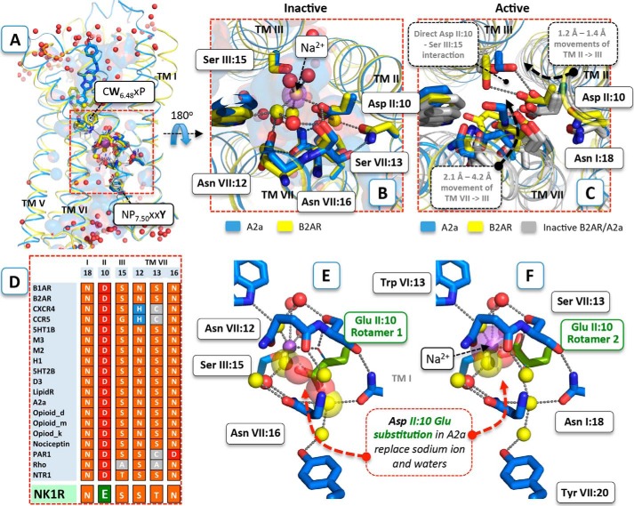
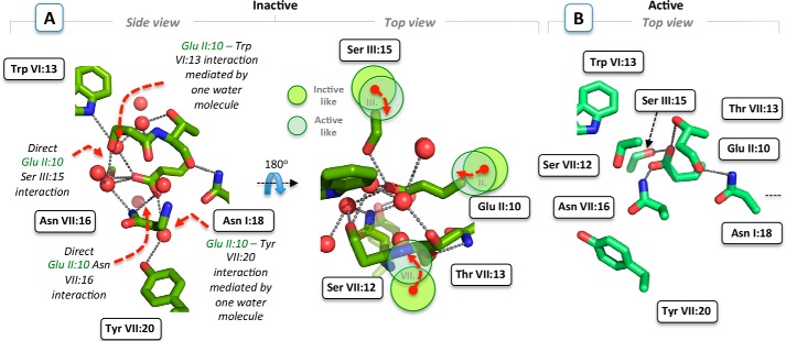
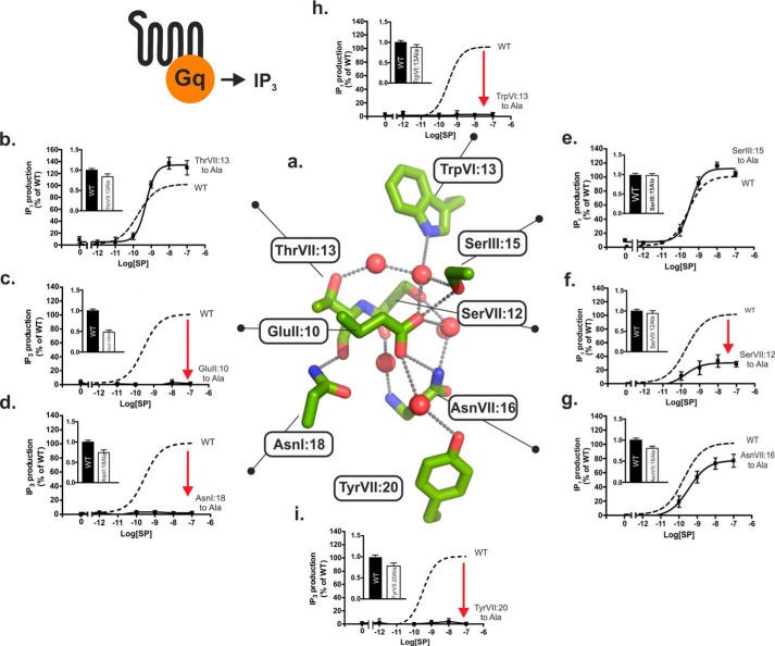
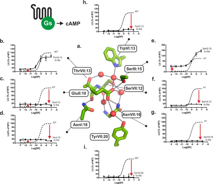
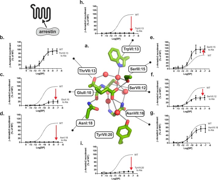
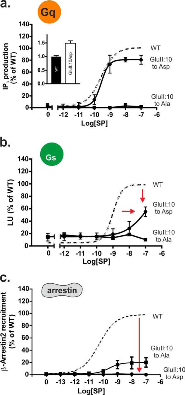
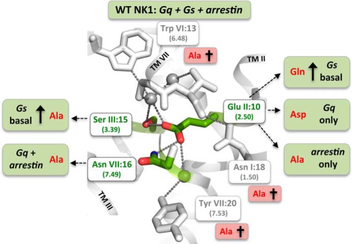
X-ray structures, molecular dynamics simulations, and mutational analysis have previously indicated that an extended water hydrogen bond network between trans-membranes I-III, VI, and VII constitutes an allosteric interface essential for stabilizing different active and inactive helical constellations during the seven-trans-membrane receptor activation. The neurokinin-1 receptor signals efficiently through Gq, Gs, and β-arrestin when stimulated by substance P, but it lacks any sign of constitutive activity. In the water hydrogen bond network the neurokinin-1 has a unique Glu residue instead of the highly conserved AspII:10 (2.50). Here, we find that this GluII:10 occupies the space of a putative allosteric modulating Na(+) ion and makes direct inter-helical interactions in particular with SerIII:15 (3.39) and AsnVII:16 (7.49) of the NPXXY motif. Mutational changes in the interface between GluII:10 and AsnVII:16 created receptors that selectively signaled through the following: 1) Gq only; 2) β-arrestin only; and 3) Gq and β-arrestin but not through Gs. Interestingly, increased constitutive Gs but not Gq signaling was observed by Ala substitution of four out of the six core polar residues of the network, in particular SerIII:15. Three residues were essential for all three signaling pathways, i.e. the water-gating micro-switch residues TrpVI:13 (6.48) of the CWXP motif and TyrVII:20 (7.53) of the NPXXY motif plus the totally conserved AsnI:18 (1.50) stabilizing the kink in trans-membrane VII. It is concluded that the interface between position II:10 (2.50), III:15 (3.39), and VII:16 (7.49) in the center of the water hydrogen bond network constitutes a focal point for fine-tuning seven trans-membrane receptor conformations activating different signal transduction pathways.

Keywords: G protein-coupled receptor (GPCR); cell signaling; functional selectivity; homology modeling; hydrogen bond network; molecular pharmacology; receptor structure-function.


Figures

FIGURE 1.
FIGURE 1.
Structural conservation of the polar interface between TM-I–III and -VII. A, structural distribution of conserved polar residues between the intracellular half of TM-I–III and -VII of 18 unique inactive GPCR structures. The residues AsnI:18 (1.50), AspII:10 (2.50), SerIII:15 (3.39), SerVII:12 (7.45), ThrVII:13 (7.46), and AsnVII:16 (7.49) of the conserved polar interface restricted by the functionally important and conserved TrpVI:13 (6.48) and TyrVII:20 (7.53) in the antagonist-bound structures of A2AAR (PDB code 4EIY solved at 1.8 Å resolution) (blue) and B2AR (PDB code 2RH1) (yellow) are shown as sticks. The corresponding residues in the other inactive structures are shown by gray lines. B, close-up view of the polar interface comprising a sodium ion (purple sphere) and water molecules (red spheres). Co-localized water molecules in the A2AAR and the B2AR structure are shown as yellow spheres. The salt bridge between sodium and AspII:10 (2.50) and water-mediated hydrogen bond interactions are shown as gray dotted lines. C, close-up view of the polar pocket in the active-like agonist-bound A2AAR/UK432,097 structure (PDB code 3QAK) (blue) and the active agonist-bound B2AR structure in complex with Gs (PDB code 3SN6). D, polar pocket alignment of the distinct inactive experimental structures compared with the conforming sequence in the NK1 receptor, which have a glutamic acid instead of the highly conserved AspII:10 (2.50). E and F, structural details of AspII:10 Glu substitution (green) in the inactive A2AAR structure (E, first rotamer conformation of AspII:10 Glu substitution; F, second rotamer conformation of AspII:10 Glu substitution). Water molecules that make steric clashes with the substituted glutamic acid are shown as big transparent spheres.
FIGURE 2.
FIGURE 2.
NK1 receptor models. A, side and top close-up view of the conserved polar interface, including AsnI:18 (1. 50), GluII:10 (2.50), SerIII:15 (3.39), SerVII:12 (7.45), ThrVII:13 (7.46), and AsnVII:16 (7.49) in the inactive NK1 receptor model restricted by the functionally important and conserved residues TrpVI:13 (6.48) and TyrVII:20 (7.53). Water molecules are shown as red spheres and hydrogen bonds as gray dotted lines. In contrast to highly conserved AspII:10, the longer GluII:10 side chain in the NK1 model is predicted to make direct rather than water-mediated interactions with SerIII:15 and AsnVII:16. Dashed red arrows show the conformational rearrangements of TM-II, -III, and -VII (illustrated by light green and transparent circles) required to obtain an “active” NK1 receptor structure consistent with common conformational changes observed in distinct active structures. B, model of the conserved polar interface of the NK1 receptor in an active state, based on the agonist-bound A2AAR structure (PDB code 3QAK).
FIGURE 3.
FIGURE 3.
Functional consequence of Ala substitution on Gq signaling of the conserved polar residues in the NK1 receptor. a, structure of the hydrogen bond network in the NK1 receptor model based on A2AAR (PDB code 4EIY) as a template structure. Key side chains are shown as sticks and water molecules as red spheres. b–h, agonist (SP)-induced IP3 production in COS-7 cells transiently transfected with either wild type NK1 (dotted lines) or mutant forms of ThrVII:13Ala (7.46) (b), GluII:10Ala (2.50) (c), AsnI:18Ala (1.50) (d), SerIII:15Ala (3.39) (e), SerVII:12Ala (7.45) (f), AsnVII:16Ala (7.49) (g), TrpVI:13Ala (6.48) (h), and TyrVII:20Ala (7.53) (i). Cell surface receptor expression measured by ELISA is shown in the column diagram insets in each panel.
FIGURE 4.
FIGURE 4.
Functional consequence of Ala substitution on Gs signaling of the conserved polar residues in the NK1 receptor. a, structure of the hydrogen bond network in the NK1 receptor model based on A2AAR (PDB code 4EIY) as a template structure. Key side chains are shown as sticks and water molecules as red spheres. b–h, agonist (SP)-induced cAMP production in COS-7 cells transiently transfected with either wild type NK1 (dotted lines) or mutant forms of ThrVII:13Ala (7.46) (b), GluII:10Ala (2.50) (c), AsnI:18Ala (1.50) (d), SerIII:15Ala (3.39) (e), SerVII:12Ala (7.45) (f), AsnVII:16Ala (7.49) (g), TrpVI:13Ala (6.48) (h), and TyrVII:20Ala (7.53) (i).
FIGURE 5.
FIGURE 5.
Functional consequence of Ala substitution on β-arrestin2 mobilization of the conserved polar residues in the NK1 receptor. a, structure of the hydrogen bond network in the NK1 receptor model based on A2AAR (PDB code 4EIY) as a template structure. Key side chains are shown as sticks and water molecules as red spheres. b–h, agonist (SP)-induced β-arrestin2 mobilization in CHOK1 cells transiently transfected with either wild type NK1 (dotted lines) or mutant forms of ThrVII:13Ala (7.46) (b), GluII:10Ala (2.50) (c), AsnI:18Ala (1.50) (d), SerIII:15Ala (3.39) (e), SerVII:12Ala (7.45) (f), AsnVII:16Ala (7.49) (g), TrpVI:13Ala (6.48) (h), and TyrVII:20Ala (7.53) (i).
FIGURE 6.
FIGURE 6.
Functional consequence of Asp substitution of the GluII:10 (2.50) residue in the NK1 receptor. a–c, agonist (SP)-induced IP3 (a), cAMP (b), or β-Arrestin2 (c) mobilization in COS-7 cells (IP3 and cAMP) or CHOK1 cells (β-arrestin2) transiently transfected with either wild type NK1 (dotted lines) or mutant GluII:10Ala/Asp. Cell surface receptor expression measured by ELISA is shown in the column diagram inset in a.
FIGURE 7.
FIGURE 7.
Residues of the water hydrogen bond network of the NK1 receptor identified to be either essential for overall signal transduction or for the fine-tuning and bias of signaling through the three different signaling pathways: Gs, Gq, and β-arrestin. Three residues were by Ala substitutions identified to be essential for all three signal transduction pathways as follows: the water-gating TrpVI:13 (6.48) of the CWXP motif and the TyrVII:20 (7.53) of the NPXXY motif as well as the ultra-highly conserved TM-VII kink-stabilizing AsnI:18 (1.50). Mutations of GluII:10 (2.50) and the two residues SerIII:15 (3.39) and AsnVII:16 (7.49), to which GluII:10 according to the molecular models makes direct hydrogen bond interactions, result in receptor mutants with biased signaling as follows: β-arrestin only (GluII:10 to Ala), Gq only (GluII:10 to Asp), high constitutive Gs signaling (GluII:10 to Gln), and Gq plus β-arrestin but not Gs (AsnVII:16 to Ala). Ala substitution of SerIII:15 selectively increases Gs signaling from zero to ∼40% of Emax.

Similar articles

Cited by

References

    1. Rosenbaum D. M., Rasmussen S. G., Kobilka B. K. (2009) The structure and function of G-protein-coupled receptors. Nature 459, 356–363 - PMC - PubMed
    1. Nygaard R., Frimurer T. M., Holst B., Rosenkilde M. M., Schwartz T. W. (2009) Ligand binding and micro-switches in 7TM receptor structures. Trends Pharmacol. Sci. 30, 249–259 - PubMed
    1. Katritch V., Cherezov V., Stevens R. C. (2013) Structure-function of the G protein-coupled receptor superfamily. Annu. Rev. Pharmacol. Toxicol. 53, 531–556 - PMC - PubMed
    1. Wisler J. W., Xiao K., Thomsen A. R., Lefkowitz R. J. (2014) Recent developments in biased agonism. Curr. Opin. Cell Biol. 27, 18–24 - PMC - PubMed
    1. Nygaard R., Valentin-Hansen L., Mokrosinski J., Frimurer T. M., Schwartz T. W. (2010) Conserved water-mediated hydrogen bond network between TM-I, -II, -VI, and -VII in 7TM receptor activation. J. Biol. Chem. 285, 19625–19636 - PMC - PubMed

Publication types

MeSH terms

LinkOut - more resources