

Establishment and characterization of cetuximab resistant head and neck squamous cell carcinoma cell lines: focus on the contribution of the AP-1 transcription factor
- PMID: 26269754
- PMCID: PMC4529614
Establishment and characterization of cetuximab resistant head and neck squamous cell carcinoma cell lines: focus on the contribution of the AP-1 transcription factor
Abstract
Background: After an initial response to EGFR targeted therapy, secondary resistance almost invariably ensues, thereby limiting the clinical benefit of the drug. Hence, it has been recognized that the successful implementation of targeted therapy in the treatment of HNSCC cancer is very much dependent on predictive biomarkers for patient selection.
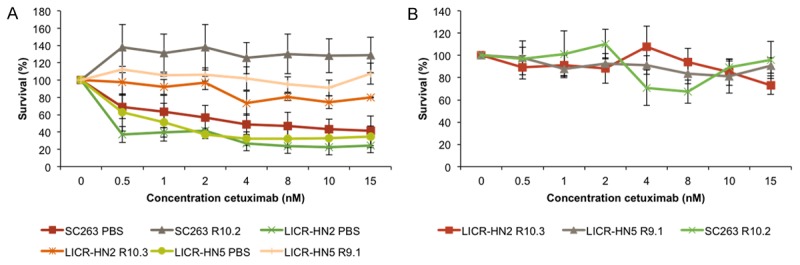
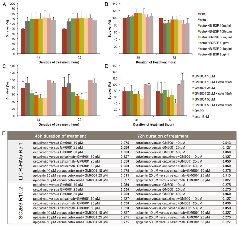
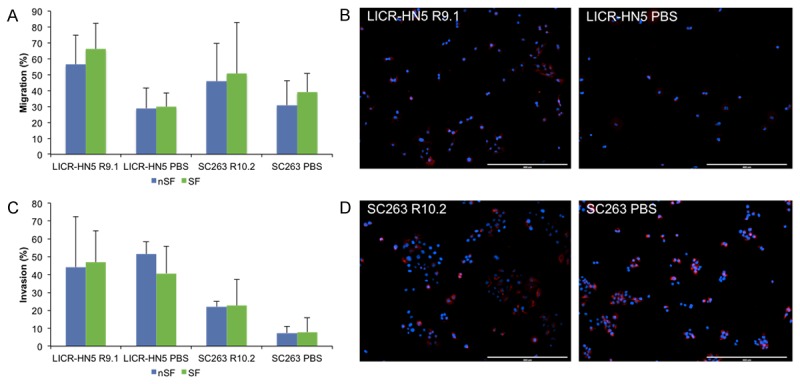
Methods: We generated an in vitro model of acquired cetuximab resistance by chronically exposing three HNSCC cell lines to increasing cetuximab doses. Gene expression profiles of sensitive parental cells and resistant daughter cells were compared using microarray analysis. Growth inhibitory experiments were performed with an HB-EGF antibody and the MMP inhibitor, both in combination with cetuximab. Characteristics of EMT were analyzed using migration and invasion assays, immunofluorescent vimentin staining and qRT-PCR for several genes involved in this process. The function of the transcription factor AP-1 was investigated using qRT-PCR for several genes upregulated or downregulated in cetuximab resistant cells. Furthermore, anchorage-independent growth was investigated using the soft agar assay.
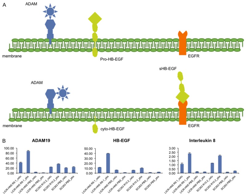
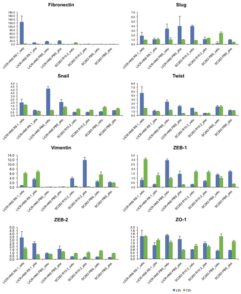
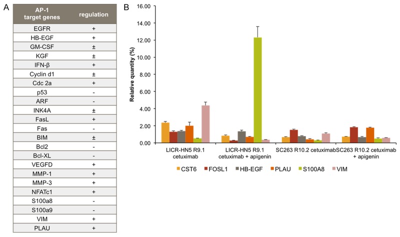
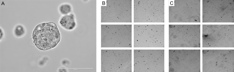
Results: Gene expression profiling shows that cetuximab resistant cells upregulate several genes, including interleukin 8, the EGFR ligand HB-EGF and the metalloproteinase ADAM19. Cytotoxicity experiments with neutralizing HB-EGF antibody could not induce any growth inhibition, whereas an MMP inhibitor inhibited cell growth in cetuximab resistant cells. However, no synergetic effects combined with cetuximab could be observed. Cetuximab resistant cells showed traits of EMT, as witnessed by increased migratory potential, increased invasive potential, increased vimentine expression and increased expression of several genes involved in EMT. Furthermore, expression of upregulated genes could be repressed by the treatment with apigenin. The cetuximab resistant LICR-HN2 R10.3 cells tend to behave differently in cell culture, forming spheres. Therefore, soft agar assay was performed and showed more and larger colonies when challenged with cetuximab compared to PBS challenged cells.
Conclusions: In summary, our results indicate that increased expression of the ligand HB-EGF could contribute to resistance towards cetuximab in our cetuximab resistant HNSCC cells. Furthermore, several genes upregulated or downregulated in cetuximab resistant cells are under control of the AP-1 transcription factor. However, more studies are warranted to further unravel the role of AP-1 in cetuximab resistance.
Keywords: ADAM19; HB-EGF; Head and neck squamous cell carcinoma (HNSCC); cetuximab; interleukin 8; resistance; transcription factor AP-1.
Figures
References
-
- Kalia M. Personalized oncology: recent advances and future challenges. Metabolism. 2013;62(Suppl 1):S11–14. - PubMed
-
- Temam S, Kawaguchi H, El-Naggar AK, Jelinek J, Tang H, Liu DD, Lang W, Issa JP, Lee JJ, Mao L. Epidermal growth factor receptor copy number alterations correlate with poor clinical outcome in patients with head and neck squamous cancer. J. Clin. Oncol. 2007;25:2164–2170. - PubMed
-
- Dittmann K, Mayer C, Rodemann HP. Nuclear EGFR as novel therapeutic target: insights into nuclear translocation and function. Strahlentherapie und Onkologie. 2010;186:1–6. - PubMed
-
- Vermorken JB, Trigo J, Hitt R, Koralewski P, Diaz-Rubio E, Rolland F, Knecht R, Amellal N, Schueler A, Baselga J. Open-label, uncontrolled, multicenter phase II study to evaluate the efficacy and toxicity of cetuximab as a single agent in patients with recurrent and/or metastatic squamous cell carcinoma of the head and neck who failed to respond to platinum-based therapy. J. Clin. Oncol. 2007;25:2171–2177. - PubMed
-
- Bonner JA, Harari PM, Giralt J, Azarnia N, Shin DM, Cohen RB, Jones CU, Sur R, Raben D, Jassem J, Ove R, Kies MS, Baselga J, Youssoufian H, Amellal N, Rowinsky EK, Ang KK. Radiotherapy plus cetuximab for squamous-cell carcinoma of the head and neck. N Engl J Med. 2006;354:567–578. - PubMed
LinkOut - more resources
Full Text Sources
Research Materials
Miscellaneous