. 2015 Aug 13;10(8):e0135680.
doi: 10.1371/journal.pone.0135680. eCollection 2015.

Lipid Binding of the Amphipathic Helix Serving as Membrane Anchor of Pestivirus Glycoprotein Erns

Affiliations

Lipid Binding of the Amphipathic Helix Serving as Membrane Anchor of Pestivirus Glycoprotein Erns

Daniel Aberle et al. PLoS One. .

Abstract

Pestiviruses express a peculiar protein named Erns representing envelope glycoprotein and RNase, which is important for control of the innate immune response and persistent infection. The latter functions are connected with secretion of a certain amount of Erns from the infected cell. Retention/secretion of Erns is most likely controlled by its unusual membrane anchor, a long amphipathic helix attached in plane to the membrane. Here we present results of experiments conducted with a lipid vesicle sedimentation assay able to separate lipid-bound from unbound protein dissolved in the water phase. Using this technique we show that a protein composed of tag sequences and the carboxyterminal 65 residues of Erns binds specifically to membrane vesicles with a clear preference for compositions containing negatively charged lipids. Mutations disturbing the helical folding and/or amphipathic character of the anchor as well as diverse truncations and exchange of amino acids important for intracellular retention of Erns had no or only small effects on the proteins membrane binding. This result contrasts the dramatically increased secretion rates observed for Erns proteins with equivalent mutations within cells. Accordingly, the ratio of secreted versus cell retained Erns is not determined by the lipid affinity of the membrane anchor.


Conflict of interest statement

Competing Interests: The authors have declared that no competing interests exist.

Figures

Fig 1
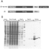
Fig 1. Test proteins for lipid pull down assay.
(A) Schematic representation of the composition of the test proteins Z2+anchor and Z2 control. His-tag: 6xHis residues; Z2-tag: single Z-domain from S. aureus protein A; TEV: tobacco etch virus proteinase cleavage site; Erns anchor: 65 C-terminal residues of the Erns protein from BVDV CP7 [41]. (B) Coomassie-stained SDS-PAGE with samples taken during expression and purification of Z2+anchor. The individual fractions analyzed here are given on top. Between Wash I and Wash II, a protein size marker (PageRuler prestained protein ladder, New England Biolabs, Schwalbach, Germany) is shown (bands represent sizes of 170, 130, 100, 70, 55, 40, 35, 25, 15, 10 kDa, top to bottom).
Fig 2
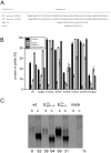
Fig 2. Lipid pull down assay.
(A) Coomassie-stained SDS-PAGE with the proteins found in the supernatant (upper part) or pellet (lower part) of pull down assays conducted in buffer or in buffer containing lipid vesicles of different compositions (composition indicated on top: DMPC/PG = 95% DMPC/ 5% DMPG; DMPC/PS = 95% DMPC/ 5% DMPS). Left part: protein Z2+anchor was used in pulldown; right part: Z2 control. For the quantification given in B and C the total amount of the detected protein was determined densitometrically and the percentage of the product found in the pellet fraction was calculated. Accordingly, the percentage of Z2+anchor in the pellet is increased in the presence of DMPC vesicles as the amount of protein in the supernatant is lower than in buffer alone (left part of left gel). (B) Quantitative results of pull down of Z2+anchor (black bars) or Z2 control (gray bars) with vesicles composed of different lipids as indicated below the X-axis. PC: 100% DMPC; Mixtures: 95% DMPC and 5% of DMPG (PC/PG), L-α-Phosphatidylinositol (PC/PI); DMPE (PC/PE), DMPA (PC/PA), DMPS (PC/PS) or sphingomyelin (PC/Sph). Buffer: control of lipid binding buffer without lipids. (C) Comparison of binding of proteins containing the Z2 tag or an alternative tag (GB = GB-carrier protein [42]) to vesicles with the indicated lipid composition or buffer without lipids. The bars in (B) and (C) give the percent of the total protein recovered in the pellet fractions and represent the mean of at least three independent experiments with the standard deviation shown by error bars.
Fig 3
Fig 3. Temperature dependence and kinetics of lipid binding of the Erns.
(A) Temperature dependence and (B) kinetics of lipid binding of the Z+anchor compared with Z control. The composition of the lipid vesicles is indicated below the X-axis and specified in detail in the legend to Fig 2B.
Fig 4
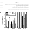
Fig 4. Comparison of lipid binding of constructs with Z2 tags fused to different amphipathic helixes.
(A) Sequences of the tested amphipathic helices are given. Erns anchor (BVDV) represents the sequence present in the standard Z2+anchor construct. Below, the corresponding sequence of the related classical swine fever virus Erns protein is shown. BMV-1a represents the sequence from the brome mosaic virus 1a protein. MinD is the amphipathic helix sequence from the MinD protein. Please note that the BVDV Erns anchor sequence contains a consensus site for N-glycosylation close to the carboxyterminus. This sequence is conserved for at least many BVDV but we have no indication that this potential site is glycosylated. Moreover, this motif is absent from the corresponding sequences of the closely related classical swine fever virus (CSFV) and border disease virus (BDV) that show equivalent features with regard to Erns secretion as BVDV. Thus, the presence of this motif in BVDV likely is not relevant for Erns membrane binding/secretion. (B) Presentation of the results of lipid pull down assays with Z2 constructs containing the anchor sequences given in (A). Vesicles with different lipid compositions were tested (see legend of Fig 2 for further information on lipids and vesicle compositions). The bars give the percentage of the total protein recovered in the pellet fractions and represent the mean of at least three independent experiments with the standard deviation shown by error bars. (C) Secretion/retention rates of Erns proteins with different carboxyterminal amphipathic helices. As a reference, wildtype Erns of BVDV strain CP7 is shown on the left (wt), next to the constructs with the Erns anchor replaced by the BMV-1a or MinD sequence. Phosphoimager based quantification of the results of immunoprecipitation experiments are given below the gel. For each construct the calculated amounts of secreted and retained protein are given in percent of the total recovered protein (sum of the values determined for supernatant and cell extract). All constructs were tested at least three times and the average values are given. The considerably different electrophoretic mobility of Erns from cell extracts and supernatant results from processing of the carbohydrate groups when the protein is transported through the Golgi apparatus during secretion. In the secreted form, more than 50% of the protein are made up by carbohydrates.
Fig 5
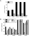
Fig 5. Effect of alterations affecting the amphipathic character of the Erns membrane anchor on lipid binding in vitro and in cells.
(A) Results of lipid pulldown assays with Z2+anchor constructs containing single alanine insertions downstream of positions 181, 194 or 204 (181A, 194A or 204A, respectively) to twist the helix, thereby interfering with the amphipathic conformation. (B) (C) Effects of replacement of individual amino acids in the anchor sequence by proline to interfere with helical folding. The individual exchanges are given in the graphs. (D) (E) Combination of the alterations tested in (A) and (B) (C) by insertion of proline residues into the anchor sequence. The prolines were inserted downstream of the positions indicated in the graphs. (A-E) show the results of lipid pull down assays with the mutated Z2 constructs tested with different lipid compositions as above (see legend of Fig 2 for further information on lipids and vesicle compositions). The bars give the percentage of the total protein recovered in the pellet fractions and represent the mean of at least three independent experiments with the standard deviation shown by error bars. (F) Secretion/retention rates of Erns proteins with the different indicated mutations in the carboxyterminal amphipathic helix. As a reference, wildtype Erns of BVDV CP7 is shown on the left (wt). Phosphoimager based quantification of the results of immunoprecipitation experiments are given below the gel. For each construct the calculated amounts of secreted and retained protein are given in percent of the total recovered protein (sum of the values determined for supernatant and cell extract). All constructs were tested at least three times and the average values are given. As pointed out in detail in the legend to Fig 4C, the increased molecular weight of secreted Erns is due to carbohydrate processing.
Fig 6
Fig 6. Effect of insertions in the amphipathic helix on lipid binding.
3 G residues (GGG) or the sequence GGGGPGGGGPGGGG (GPG) were inserted between residues 210 and 211 of the Erns anchor. The insertions divide the amphipathic helix into an N-terminal and C-terminal part. The mutants were tested in lipid pull down assays with different lipid compositions as above (see legend of Fig 2 for further information on lipids and vesicle compositions). The bars give the percentage of the total protein recovered in the pellet fractions and represent the mean of at least three independent experiments with the standard deviation shown by error bars.
Fig 7
Fig 7. Influence of N-terminal truncation on lipid binding of the Erns anchor.
(A) Sequences of wt (Erns-anchor) and N-terminally truncated anchor sequences. (B) Results of lipid pull-down experiments with the constructs containing the sequences given in (A).
Fig 8
Fig 8. Lipid binding efficiency of internal fragments of the Erns membrane anchor.
(A) Sequences of wt (Erns-anchor) and N-terminally truncated anchor sequences. (B) Results of lipid pull-down experiments with the constructs containing the sequences given in (A).
Fig 9
Fig 9. Effect of C-terminal truncation on lipid binding of an Erns anchor fragment.
(A) Sequences of the Erns-anchor fragment Erns-1 (C-terminal 35 amino acids of the anchor) and C-terminally truncated forms thereof. (B) Results of lipid pull-down experiments with the constructs containing the sequences given in (A).
Fig 10
Fig 10. Determinants of intracellular location of Erns.
In (A), binding of Z2+achor to lipid vesicles with lipid compositions mimicking those of cellular membranes is shown. ER (ER-like), the Golgi apparatus (Golgi-like) or the plasma membrane (PM-like). The percentage of DMPC (PC), DMPE (PE), DMPS (PS), PI (PI), DMPG (PG) und sphingosine (Sph) are given in Table 1. (B) Influence of mutations shown before to influence intracellular retention of Erns [25]. The tested exchanges are indicated in the graph.

References

    1. Thiel HJ, Plagemann PGW, Moennig V. Pestiviruses In: Fields BN, Knipe DM, Howley PM, editors. Fields Virology. 3rd Philadelphia, New York: Lippincott—Raven Publishers; 1996. p. 1059–73.
    1. Simmonds P, Becher P, Collett MS, Gould EA, Heinz FX, Meyers G, et al. Flaviviridae In: King AMQ, Lefkowitz E, Adams MJ, Carstens EB, Fauquet CM, editors. Virus Taxonomy Ninth Report of the International Committee on Taxonomy of Viruses. San Diego, USA: Academic Press; 2012. p. 1003–20.
    1. Lindenbach BD, Thiel HJ, Rice CM. Flaviviridae: The Viruses and Their Replication In: Knipe DM, Howley PM, editors. Fields Virology. 5th Philadelphia, New York: Lippincott—Raven Publishers; 2007. p. 1101–52.
    1. Bauhofer O, Summerfield A, Sakoda Y, Tratschin JD, Hofmann MA, Ruggli N. Classical swine fever virus Npro interacts with interferon regulatory factor 3 and induces its proteasomal degradation. JVirol. 2007;81(7):3087–96. - PMC - PubMed
    1. Chen Z, Rijnbrand R, Jangra RK, Devaraj SG, Qu L, Ma Y, et al. Ubiquitination and proteasomal degradation of interferon regulatory factor-3 induced by Npro from a cytopathic bovine viral diarrhea virus. Virology. 2007;366(2):277–92. - PMC - PubMed

Publication types

LinkOut - more resources