. 2015 Aug 13;10(8):e0135804.
doi: 10.1371/journal.pone.0135804. eCollection 2015.

A Comparison of Ci/Gli Activity as Regulated by Sufu in Drosophila and Mammalian Hedgehog Response

Affiliations

A Comparison of Ci/Gli Activity as Regulated by Sufu in Drosophila and Mammalian Hedgehog Response

Sekyung Oh et al. PLoS One. .

Abstract

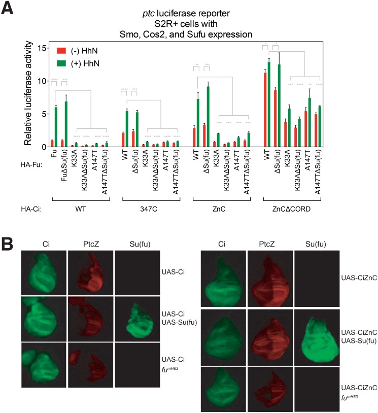
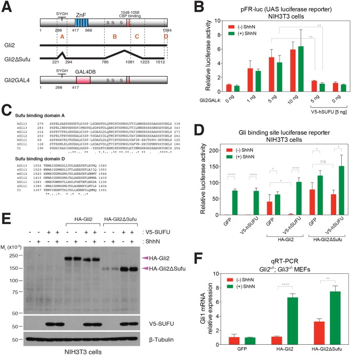
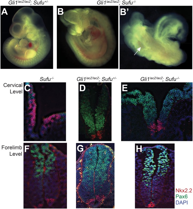
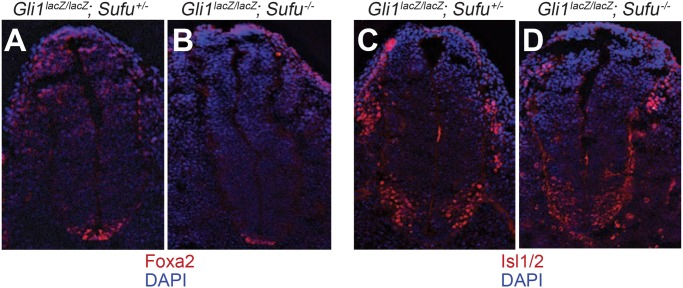
Suppressor of fused (Su(fu)/Sufu), one of the most conserved components of the Hedgehog (Hh) signaling pathway, binds Ci/Gli transcription factors and impedes activation of target gene expression. In Drosophila, the Su(fu) mutation has a minimal phenotype, and we show here that Ci transcriptional activity in large part is regulated independently of Su(fu) by other pathway components. Mutant mice lacking Sufu in contrast show excessive pathway activity and die as embryos with patterning defects. Here we show that in cultured cells Hh stimulation can augment transcriptional activity of a Gli2 variant lacking Sufu interaction and, surprisingly, that regulation of Hh pathway targets is nearly normal in the neural tube of Sufu-/- mutant embryos that also lack Gli1 function. Some degree of Hh-induced transcriptional activation of Ci/Gli thus can occur independently of Sufu in both flies and mammals. We further note that Sufu loss can also reduce Hh induction of high-threshold neural tube fates, such as floor plate, suggesting a possible positive pathway role for Sufu.


Conflict of interest statement

Competing Interests: The authors have declared that no competing interests exist.

Figures

Fig 1
Fig 1. Functional characterization of Su(fu) phosphorylation in Drosophila.
(A) Hh-dependent transcriptional activation was measured by activity of a ptc-luciferase reporter normalized to constitutively expressed control Renilla luciferase. Cells were stimulated with or without conditioned medium containing HhN, the N-terminal signaling domain of the Hh protein. (B) Identification of phosphorylation sites in Su(fu). A schematic diagram of Su(fu) is shown and phosphoserine residues identified by mass spectrometry in endogenous Su(fu) purified from HhN-stimulated Drosophila cl-8 cells are marked with “P”. (C) Western blot analysis of Ala or Glu substitution of Su(fu) phosphoresidues. (D) Endogenous Su(fu) mRNA was targeted using dsRNA corresponding to the 3’ untranslated region (3’UTR), and expression constructs encoding Su(fu) but lacking the 3’UTR sequence were used to test for function of Su(fu) variants, with pathway activity measured as in (A). A representative experiment from at least three independent experiments is shown. Error bars show mean +/- standard deviation. Statistical significance was measured by Student’s t-test: **** (P<0.0001), *** (0.0001<P<0.001), and ns (not significant, P>0.05).
Fig 2
Fig 2. Physical and functional characterization of Fu interaction with Sufu and Cos2.
(A) Schematic representation of Fu variants and their interactions with Su(fu) and Cos2. (B) Distinct regions in Fu bind to Su(fu) and Cos2. S2R+ cells were transiently co-transfected with HA-tagged Fu variants (A) and Myc-tagged Cos2 and V5-tagged Su(fu) constructs. Green arrows denote phosphorylated forms of Su(fu). (C) Su(fu) phosphorylation (green arrows) does not increase in response to Hh in the presence of either kinase-inactive Fu or Fu lacking Su(fu) binding determinants (Δ361–390). (D) Fu kinase functions in Hh-dependent transcriptional activation in cl-8 cells independently of Su(fu) binding determinants. Expression constructs encoding Fu but lacking the 3’UTR sequence were used to test for rescue of Fu function. (E) Fu variants containing Cos2-interacting determinants but lacking kinase activity suppress Hh-dependent transcriptional activity. A representative experiment from at least three independent experiments is shown. Error bars show mean +/- standard deviation. Statistical significance was measured by Student’s t-test: **** (P<0.0001), *** (0.0001<P<0.001), ** (0.001<P<0.01), * (0.01<P<0.05), and ns (not significant, P>0.05). P values in (E) derive from the HhN-induced mediated by each Fu variant as compared to luciferase activity with no Fu expression (blue dotted line).
Fig 3
Fig 3. Reconstitution of Hh signaling in S2R+ Cells.
(A) Western blot analysis of exogenously introduced pathway components. Permuted combinations of pathway components reveal their effects on posttranslational modifications of themselves or other components. Hh-induced phosphorylation of pathway components are indicated by green arrows. β-Tubulin is used as a loading control. (B) Hh pathway activity was measured by expression of luciferase under control of the Hh-responsive ptc promoter (ptc-luciferase) and of the control Renilla luciferase under the ubiquitous copia promoter (copia-Renilla luciferase). Gray numbers indicate fold induction of luciferase activity in response to HhN stimulation. (C) Fu kinase-dependent Ci activation in response to HhN occurs in the context of a Hh-responsive complex comprising Smo, Cos2, Fu and Ci. A representative experiment from at least three independent experiments is shown. Error bars equal to mean +/- standard deviation. Statistical significance was measured by Student’s t-test: **** (P<0.0001) and ns (not significant, P>0.05).
Fig 4
Fig 4. Physical and functional characterization of Su(fu) and Cos2 interactions with Ci.
(A) Schematic representation of Ci variants and their interactions with Su(fu) and Cos2. (B) Su(fu) represses Ci activity through binding to a.a. 1–346 of Ci. (Top) S2R+ cells were transfected with indicated expression constructs, and with dsRNA against Cos2 to deplete endogenous Cos2, which could indirectly bring V5-Su(fu) to FLAG-Ci constructs through endogenous Fu. (Bottom) Su(fu) binding is required for Ci suppression by Su(fu). Increasing amounts of V5-Su(fu) (0, 25, 50, 125 ng) and fixed amount of FLAG-Ci constructs (25 ng) were transfected in S2R+ cells with Cos2 RNAi to deplete endogenous Cos2. (C) CDN and CORD mediate most but not all of the Cos2 binding and repression of Ci. (Top) S2R+ cells were transfected with dsRNA against Su(fu) as well as indicated expression constructs, and subjected to either anti-FLAG or anti-Myc IP. (Bottom) Increasing amounts of Myc-Cos2 (0, 25, 50, 125 ng) and fixed amount of FLAG-Ci constructs (25 ng) were expressed in S2R+ cells with Su(fu) RNAi to deplete endogenous Su(fu). A representative experiment from at least three independent experiments is shown. Error bars show mean +/- standard deviation. Statistical significance was measured by Student’s t-test: **** (P<0.0001), *** (0.0001<P<0.001), ** (0.001<P<0.01), * (0.01<P<0.05), and ns (not significant, P>0.05). P values in (B) and (C) were calculated for pairs of the same DNA mass of V5-Sufu (B) or Myc-Cos2 (C) construct between WT Ci-expressing cells and a Ci deletion/truncation variant-expressing cells.
Fig 5
Fig 5. Fu kinase activates Ci independently of Ci suppression by Su(fu).
(A) S2R+ cell reconstitution assay to test activity of Ci variants. Exogenous expression of core pathway components, Smo, Cos2, Fu, and Su(fu), with Ci produced a reliable Hh-dependent ptc-luciferase activity. (B) Su(fu)-independent activation of Hh-dependent transcriptional targets in the Drosophila wing imaginal disc, as monitored by the ptc-lacZ reporter. Activities of UAS-Ci or UAS-CiZnC, expressed throughout the wing disc under control of the C765-Gal4 driver, were monitored by immunostaining for β-galactosidase (red) and Ci (green). A representative experiment from at least three independent experiments is shown. Error bars show mean +/- standard deviation. Statistical significance was measured by Student’s t-test: **** (P<0.0001), *** (0.0001<P<0.001), ** (0.001<P<0.01), and * (0.01<P<0.05).
Fig 6
Fig 6. Physical and functional characterization of Gli2 interaction with Sufu.
(A) Schematic representation of Gli2 variants used in this study. Gli2 determinants that confer interaction with Sufu is shown in a Gli2 deletion variant Gli2ΔSufu, in which regions A-D indicate Sufu-binding regions (S2 Fig). The SYGH motif is a conserved portion of Sufu binding region A [29]. Gli2GAL4 is a Gli2 variant in which the GAL4 DNA-binding domain replaces the zinc finger domain. (B) NIH3T3 cells transfected for expression of Gli2GAL4 construct with UAS-luciferase reporter (pFR-luc), SV40-Renilla and GFP or hSUFU show suppression of luciferase expression by Sufu, but not Shh induction. Cells were stimulated with or without conditioned medium contatining ShhN, the N-terminal signaling domain of the Sonic Hedgehog (Shh) protein. (C) Amino acid sequence alignment of Sufu-binding regions A and D of Gli/Ci proteins. (D) The Gli2ΔSufu variant lacking Sufu-binding regions is resistant to suppression by Sufu overexpression. NIH3T3 cells were transiently transfected with Gli-luc, SV40-Renilla, and GFP, Gli2 or Gli2ΔSufu, either alone or in combination with either GFP or hSUFU. (E) Exogenous Sufu expression increases Gli2ΔSufu expression level via a mechanism that is not dependent on direct Gli2/Sufu interaction or ShhN stimulation. HA-Gli2 and HA-Gli2ΔSufu expression in NIH3T3 cells with or without ShhN stimulation was examined by co-transfecting with V5-SUFU construct and Western blot analysis. β-Tubulin is used as a loading control. (F) The Gli2ΔSufu variant lacking Sufu-binding regions has high basal pathway activity but is still inducible upon ShhN-stimulation in Gli2 -/- ; Gli3 -/- double mutant MEFs. Gli2 -/- ; Gli3 -/- double mutant MEFs were infected with HA-tagged Gli2 or Gli2ΔSufu coding sequences fused to IRES-GFP in a retroviral expression construct and sorted by GFP intensity. Gli1 mRNA expression was measured using qRT-PCR to indicate Hh pathway activity. A representative experiment from at least three independent experiments is shown. Error bars show mean +/- standard deviation. Statistical significance was measured by Student’s t-test: **** (P<0.0001), *** (0.0001<P<0.001), ** (0.001<P<0.01), * (0.01<P<0.05), and ns (not significant, P>0.05).
Fig 7
Fig 7. Ciliary trafficking of Gli2 in the absence of Sufu function.
Ciliary localization of endogenous Gli2 is undetectable in Sufu -/- MEFs even though Gli2 shuttles in and out of cilia. (A) Immunofluorescence staining of Sufu -/- MEFs and Sufu -/- MEFs rescued with human SUFU (hSUFU) for Gli2 (green) and acetylated Tubulin (AcTub, red) and DAPI (blue). (B) Immunofluorescence staining of Sufu -/- MEFs stably transfected for expression of Dync2hc or control shRNA with antibodies against Gli2 (green) and AcTub (red). (C) Unlike Gli2, a block of retrograde transport in primary cilia by Dync2hc shRNA does not increase ciliary accumulation of Sufu. NIH3T3 cells stably expressing HA-Gli2 with shRNA targeting either Dync2h1 or control were stained with antibodies against AcTub (green) and HA or Sufu (red). Arrowheads denote ciliary tip localization. A representative experiment from at least three independent experiments is shown. Ciliary tip localization of Gli2, Sufu, and HA-Gli2 was scored from more than 50 cells per each replicate and plotted in graphs right to immunofluorescence images. Error bars show mean +/- standard deviation. Statistical significance was measured by Student’s t-test: **** (P<0.0001), *** (0.0001<P<0.001), and ns (not significant, P>0.05).
Fig 8
Fig 8. Loss of Gli1 partially rescues the Sufu mutant phenotype.
(A-B’) Gli1 lacZ/lacZ; Sufu +/- (A) and Gli1 lacZ/lacZ; Sufu -/- (B and B’) E10.5 embryos. Gli1 lacZ/lacZ; Sufu -/- embryos survive beyond E10.5, whereas Sufu -/- embryos die around E9.5. Gli1 lacZ/lacZ; Sufu -/- shows an open neural tube phenotype (B’) (indicated by a white arrow). (C-E) Transverse sections of the neural tube at the cervical level of E9.5 Sufu-/- (C), E10.5 Gli1 lacZ/lacZ; Sufu +/- (D), and Gli1 lacZ/lacZ; Sufu -/- embryos (E) immunostained for markers of neural fate (Nkx2.2: red, Pax6: green, DAPI: blue). In Sufu mutants that survive to E9.5, the ventral spinal cord marker Nkx2.2 expands dorsally (C). However, loss of Gli1 function from Sufu -/- mutant greatly reduced the expression of Nkx2.2 and restored the expression of Pax6 (E). (F-H) Neural tube sections at the level of the forelimb were immunostained for markers of neural fate (Nkx2.2: red, Pax6: green, and blue: DAPI). Embryos were Sufu -/- (E9.5) (F), Gli1 lacZ/lacZ; Sufu +/- (E10.5) (G), and Gli1 lacZ/lacZ; Sufu -/- (E10.5) (H). Note that expression of Nkx2.2 at the level of the forelimb does not expand to more dorsal regions in the Sufu -/- mutant. The expression of Nkx2.2 in Gli1 lacZ/lacZ; Sufu -/- embryos is reduced compared to that in Gli1 lacZ/lacZ; Sufu +/- embryos. A representative experiment from at least three independent experiments is shown.
Fig 9
Fig 9. A positive role for Sufu in Hh signaling in neural tube patterning at forelimb level.
Neural tube sections from E10.5 embryos at the level of the forelimb were immunostained for markers of neural fate (red: Foxa2 (A and B) or Isl1/2 (C and D), blue: DAPI) in Gli1 lacZ/lacZ; Sufu +/- (A and C), and Gli1 lacZ/lacZ; Sufu -/- embryos (B and D) Note that in the Gli1 -/- background, floor plate formation (marked by expression of Foxa2) is reduced by the absence of Sufu function. A representative experiment from at least three independent experiments is shown.

References

    1. Briscoe J, Thérond PP. The mechanisms of Hedgehog signalling and its roles in development and disease. Nat Rev Mol Cell Biol. 2013;14: 416–429. 10.1038/nrm3598 - DOI - PubMed
    1. Varjosalo M, Taipale J. Hedgehog: functions and mechanisms. Genes Dev. 2008;22: 2454–2472. 10.1101/gad.1693608 - DOI - PubMed
    1. Ingham PW, Nakano Y, Seger C. Mechanisms and functions of Hedgehog signalling across the metazoa. Nat Rev Genet. 2011;12: 393–406. 10.1038/nrg2984 - DOI - PubMed
    1. Teglund S, Toftgård R. Hedgehog beyond medulloblastoma and basal cell carcinoma. Biochim Biophys Acta. 2010;1805: 181–208. 10.1016/j.bbcan.2010.01.003 - DOI - PubMed
    1. Hui C-C, Angers S. Gli proteins in development and disease. Annu Rev Cell Dev Biol. 2011;27: 513–537. 10.1146/annurev-cellbio-092910-154048 - DOI - PubMed

Publication types

MeSH terms

LinkOut - more resources