. 2015 Dec 15;21(24):5499-5510.
doi: 10.1158/1078-0432.CCR-14-3091. Epub 2015 Aug 13.

MEK plus PI3K/mTORC1/2 Therapeutic Efficacy Is Impacted by TP53 Mutation in Preclinical Models of Colorectal Cancer

Affiliations

MEK plus PI3K/mTORC1/2 Therapeutic Efficacy Is Impacted by TP53 Mutation in Preclinical Models of Colorectal Cancer

Celina García-García et al. Clin Cancer Res. .

Erratum in

Abstract

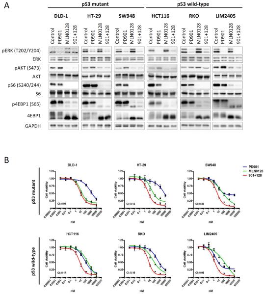
Purpose: PI3K pathway activation occurs in concomitance with RAS/BRAF mutations in colorectal cancer, limiting the sensitivity to targeted therapies. Several clinical studies are being conducted to test the tolerability and clinical activity of dual MEK and PI3K pathway blockade in solid tumors.

Experimental design: In the present study, we explored the efficacy of dual pathway blockade in colorectal cancer preclinical models harboring concomitant activation of the ERK and PI3K pathways. Moreover, we investigated if TP53 mutation affects the response to this therapy.

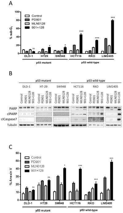
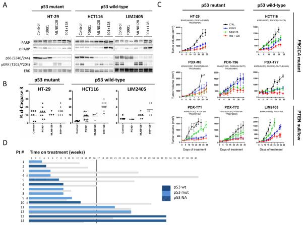
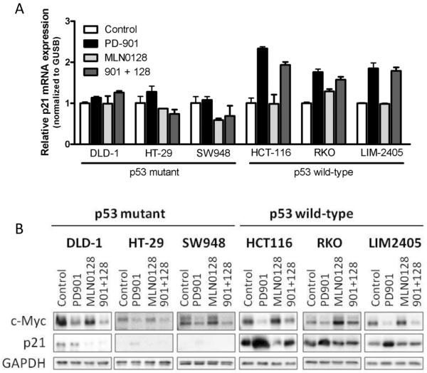
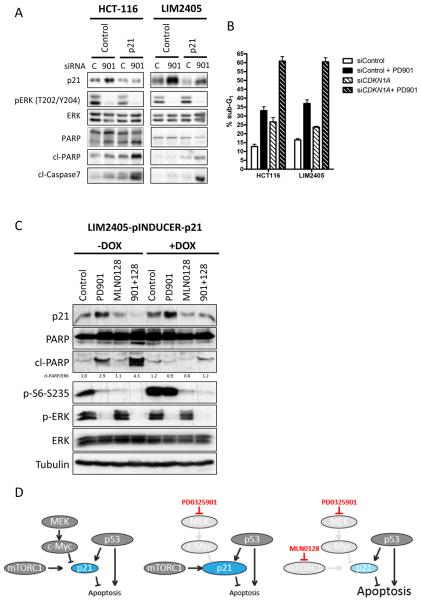
Results: Dual MEK and mTORC1/2 blockade resulted in synergistic antiproliferative effects in cell lines bearing alterations in KRAS/BRAF and PIK3CA/PTEN. Although the on-treatment cell-cycle effects were not affected by the TP53 status, a marked proapoptotic response to therapy was observed exclusively in wild-type TP53 colorectal cancer models. We further interrogated two independent panels of KRAS/BRAF- and PIK3CA/PTEN-altered cell line- and patient-derived tumor xenografts for the antitumor response toward this combination of agents. A combination response that resulted in substantial antitumor activity was exclusively observed among the wild-type TP53 models (two out of five, 40%), but there was no such response across the eight mutant TP53 models (0%). Interestingly, within a cohort of 14 patients with colorectal cancer treated with these agents for their metastatic disease, two patients with long-lasting responses (32 weeks) had TP53 wild-type tumors.

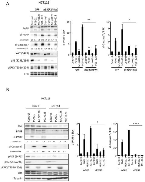
Conclusions: Our data support that, in wild-type TP53 colorectal cancer cells with ERK and PI3K pathway alterations, MEK blockade results in potent p21 induction, preventing apoptosis to occur. In turn, mTORC1/2 inhibition blocks MEK inhibitor-mediated p21 induction, unleashing apoptosis. Clin Cancer Res; 21(24); 5499-510. ©2015 AACR.


Figures

Figure 1
Figure 1. MEK- and mTOR-inhibition suppress pathway activation and proliferation irrespective of p53 mutational status
A. The indicated CRC cell lines were treated with DMSO (Control), 50 nM PD901, 50 nM MLN0128 or the combination of both inhibitors (901 + 128). Whole-cell protein extracts were analyzed by Western blotting with the indicated antibodies. ERK antibody was used as loading control. Figures are representative of three independent experiments. B. The indicated CRC cell lines were treated with 1:10 serial dilutions of PD901, MLN0128 or the combination. Proliferation was measured after 4 days of treatment with a cell viability assay (Cell Titer-Glo, Promega). Proliferation curves were graphed using GraphPad Prism software. Combination index (CI) was calculated using CompuSyn computer software (Fa=0.5). Data are expressed as mean ± SE from 3 independent experiments.
Figure 2
Figure 2. MEK- and mTOR-inhibition induces apoptosis in p53 wild-type CRC cells
The indicated CRC cell lines were treated with DMSO (Control), 50 nM PD901, 50 nM MLN0128 or the combination of both inhibitors (901 + 128). A. Apoptosis was measured after 72 hours of treatment as the percentage of cells with sub-G1 DNA content by flow cytometry and analyzed with FCS Express 4 Flow software. Data are expressed as mean ± S.E from three independent experiments. B. Whole-cell protein extracts were analyzed after 24 hours of treatment by Western blotting with the indicated antibodies. Tubulin antibody was used as loading control. Figures are representative of three independent experiments. C. Apoptosis was measured after 72 hours of treatment quantification of the Annexin V positive cells (Guava Nexin Reagent, Millipore). n.s., not significant, *p<0.5, ***p<0.001.
Figure 3
Figure 3. p53 wild-type CRC tumors benefit from combined MEK- and mTOR- inhibition
Mice bearing HT-29, HCT116 or LIM2405 tumors were treated with 3 consecutive doses of vehicle control (Control, C), PD901 (901, 2mg/kg, 6QD/W), MLN0128 (128, 0.3 mg/kg, 6QD/W) or the combination of both inhibitors (901+ 128). A. Tumors were collected and analyzed by western blot with the indicated antibodies. B. Tumors were collected and analyzed by immunohistochemistry with cleaved caspase 3 antibody. The images were quantified by a pathologist blinded to the identity of the samples. C. Mice bearing HT-29, HCT116, LIM2405, PDX-M6, PDX-T71, PDX-T72, PDX-T77, PDX-T96 were treated as indicated for 20–60 days. Measurements are displayed as mean ± S.E. D. Time on MEK- plus PI3K-therapy of 14 patients (blue) compared to the time on treatment for the previous chemotherapy-containing regimen (grey). The vertical line stands for the 16-week threshold (second response evaluation by computer tomography). The p53 status (sequencing and IHC) is provided in dark blue for wild-type p53, light blue for mutant p53 and shaded blue for non-available data. #, Patients exiting MEK- plus PI3K-therapeutic regimen because of toxicity.
Figure 4
Figure 4. p53 proficiency is required to induce apoptosis in CRC tumors upon anti MEK- and mTOR-inhibition
A. Stable GFP or p53(R248W) HCT116 cells were treated with DMSO (Control), 50 nM PD901, 50 nM MLN0128 or both inhibitors (901 + 128) for 24 hours. Whole-cell protein extracts were analyzed with the indicated antibodies. Figures are representative of three independent experiments, and quantified by densitometry (Image J). B. Stable shGFP or shTP53 HCT116 cells were treated with DMSO (Control), 50 nM PD901, 50 nM MLN0128 or both inhibitors (901 + 128) for 24 hours and resolved as in panel 4A. Figures are representative of three independent experiments, and quantified. * p<0.05. **, p<0.01. ****, p<0.0001
Figure 5
Figure 5. MEK-inhibition induces p21 upregulation in p53 proficient CRC cells
A. CRC cell lines were treated with DMSO (Control), 50 nM PD901, 50 nM MLN0128 or the combination of both for 16 hours. p21 mRNA levels were analyzed by qRT-PCR, normalized to GUSB mRNA levels and expressed as fold change compared to control. B. CRC cell lines were treated with DMSO (Control), 50 nM PD901, 50 nM MLN0128 or the combination of both for 24 hours and whole-cell protein extracts were analyzed by Western blotting with the indicated antibodies. GAPDH antibody was used as loading control. Figures are representative of three independent experiments.
Figure 6
Figure 6. Induction of p21 by MEK-inhibition inhibits apoptosis in p53 wild-type CRC cells
A. HCT116 and LIM2405 cell lines cells were transfected with either a siRNA oligonucleotide targeting p21 (CDKN1A) or a control siRNA oligonucleotide as described. After 24 hours of treatment with DMSO (C) or 50 nM PD901 (901), whole-cell protein extracts were analyzed with the indicated antibodies. Figures are representative of three independent experiments. B. Apoptosis was measured after 72 hours of treatment as percentage of cells with sub-G1 DNA content by flow cytometry and analyzed with FCS Express 4 Flow software. Data are expressed as mean ± S.E from three independent experiments. C. LIM2405 cell line was stably transfected with an inducible p21 vector or with control EGFP. Cells were treated as indicated and after 24 h whole-cell protein extracts were analyzed with the indicated antibodies. D. Model depicting the proposed mechanism of action for the combination of anti-MEK and – mTOR therapy in KRAS/BRAF and PI3K/PTEN mutated CRC in p53 wild-type cells. In wild-type p53 backgrounds, p21 is under transcriptional and translational control of p53, c-Myc and mTORC1. PD0325901 blocks the negative transcriptional control of p21 by MEK/c-Myc and enhances the p21 levels, which inhibit apoptosis induction. Concomitant blockade of mTORC1 prevents translation of p21, thereby enabling apoptosis.

References

    1. Saltz LB, Clarke S, Diaz-Rubio E, Scheithauer W, Figer A, Wong R, et al. Bevacizumab in combination with oxaliplatin-based chemotherapy as first-line therapy in metastatic colorectal cancer: a randomized phase III study. J Clin Oncol. 2008;26:2013–9. - PubMed
    1. Tol J, Koopman M, Cats A, Rodenburg CJ, Creemers GJ, Schrama JG, et al. Chemotherapy, bevacizumab, and cetuximab in metastatic colorectal cancer. N Engl J Med. 2009;360:563–72. - PubMed
    1. Grothey A, Van Cutsem E, Sobrero A, Siena S, Falcone A, Ychou M, et al. Regorafenib monotherapy for previously treated metastatic colorectal cancer (CORRECT): an international, multicentre, randomised, placebo-controlled, phase 3 trial. Lancet. 2013;381:303–12. - PubMed
    1. Benvenuti S, Sartore-Bianchi A, Di Nicolantonio F, Zanon C, Moroni M, Veronese S, et al. Oncogenic activation of the RAS/RAF signaling pathway impairs the response of metastatic colorectal cancers to anti-epidermal growth factor receptor antibody therapies. Cancer Res. 2007;67:2643–8. - PubMed
    1. Sartore-Bianchi A, Martini M, Molinari F, Veronese S, Nichelatti M, Artale S, et al. PIK3CA mutations in colorectal cancer are associated with clinical resistance to EGFR-targeted monoclonal antibodies. Cancer Res. 2009;69:1851–7. - PubMed

Publication types

MeSH terms

Substances