. 2015 Aug 14;6(1):142.
doi: 10.1186/s13287-015-0135-9.

Early severe impairment of hematopoietic stem and progenitor cells from the bone marrow caused by CLP sepsis and endotoxemia in a humanized mice model

Affiliations

Early severe impairment of hematopoietic stem and progenitor cells from the bone marrow caused by CLP sepsis and endotoxemia in a humanized mice model

Tomasz Skirecki et al. Stem Cell Res Ther. .

Abstract

Introduction: An effective immune response to severe bacterial infections requires a robust production of the innate immunity cells from hematopoietic stem and progenitor cells (HSPCs) in a process called emergency myelopoiesis. In sepsis, an altered immune response that leads to a failure of bacterial clearance is often observed. In this study, we aimed to evaluate the impact of sepsis on human HSPCs in the bone marrow (BM) microenvironment of humanized mice subjected to acute endotoxemia and polymicrobial sepsis.

Methods: Humanized mice (hu-NSG) were generated by transplanting NOD.Cg-Prkdc/scidIL2rγ (NSG) mice with the human cord blood CD34(+) cells. Eight weeks after the transplantation, hu-NSG mice were subjected to sepsis induced by endotoxemia-Escherichia coli lipopolysaccharide (LPS)-or by cecal ligation and puncture (CLP). Twenty-four hours later, HSPCs from BM were analyzed by flow cytometry and colony-forming unit (CFU) assay. CLP after inhibition of Notch signaling was also performed. The effects of LPS on the in vitro proliferation of CD34(+) cells from human BM were tested by CellTrace Violet dye staining.

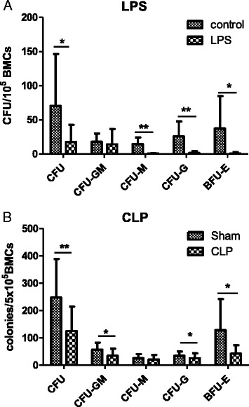
Results: The expression of Toll-like receptor 4 receptor was present among engrafted human HSPCs. Both CLP and endotoxemia decreased (by 43 % and 37 %) cellularity of the BM. In addition, in both models, accumulation of early CD34(+) CD38(-) HSCs was observed, but the number of CD34(+) CD38(+) progenitors decreased. After CLP, there was a 1.5-fold increase of proliferating CD34(+) CD38(-)Ki-67(+) cells. Moreover, CFU assay revealed a depressed (by 75 % after LPS and by 50 % after CLP) production of human hematopoietic colonies from the BM of septic mice. In contrast, in vitro LPS stimulated differentiation of CD34(+) CD38(-) HSCs but did not induce proliferation of these cells in contrast to the CD34(+) CD38(+) progenitors. CLP sepsis modulated the BM microenvironment by upregulation of Jagged-1 expression on non-hematopoietic cells, and the proliferation of HSCs was Notch-dependent.

Conclusions: CLP sepsis and endotoxemia induced a similar expansion and proliferation of early HSCs in the BM, while committed progenitors decreased. It is suggestive that the Notch pathway contributed to this effect. Targeting early hematopoiesis may be considered as a viable alternative in the existing arsenal of supportive therapies in sepsis.


Figures

Fig. 1
Fig. 1
Development of human hematopoietic cells in the bone marrow of NSG mice 8 weeks after transplantation of human CD34+ cells. Representative dot plots show flow cytometry analysis of bone marrow cells with specific anti-human monoclonal antibodies. a Diagram showing mean frequency of human leukocyte subsets in the bone marrow of humanized mice. b Staining of bone marrow from non-humanized NSG mouse with anti-human CD45. c Morphology of bone marrow cells from hu-NSG. d Expression of pan-hematopoietic CD45 antigen in cells gated in R1. e Analysis of monocytes. f Analysis of B cells. g Analysis of T cells. h Analysis of myeloid cells. i Analysis of hematopoietic stem and progenitor cells (gated from R1). j Histogram comparing expression of TLR4 receptor on HSCs and progenitor cells (gated from i). k Expression of TLR4 is present on both undifferentiated Lineage cells and mature Lineage+ cells. The values on graphs present percentages of a given population from a maternal gate. HSC hematopoietic stem cell, hu-NSG humanized NOD.Cg-Prkdc/scidIL2rγ, NSG NOD.Cg-Prkdc/scidIL2rγ, TLR Toll-like receptor
Fig. 2
Fig. 2
Analysis of human HSPC subpopulations in the murine bone marrow after induction of experimental sepsis. Eight weeks after transplantation of human umbilical cord blood CD34+ cells, mice were subjected to endotoxemia (40 μg of LPS intravenous) or cecal ligation and puncture surgery (CLP). Twenty-four hours later, bone marrow cells were harvested, counted, and stained with anti-human antibodies for flow cytometry analysis. Graphs in the upper panel (a, b, c, d) present results obtained in the model of endotoxemia, and graphs in the lower panel (e, f, g, h) impact of CLP on the human HSCPs. n = 6, *P < 0.05, **P < 0.01. C control, HSPC hematopoietic stem and progenitor cell, LPS lipopolysaccharide, TCC total cell count
Fig. 3
Fig. 3
Analysis of the growth of human hematopoietic colonies from bone marrow cells isolated from humanized mice 24 h after induction of experimental sepsis. Twenty-four hours after induction of experimental sepsis, bone marrow cells (5 × 105) from hu-NSG were seeded on methylocellulose medium supplemented with human growth factors. After 20 days of culture, the colonies formed by human progenitor cells were counted. a Results from the model of endotoxemia. b Results from the CLP-induced sepsis model. n = 6, *P < 0.05, **P < 0.01. BMC bone marrow cell, CLP cecal ligation and puncture surgery, hu-NSG humanized NOD.Cg-Prkdc/scidIL2rγ
Fig. 4
Fig. 4
Modulation of the expression of TLR4 and CXCR4 receptors on human HSCs. Changes of TLR4 (a, c) and CXCR4 (e) expression on CD34+ CD38 cells 24 h after single LPS injection. Impact of CLP model on the modulation of TLR4 receptor expression (b, d) and CXCR4 expression (f). Representative histograms of TLR4 (g) and CXCR4 (h) expression on CD34+ CD38 HSCs from control and CLP mice are presented. n = 6, *P < 0.05, **P < 0.01. CLP cecum ligation and puncture, GMF geometric mean fluorescence, HSC hematopoietic stem cell, LPS lipopolysaccharide, TLR Toll-like receptor
Fig. 5
Fig. 5
Impact of LPS on the differentiation and proliferation of purified CD34+ human BM cells in vitro. a Effect of LPS on the frequency of the CD34+ CD38 subpopulation after 9 days of culture. b Effect of LPS on the proliferative index of CD34+ CD38+ subpopulation. c Proliferation of CD34+ CD38+ cells in the presence of LPS. Impact of LPS on the frequency of CD34+ CD38 cells (d) and CD34+ CD38+ cells (e) within first, second, and third generations of daughter cells (fewer than four generations). All results obtained after 9 days of culture in atmosphere of 1 % O2. n = 6, *P < 0.05, **P < 0.001. BM bone marrow, LPS lipopolysaccharide
Fig. 6
Fig. 6
Endotoxemia modulates expression of Jagged-1, a Notch-1 ligand on the bone marrow endothelial cells. Twelve hours after injection of LPS (40 μg), murine femurs were crushed and digested with collagenase II, and then cells were stained with anti-mouse CD45, anti-mouse CD31, and anti-mouse Jagged-1 antibodies. Expression of Jagged-1 on murine CD45CD31+ endothelial cells was measured (a). b Representative histograms overlay of the expression of Jagged-1 after LPS injection and in control mice. n = 6, *P < 0.05. BMC bone marrow cell, GMF geometric mean fluorescence, LPS lipopolysaccharide
Fig. 7
Fig. 7
Effects of Notch pathway inhibition in human HSPCs 24 h after induction of CLP sepsis in hu-mice. Pretreatment with DAPT (60 mg/kg) 2 h before CLP surgery inhibited activation of Notch-1 assessed by fluorescence intensity of NICD (a). Impact of the inhibition of Notch signaling on the CLP-induced expansion of CD34+ CD38 HSCs (b, c) and effect on CD34+ CD38+ progenitors (d, e). Influence of DAPT treatment on the frequency of proliferating HSCs (f), and the rate of apoptosis of CD34+ CD38 cells (g). Effect of DAPT on the expression of G-CSFR on HSPCs after CLP (h), and the phosphorylation of STAT3 (i). n = 3–5. *P < 0.05, **P < 0.001, ***P < 0.001. CLP cecum ligation and puncture, G-CSFR granulocyte colony-stimulating factor receptor, GMF geometric mean fluorescence, HSC hematopoietic stem cell, HSPC hematopoietic stem and progenitor cell, NICD Notch-1 intracellular domain

References

    1. Dombrovskiy VY, Martin AA, Sunderram J, Paz HL. Rapid increase in hospitalization and mortality rates for severe sepsis in the United States: a trend analysis from 1993 to 2003. Crit Care Med. 2007;35:1244–50. doi: 10.1097/01.CCM.0000261890.41311.E9. - DOI - PubMed
    1. Levy MM, Artigas A, Phillips GS, Rhodes A, Beale R, Osborn T, et al. Outcomes of the Surviving Sepsis Campaign in intensive care units in the USA and Europe: a prospective cohort study. Lancet Infect Dis. 2012;12:919–24. doi: 10.1016/S1473-3099(12)70239-6. - DOI - PubMed
    1. Dellinger RP, Carlet JM, Masur H, Gerlach H, Calandra T, Cohen J, et al. Surviving Sepsis Campaign guidelines for management of severe sepsis and septic shock. Intensive Care Med. 2004;30:536–55. doi: 10.1007/s00134-004-2398-y. - DOI - PubMed
    1. Manz MG, Boettcher S. Emergency granulopoiesis. Nat Rev Immunol. 2014;14:302–14. doi: 10.1038/nri3660. - DOI - PubMed
    1. Nagai Y, Garrett KP, Ohta S, Bahrun U, Kouro T, Akira S, et al. Toll-like receptors on hematopoietic progenitor cells stimulate innate immune system replenishment. Immunity. 2006;24:801–12. doi: 10.1016/j.immuni.2006.04.008. - DOI - PMC - PubMed

Publication types