Review
. 2013 Dec 31;1(4):311-22.
doi: 10.4248/BR201304002. eCollection 2013 Dec.

Skeletal Blood Flow in Bone Repair and Maintenance

Affiliations
Review

Skeletal Blood Flow in Bone Repair and Maintenance

Ryan E Tomlinson et al. Bone Res. .

Abstract

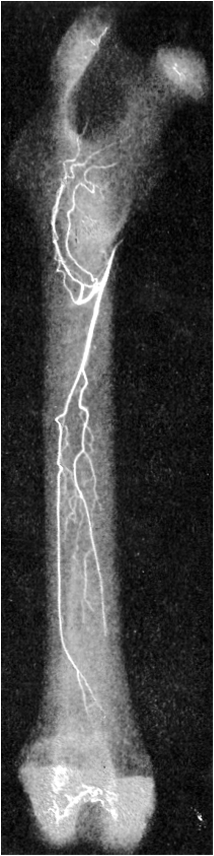
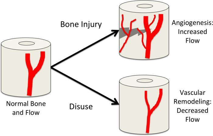
Bone is a highly vascularized tissue, although this aspect of bone is often overlooked. In this article, the importance of blood flow in bone repair and regeneration will be reviewed. First, the skeletal vascular anatomy, with an emphasis on long bones, the distinct mechanisms for vascularizing bone tissue, and methods for remodeling existing vasculature are discussed. Next, techniques for quantifying bone blood flow are briefly summarized. Finally, the body of experimental work that demonstrates the role of bone blood flow in fracture healing, distraction osteogenesis, osteoporosis, disuse osteopenia, and bone grafting is examined. These results illustrate that adequate bone blood flow is an important clinical consideration, particularly during bone regeneration and in at-risk patient groups.

Keywords: angiogenesis; blood flow; bone repair; fracture; vascular remodeling.


Figures

Figure 1
Figure 1
Femoral blood supply. The principal nutrient artery and its main medullary branches are shown in this angiograph of a rabbit femur (10).
Figure 2
Figure 2
The vascular role in fracture. Blood flow rate was quantified using PET imaging after tibial fracture (58). A) In an uninjured limb, blood flow is highest in the muscle (M) near the tibia (T). B) In contrast, blood flow rate in and around the tibia (T) is markedly increased following fracture. This increase in blood flow rate is due in part to angiogenesis. Recent work has shown that these new blood vessels coinvade the cartilaginous template along with osteoblast precursors during endochondral fracture repair (71). C) In the periosteal fracture callus, Osx-expressing osteoprogenitors (green) are intimately associated with new vasculature (red) seven days after fracture. Pockets of avascular cartilage are labeled with a *. Figures used with permission (License Number: 3266071128303).
Figure 3
Figure 3
Rapid angiogenesis after bone injury. Damaging mechanical loading that culminates in an ulnar stress fracture is associated with significant angiogenesis to increase blood flow rate following injury (91). Microfil vascular perfusion was used to visualize periosteal vascularity in control (A) and fractured ulnae (B). Figures used with permission (License Number: 3260231064494).
Figure 4
Figure 4
Vascular remodeling after unloading. Following cannulation with micropipettes, the diameter of the primary nutrient artery was measured in A) control and B) 14 day hindlimb unloaded animals (35). These results demonstrate that blood flow is decreased following chronic skeletal unloading by vascular remodeling. Figures used with permission (License Number: 3260231186916).
Figure 5
Figure 5
Vascular responses in bone. In this schematic, normal bone blood flow is represented on the left. In response to bone injury, such as a fracture, robust angiogenesis occurs to relieve oxygen tension and transport osteoprogenitor cells for repair. In contrast, bone disuse can result in decreased blood flow by vascular remodeling.

References

    1. Brandi ML, Collin-Osdoby P. Vascular biology and the skeleton. J Bone Miner Res. 2006;21:183–192. - PubMed
    1. McCarthy I. The physiology of bone blood flow: a review. J Bone Joint Surg Am. 2006;88 Suppl 3:4–9. - PubMed
    1. Marenzana M, Arnett TR. The key role of the blood supply to bone. Bone Res. 2013;3:203–215. - PMC - PubMed
    1. Maes C, Carmeliet G, Schipani E. Hypoxia-driven pathways in bone development, regeneration and disease. Nat Rev Rheumatol. 2012;8:358–366. - PubMed
    1. Burkhardt R, Kettner G, Bohm W, Schmidmeier M, Schlag R, Frisch B, Mallmann B, Eisenmenger W, Gilg T. Changes in trabecular bone, hematopoiesis and bone marrow vessels in aplastic anemia, primary osteoporosis, and old age: a comparative histomorphometric study. Bone. 1987;8:157–164. - PubMed