. 2015 Aug 14;10(8):e0135374.
doi: 10.1371/journal.pone.0135374. eCollection 2015.

Absence of Vitamin K-Dependent γ-Carboxylation in Human Periostin Extracted from Fibrotic Lung or Secreted from a Cell Line Engineered to Optimize γ-Carboxylation

Affiliations

Absence of Vitamin K-Dependent γ-Carboxylation in Human Periostin Extracted from Fibrotic Lung or Secreted from a Cell Line Engineered to Optimize γ-Carboxylation

Douglas S Annis et al. PLoS One. .

Abstract

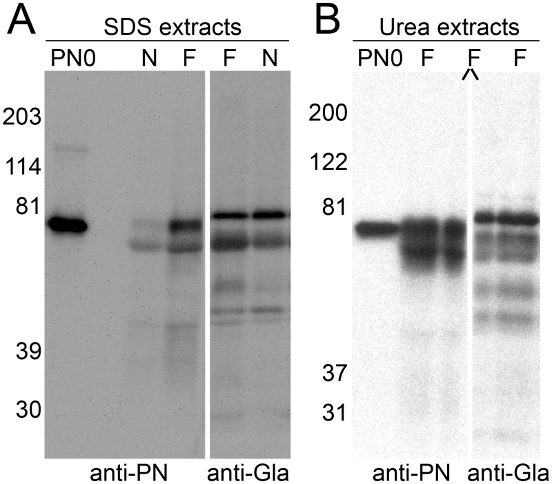
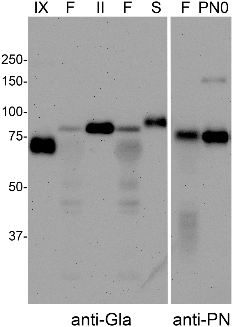
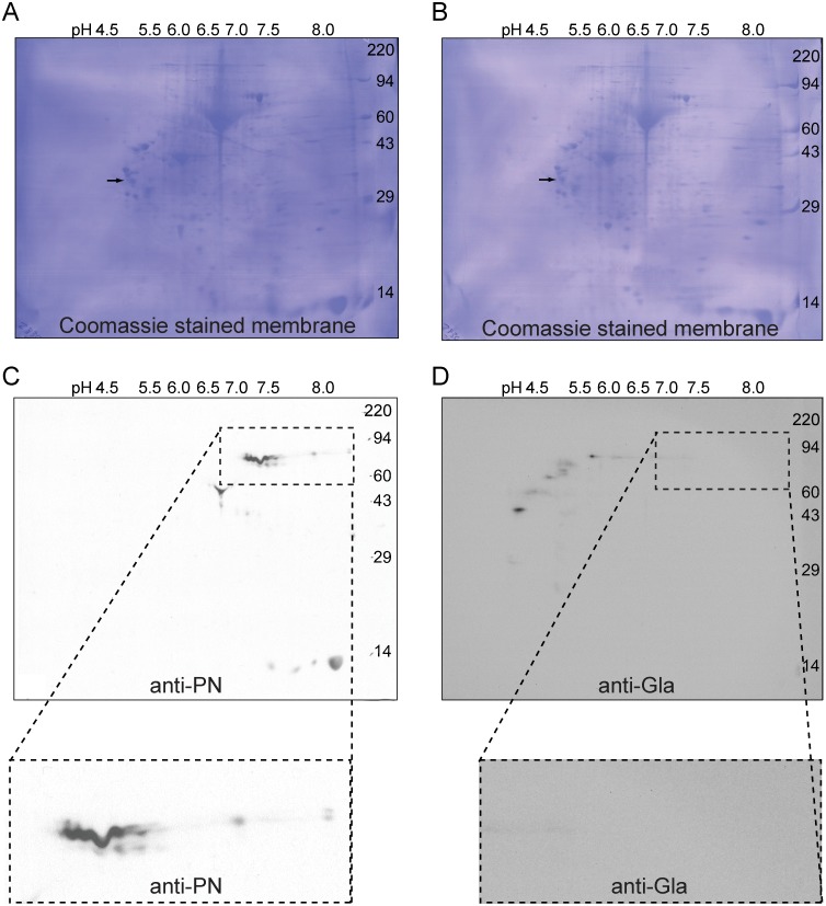
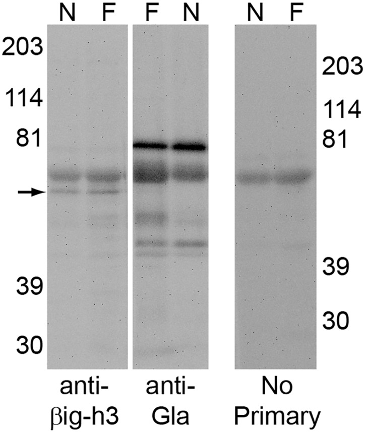
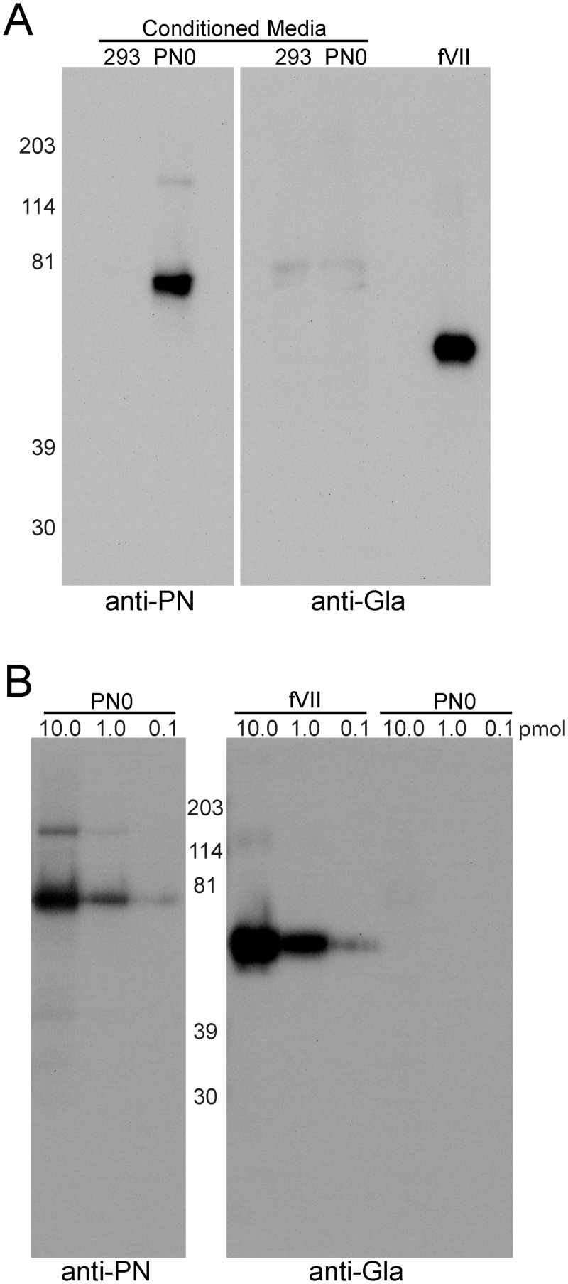
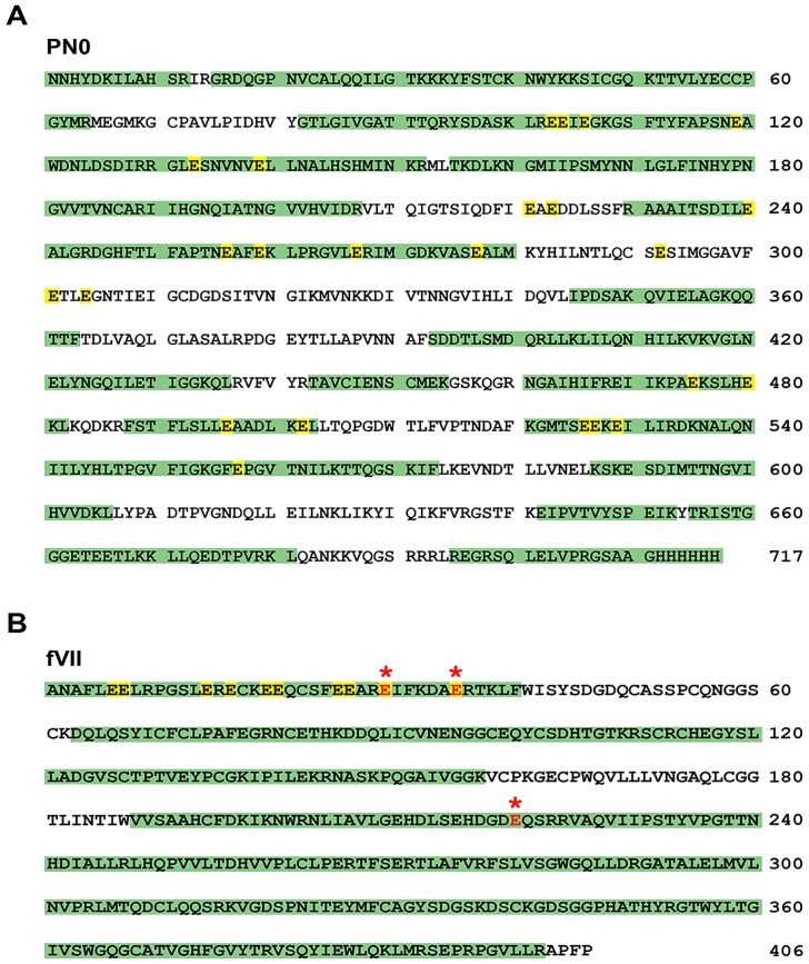
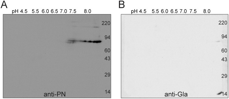
Periostin (PN, gene name POSTN) is an extracellular matrix protein that is up-regulated in bronchial epithelial cells and lung fibroblasts by TH-2 cytokines. Its paralog, TGF-β-induced protein (βig-h3, gene name TGFBI), is also expressed in the lung and up-regulated in bronchial myofibroblasts by TGF-β. PN and βig-h3 contain fasciclin 1 modules that harbor putative recognition sequences for γ-glutamyl carboxylase and are annotated in UniProt as undergoing vitamin K-dependent γ-carboxylation of multiple glutamic acid residues. γ-carboxylation profoundly alters activities of other proteins subject to the modification, e.g., blood coagulation factors, and would be expected to alter the structure and function of PN and βig-h3. To analyze for the presence of γ-carboxylation, proteins extracted from fibrotic lung were reacted with monoclonal antibodies specific for PN, βig-h3, or modification with γ-carboxyglutamic acid (Gla). In Western blots of 1-dimensional gels, bands stained with anti-PN or -βig-h3 did not match those stained with anti-Gla. In 2-dimensional gels, anti-PN-positive spots had pIs of 7.0 to >8, as expected for the unmodified protein, and there was no overlap between anti-PN-positive and anti-Gla-positive spots. Recombinant PN and blood coagulation factor VII were produced in HEK293 cells that had been transfected with vitamin K 2, 3-epoxide reductase C1 to optimize γ-carboxylation. Recombinant PN secreted from these cells did not react with anti-Gla antibody and had pIs similar to that found in extracts of fibrotic lung whereas secreted factor VII reacted strongly with anti-Gla antibody. Over 67% coverage of recombinant PN was achieved by mass spectrometry, including peptides with 19 of the 24 glutamates considered targets of γ-carboxylation, but analysis revealed no modification. Over 86% sequence coverage and three modified glutamic acid residues were identified in recombinant fVII. These data indicate that PN and βig-h3 are not subject to vitamin K-dependent γ-carboxylation.


Conflict of interest statement

Competing Interests: The authors have declared that no competing interests exist.

Figures

Fig 1
Fig 1. Fibrotic lung extracts have increased amounts of PN, and alignment of PN-positive and Gla-positive bands is inconclusive.
(A) A representative Western blot of proteins extracted with SDS from pulverized lung tissue (10mg tissue per 0.1 mL extraction buffer). SDS extracts (2.5 μl/lane) of fibrotic (F) and normal (N) lung sample, were blotted with mouse anti-PN (left) and anti-Gla (right). PN-positive bands in lung extracts migrate at approximately 79 kDa, similar to the migration of PN0 expressed in HEK293VKOR cells (PN0). (B) Urea/EDTA extracts (10.0 μl/lane) of fibrotic lung (F) have protein patterns similar to those seen with the SDS extracts. A lane has been split (side slashes) and probed with both anti-PN (left) and anti-Gla (right) antibodies. The anti-PN- and anti-Gla-positive bands do not align exactly but bands do overlap. PN0 was expressed in insect cells with baculovirus.
Fig 2
Fig 2. Gla positive bands in fibrotic lung extracts co-migrate with fII and fIX.
SDS-extracts of fibrotic lung (F) and purified fIX (IX), fII (II), and protein S (S) were immunoblotted with anti-Gla (left). Gla positive bands in fibrotic lung extracts have similar migration and band shape as fII and fIX. Protein S migrated slightly slower than fII. Periostin from fibrotic lung extracts is detected in a 79 kDa band between fIX and fII, and has a similar migration to recombinant PN0 produced in HEK293VKOR cells (PN0) when stained with anti-PN (right).
Fig 3
Fig 3. PN extracted from fibrotic human lung has the pI expected of un-modified protein and does not react with anti-Gla.
Approximately 200 μg of protein extracted with SDS from fibrotic lung samples were subjected in parallel to 2-dimensional isoelectric focusing/electrophoresis, transferred to PVDF membranes, and Western blotted. The membranes were stained with Coomassie blue (A and B) and probed with (C) mouse anti-PN or (D) mouse anti-Gla mAbs. Enlarged views of the PN-positive region are shown in the insets for both blots. Isoelectric points of PN-positive spots ranged between pH 7.0 and > pH 8.0. Gla-positive spots were more acidic and did not overlap with PN positive spots. An internal isoelectric focusing standard, tropomyosin, migrated as a doublet with the lower polypeptide spot (arrows) of MW 33 kDa and pI 5.2 on the parallel 10% gels.
Fig 4
Fig 4. βig-h3 extracted from lung does not react with anti-Gla.
Pulverized fibrotic (F) and normal (N) lung samples were extracted with SDS and probed with anti-βig-h3 (left) and anti-Gla (middle). βig-h3 positive bands (arrow) were observed in both normal and fibrotic lung extractions. The βig-h3 band was not detected with the anti-Gla antibody. A duplicate blot, from a different gel, had primary antibody omitted (right), and shows a band attributed to the non-specific binding of the secondary antibody to the lung extracts. This band is seen in both the anti-βig-h3 (left), and the anti-Gla (middle) blots.
Fig 5
Fig 5. Recombinant Vitamin K-dependent coagulation factor VII produced in HEK293VKOR cells is γ-carboxylated whereas PN is not.
(A) Conditioned media containing 10 μg/mL vitamin K from HEK293VKOR un-transfected (293) or transfected with PN0 (PN0), 30 μl, or 0.5 pmol of purified factor VII (fVII) were examined in Western blot. Mouse anti-PN (left) reacted strongly with media of cells transfected with PNO but not with control media. Both media were negative when probed with the mouse anti-Gla (right), but purified fVII reacted strongly. (B) Western blot analysis of purified fVII and PN0 expressed by HEK293VKOR cells in the presence of vitamin K. Varying amounts, 10.0, 1.0 and 0.1 pmol, of recombinant protein were probed with anti-PN (left) and anti-Gla (right). The anti-Gla antibody reacted with all inputs tested of fVII but not with PN.
Fig 6
Fig 6. Mass Spec sequence coverage for PN0 and FVII.
Combined ETD and AI-ETD sequence coverage of tryptic and chymotryptic digests of purified proteins. Mass spectrometry analysis obtained (A) 68% sequence coverage of PN0 and (B) 86% coverage of fVII. Identified peptides are shown in green, with Uniprot annotated γ-carboxylation sites highlighted in yellow. Experimentally verified γ-carboxylation sites for fVII are shown highlighted in red. Sequence and numbering are of the recombinantly expressed and secreted proteins and are therefore without the 21-residue signal sequence of PN or the 20-residue signal and 40-residue propeptide sequences of fVII.
Fig 7
Fig 7. Recombinant PN expressed in HEK293VKOR cells has the expected pIs of non- γ-carboxylated protein.
Purified PN0, 2.3 μg produced in the presence of 10 μg/mL vitamin K by HEK293VKOR cells was run on two-dimensional isoelectric focusing/electrophoresis and Western blotting gels. The parallel 8% gels were transferred to PVDF membranes and probed with (A) mouse anti-PN, or (B) mouse anti-Gla. Isoelectric points of PN positive spots ranged between pH 7.0 and > pH 8.0, consistent with unmodified protein. No anti-Gla-positive spots were detected.

References

    1. Takeshita S, Kikuno R, Tezuka K, Amann E. Osteoblast-specific factor 2: cloning of a putative bone adhesion protein with homology with the insect protein fasciclin I. Biochem J. 1993;294 (Pt 1):271–8. - PMC - PubMed
    1. Skonier J, Neubauer M, Madisen L, Bennett K, Plowman GD, Purchio AF. cDNA cloning and sequence analysis of beta ig-h3, a novel gene induced in a human adenocarcinoma cell line after treatment with transforming growth factor-beta. DNA and cell biology. 1992;11(7):511–22. . - PubMed
    1. Coutu DL, Wu JH, Monette A, Rivard GE, Blostein MD, Galipeau J. Periostin, a member of a novel family of vitamin K-dependent proteins, is expressed by mesenchymal stromal cells. J Biol Chem. 2008;283(26):17991–8001. Epub 2008/05/03. doi: M708029200 [pii]10.1074/jbc.M708029200 . - DOI - PubMed
    1. Mann KG, Nesheim ME, Church WR, Haley P, Krishnaswamy S. Surface-dependent reactions of the vitamin K-dependent enzyme complexes. Blood. 1990;76(1):1–16. . - PubMed
    1. Berkner KL. Vitamin K-dependent carboxylation. Vitamins and hormones. 2008;78:131–56. 10.1016/S0083-6729(07)00007-6 . - DOI - PubMed

Publication types