

Comparative Study
doi: 10.1165/rcmb.2015-0147LE.
Flow Cytometry Reveals Similarities Between Lung Macrophages in Humans and Mice
Affiliations
- PMID: 26274047
- PMCID: PMC4742931
- DOI: 10.1165/rcmb.2015-0147LE
Item in Clipboard
Comparative Study
Flow Cytometry Reveals Similarities Between Lung Macrophages in Humans and Mice
Am J Respir Cell Mol Biol.
2016 Jan.
No abstract available
Figures
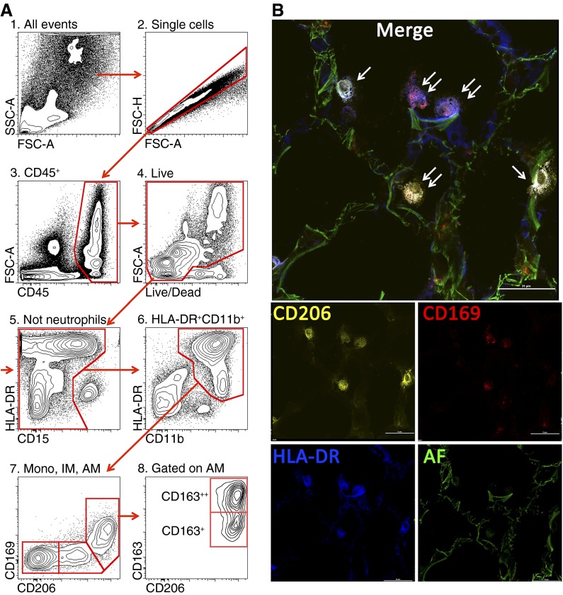
(A) Gating strategy used to identify lung monocytes and macrophages. After excluding doublets (2), cells of hematopoietic origin were identified as CD45+ (3), followed by exclusion of the dead cells (precautions were taken not to gate out highly autofluorescent alveolar macrophages) (4). Neutrophils were identified as CD11b++CD15+CD16+HLA-DR− cells and excluded from analysis (5). We then gated on CD11b++HLA-DR+ cells (6). This allows separation from natural killer (NK) cells (CD11b+HLA-DR−CD56+) and highly autofluorescent eosinophils (CD11b+/−HLA-DR−Siglec 8+). Finally, using CD206 and CD169, cells were separated into three subpopulations: alveolar macrophages (CD11b+HLA-DR++CD206++CD169+FSChighSSChigh, AM), interstitial macrophages (CD11b+HLA-DR++CD206+CD169−, IM), and monocytes (CD11b+HLA-DR+CD206−CD169−, mono) (7). CD163 identifies two subpopulations of alveolar macrophages (8). (B) Immunofluorescent microscopy on a normal human lung. Orange is CD206 PE, red is CD169 AF647, blue is HLA-DR BV421, and green is autofluorescence in fluorescein isothiocyanate channel. Single arrows indicate HLA-DR+CD206+CD169− cells (interstitial macrophages), and double arrows indicate HLA-DR+CD206+CD169+ cells (alveolar macrophages). Scale bar is 31 μm.
Comment in
-
Pulmonary Macrophages: Overlooked and Underappreciated.Am J Respir Cell Mol Biol. 2016 Jan;54(1):1-2. doi: 10.1165/rcmb.2015-0270ED. Am J Respir Cell Mol Biol. 2016. PMID: 26720905 No abstract available.
References
Publication types
MeSH terms
Substances
Grants and funding
- ES013995/ES/NIEHS NIH HHS/United States
- R01 ES013995/ES/NIEHS NIH HHS/United States
- AR061593/AR/NIAMS NIH HHS/United States
- K08 HL125940/HL/NHLBI NIH HHS/United States
- HL071643/HL/NHLBI NIH HHS/United States
- P01 HL108795/HL/NHLBI NIH HHS/United States
- HL108795/HL/NHLBI NIH HHS/United States
- R03 AR061593/AR/NIAMS NIH HHS/United States
- K23 HL093302/HL/NHLBI NIH HHS/United States
- P01 AG049665/AG/NIA NIH HHS/United States
- P01 HL071643/HL/NHLBI NIH HHS/United States
- R01 AR064546/AR/NIAMS NIH HHS/United States
- AR050250/AR/NIAMS NIH HHS/United States
- P30 CA060553/CA/NCI NIH HHS/United States
- HL079190 09/HL/NHLBI NIH HHS/United States
- HL124664 01/HL/NHLBI NIH HHS/United States
- R21 AI092490/AI/NIAID NIH HHS/United States
- AR064546/AR/NIAMS NIH HHS/United States
- R01 AR050250/AR/NIAMS NIH HHS/United States
- AG049665/AG/NIA NIH HHS/United States
- R01 HL124664/HL/NHLBI NIH HHS/United States
- R01 HL079190/HL/NHLBI NIH HHS/United States
- AI092490/AI/NIAID NIH HHS/United States
LinkOut - more resources
Full Text Sources
Other Literature Sources
Medical
