Comparative Study
. 2015 Aug 14;10(8):e0134247.
doi: 10.1371/journal.pone.0134247. eCollection 2015.

Spatial and Temporal Control of Cavitation Allows High In Vitro Transfection Efficiency in the Absence of Transfection Reagents or Contrast Agents

Affiliations
Comparative Study

Spatial and Temporal Control of Cavitation Allows High In Vitro Transfection Efficiency in the Absence of Transfection Reagents or Contrast Agents

Kamel Chettab et al. PLoS One. .

Abstract

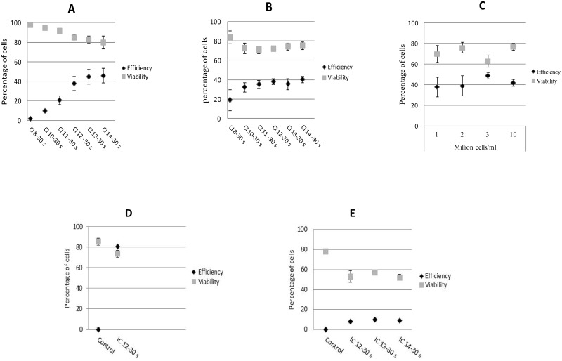
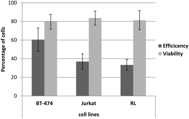
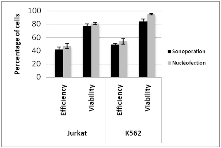
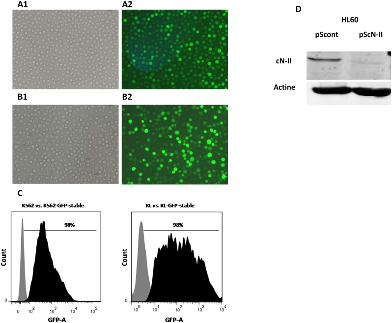
Sonoporation using low-frequency high-pressure ultrasound (US) is a non-viral approach for in vitro and in vivo gene delivery. In this study, we developed a new sonoporation device designed for spatial and temporal control of ultrasound cavitation. The regulation system incorporated in the device allowed a real-time control of the cavitation level during sonoporation. This device was evaluated for the in vitro transfection efficiency of a plasmid coding for Green Fluorescent Protein (pEGFP-C1) in adherent and non-adherent cell lines. The transfection efficiency of the device was compared to those observed with lipofection and nucleofection methods. In both adherent and non-adherent cell lines, the sonoporation device allowed high rate of transfection of pEGFP-C1 (40-80%), as determined by flow cytometry analysis of GFP expression, along with a low rate of mortality assessed by propidium iodide staining. The transfection efficiency and toxicity of sonoporation on the non-adherent cell lines Jurkat and K562 were similar to those of nucleofection, while these two cell lines were resistant to transfection by lipofection. Moreover, sonoporation was used to produce three stably transfected human lymphoma and leukemia lines. Significant transfection efficiency was also observed in two fresh samples of human acute myeloid leukemia cells. In conclusion, we developed a user-friendly and cost-effective ultrasound device, well adapted for routine in vitro high-yield transfection experiments and which does not require the use of any transfection reagent or gas micro-bubbles.


Conflict of interest statement

Competing Interests: The authors declare the following financial competing interests. KC was a paid employee by Caviskills SAS. CL and JLM own shares of Caviskills SAS and patent applications licensed to the company and are consultants of the company. These elements do not alter their adherence to PLOS ONE policies on sharing data and materials.

Figures

Fig 1
Fig 1. Experimental setup, cavitation control and pressure application.
(A) Experimental setup. (B) Pressure field (dB) at the focal point (C) Relationship between the measured fluorescence levels in 550 μL of terephthalic acid medium (20 mM) and the cavitation index (sonication time: 60 seconds).
Fig 2
Fig 2. Inertial cavitation characterization and control.
(A) CI responses using cavitation control process without feedback loop power (constant peak electric power applied: 74 W; corresponding peak pressure: ~3MPa). (B) and (C), 30 seconds sonication in 650 μL of water in a 2mL tube. (D) Zoom of Fig 2C from 13 to 14 seconds. The power was controlled to maintain CI at values of 12 and 14, respectively. The peak pressure was calculated from the monitored electrical power.
Fig 3
Fig 3. Effect of ultrasound on plasmid integrity.
Agarose gel electrophoresis of peGF-C1 plasmid. (A) Ultrasound exposure for 5–80 seconds at CI level 12 did not affect plasmid integrity. (B) Plasmid exposure to different CI levels (10, 12, 14 and 16) for 30 seconds did not damage the plasmid integrity. L, Low DNA Ladder (Life Technologies, Rockville, MA, USA). The size and length of bands were the same between exposed and non-exposed plasmids, indicating that ultrasound irradiation does not affect plasmid integrity.
Fig 4
Fig 4. Influence of CI level and cell density on transfection efficiency and cell viability.
Twenty-four hours post-sonoporation, the percentages of GFP-positive cells and levels of cytotoxicity were assessed by flow cytometry and PI staining. Two non-adherent cell lines Jurkat (A) and K562 (B) were exposed to CI values ranging between 8 and 14 during 30 seconds. The sonoporation with peGFP-C1 was performed in a 2 ml Eppendorf tube containing 10 μg of pEGFP-C1 in 550 μl of Opti-MEM medium. The cell concentration was set to 2.106 cells/ml. (C) Transfection of 10 μg peGFP-C1 in Jurkat cells at four different cell densities (1.106, 2.106, 3.106, and 10.106 cells/ml). Percentage of GFP-positive cells and dead cells were determined at different cell densities using CI of 12 for 30 seconds. (D) Transfection efficiency and cell viability of the A549 cell line (10 μg peGFP-C1, 10.106 cells/ml, and CI 12–30 seconds). (E) Transfection efficiency and cell viability of fresh AML cells. The error bars represent the mean (± standard deviation, SD) of triplicates and two measurements for Figs A-D and E, respectively.
Fig 5
Fig 5. Biological reproducibility of the device.
Assays were conducted on two non-adherent cell lines cell lines (RL and Jurkat) and one adherent cell line (BT474). Sonoporation was performed using the optimal parameters (10.106 cells/ml, IC of 12 for 30 seconds and 10 μg of pEGFP-C1). Twenty-four hours post-transfection cells were collected and analyzed for transfection efficiency (% GFP+ cells) and for viability (% of cells excluding PI). All experiments were performed at least in triplicate and repeated independently three times for each cell line. Statistical analysis using a non-parametric Wilcoxon test demonstrated no significant difference in transfection efficiency and in cell viability.
Fig 6
Fig 6. Comparison of the transfection efficacy of pEGFP-C1 with sonoporation and nucleofection.
Transfection efficiency (black bar) and cell viability (grey bar) were determined by flow cytometry analysis 24 hours post-sonoporation and nucleofection of non-adherent JURKAT and K562 cell lines. The error bars represent the mean (± standard deviation, SD) of triplicate measurements.
Fig 7
Fig 7. Production of stably transfected K562, RL and HL60 cells by sonoporation.
K562 and RL cell lines were transfected with pEGFP-C1 plasmid and cultured in selection medium containing 1.2 and 1.5 mg/ml of G418, respectively. The microscopy pictures (objective 10×) correspond to light transmission (A1 and B1) and fluorescence images of RL-pEGFP-C1 (A2) and K562-pEGFP-C1 (B2) stable clones. (C) The GFP channel histograms obtained from flow cytometry are shown. Grey indicates non-transfected cell lines and black indicates GFP-transfected cell lines. Flow cytometry analysis revealed 98% GFP positive cells in the stable clones. (D) HL-60 cell line transfected with expression vectors coding for shRNA against cN-II (pScN-II) and control sequence (pScont) were selected with continuous exposure to 1 mg/ml (HL60-pScN-II and HL60-pScont cells) of G418. Western blot analysis clearly showed strong inhibition of cN-II expression in HL60-pScN-II cells compared with cells expressing a non-silencing shRNA. Results are representative of three independent western blot analyses.

Similar articles

Cited by

References

    1. Marshall E (1999) Gene therapy death prompts review of adenovirus vector. Science 286: 2244–2245. - PubMed
    1. Baum C, Kustikova O, Modlich U, Li Z, fehse B (2006) Mutagenesis and oncogenesis by chromosomal insertion of gene transfer vectors. Hum Gene Ther 17: 253–263. - PubMed
    1. Waehler R, Russell SJ, Curiel DT (2007) Engineering targeted viral vectors for gene therapy. Nature 8: 573–587. - PMC - PubMed
    1. Bouard D, Alazard-Dany N, Cosset FL (2009) Viral vectors: from virology to transgene expression. Br J Pharmacol 157: 153–165. 10.1038/bjp.2008.349 - DOI - PMC - PubMed
    1. Pack DW, Hoffman AS, Pun S, Stayton PS (2005) Design and development of polymers for gene delivery. Nat Rev Drug Discov 5: 581–593. - PubMed

Publication types

LinkOut - more resources