. 2015 Sep 4;349(6252):1115-20.
doi: 10.1126/science.aac7049. Epub 2015 Jul 23.

RNA editing by ADAR1 prevents MDA5 sensing of endogenous dsRNA as nonself

Affiliations

RNA editing by ADAR1 prevents MDA5 sensing of endogenous dsRNA as nonself

Brian J Liddicoat et al. Science. .

Abstract

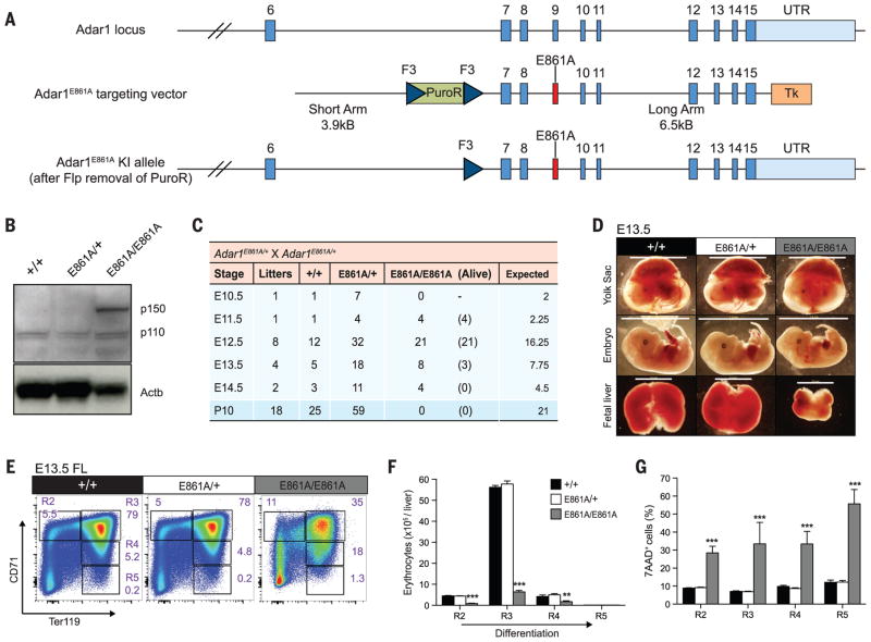
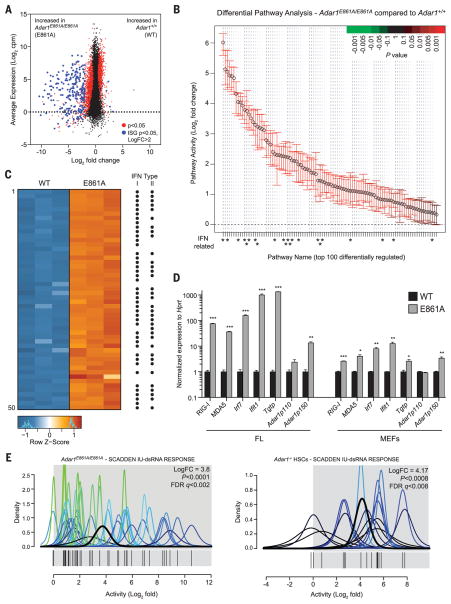
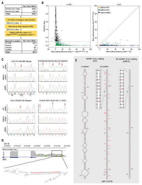
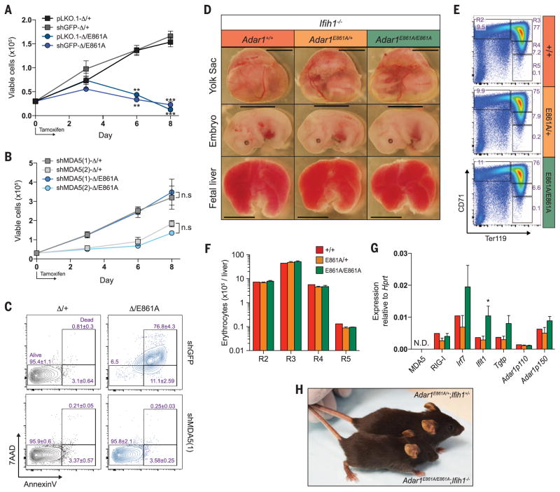
Adenosine-to-inosine (A-to-I) editing is a highly prevalent posttranscriptional modification of RNA, mediated by ADAR (adenosine deaminase acting on RNA) enzymes. In addition to RNA editing, additional functions have been proposed for ADAR1. To determine the specific role of RNA editing by ADAR1, we generated mice with an editing-deficient knock-in mutation (Adar1(E861A), where E861A denotes Glu(861)→Ala(861)). Adar1(E861A/E861A) embryos died at ~E13.5 (embryonic day 13.5), with activated interferon and double-stranded RNA (dsRNA)-sensing pathways. Genome-wide analysis of the in vivo substrates of ADAR1 identified clustered hyperediting within long dsRNA stem loops within 3' untranslated regions of endogenous transcripts. Finally, embryonic death and phenotypes of Adar1(E861A/E861A) were rescued by concurrent deletion of the cytosolic sensor of dsRNA, MDA5. A-to-I editing of endogenous dsRNA is the essential function of ADAR1, preventing the activation of the cytosolic dsRNA response by endogenous transcripts.


Figures

Fig. 1
Fig. 1. Adar1E861A/E861A embryos die in utero
(A) Schematic of Adar1E861A knock-in allele. (B) ADAR1 protein expression in whole E12.5 embryos of the indicated genotypes. (C) Survival data at the indicated stages. (D) Images of viable E13.5 yolk sacs, embryos, and FL. Scale: yolk sac and embryo, 1 cm; FL, 2 mm. Representative (E) fluorescence-activated cell sorting (FACS) profiles, (F) cell numbers, and (G) frequency of 7AAD+ FL erythroblasts at E13.5. Results are mean ± SEM (+/+, n = 5 embryos; E861A/+, n = 18; E861A/E861A, n = 3). **P < 0.005 and ***P < 0.0005 compared with Adar1+/+. R2 to R5 denote erythroblast populations.
Fig. 2
Fig. 2. Absence of editing transcriptionally phenocopies loss of ADAR1
(A) MA plot comparing gene expression in WT and E861A E12.5 FL. Red dots, differentially expressed genes; blue dots, differentially expressed ISGs. (B) QuSAGE analysis of the top 100 differential pathway signatures ranked by fold enrichment and P value. (C) Heat map of the 50 most differentially expressed genes. Black dots indicate known ISGs. (D) qRT-PCR of ISGs in E861A compared with controls in FL and MEFs. Results are mean ± SEM (n = 3). *P < 0.05, **P < 0.005, and ***P < 0.0005 compared with Adar1+/+. (E) IU-dsRNA response gene set in E861A compared with WT samples (left) and Adar1−/− HSCs compared with controls (right).
Fig. 3
Fig. 3. Defining the ADAR1 FL editome
(A) Summary of A-to-I editing site analysis in FL. SNPs, single-nucleotide polymorphisms; ANOVA, analysis of variance; ncRNA, noncoding RNA. (B) Mean editing difference for sites with >20 reads (left, n = 1634). Mean editing frequency of differentially edited sites between E861A and controls (right, n = 673). (C) Genomic DNA (gDNA) (bottom) and complementary DNA (cDNA) (top and middle) Sanger sequencing validation of editing sites. Red arrows highlight edited adenosine. (D) Integrative Genomics Viewer image of Klf1 in WT E12.5 FL and predicted secondary structure of 3′UTR. The red line denotes the region depicted in (E). (E) Predicted secondary structure ofa 212–base pairdsRNA stem loop from the 3′UTRof Klf1, with inosine (IU-dsRNA, left) and guanosine (GU-dsRNA, middle) in place of adenosine at the edited sites. Right, predicted secondary structure with no A-to-I editing.
Fig. 4
Fig. 4. Loss of MDA5 rescues Adar1E861A/E861Aviability
(A) LKS+ cells isolated from Rosa26CreERT2 Adar1fl/+ (Δ/+) and Rosa26CreERT2 Adar1fl/E861A (Δ/E861A) infected with pLKO.1 empty vector, shGFP, or (B) two independent shMDA5 [shMDA5(1) and shMDA5(2)] were cultured for 8 days. Results are mean ± SEM (n = 3). **P < 0.005 and ***P < 0.0005 compared with Δ/+. n.s, not significant. (C) Analysis of apoptosis on day 8. (D) Images of viable E13.5 yolk sac, embryo, and FL of the indicated genotype (all Ifih1−/−). Scale: yolk sac and embryo, 5 mm; FL, 1.6 mm. Representative (E) FACS profiles and (F) numbers of FL erythrocytes at E13.5. (G) E13.5 FL qRT-PCR of ISGs. Results are mean ± SEM (+/+, n = 2; E861A/+, n = 8; E861A/E861A, n = 4). *P < 0.05 compared with Adar1+/+Ifih1−/− controls. (H) Photo of an Adar1E861A/E861A Ifih1−/− mouse and an Adar1E861A/+Ifih1+/− littermate at 26 days of age.

Comment in

References

    1. Danecek P, et al. Genome Biol. 2012;13:R26. - PubMed
    1. Li JB, et al. Science. 2009;324:1210–1213. - PubMed
    1. Ramaswami G, et al. Nat Methods. 2012;9:579–581. - PMC - PubMed
    1. Neeman Y, Levanon EY, Jantsch MF, Eisenberg E. RNA. 2006;12:1802–1809. - PMC - PubMed
    1. Patterson JB, Samuel CE. Mol Cell Biol. 1995;15:5376–5388. - PMC - PubMed

Publication types

MeSH terms

Associated data