. 2015 Aug 13;162(4):727-37.
doi: 10.1016/j.cell.2015.07.019.

Plasmodium Infection Promotes Genomic Instability and AID-Dependent B Cell Lymphoma

Affiliations

Plasmodium Infection Promotes Genomic Instability and AID-Dependent B Cell Lymphoma

Davide F Robbiani et al. Cell. .

Abstract

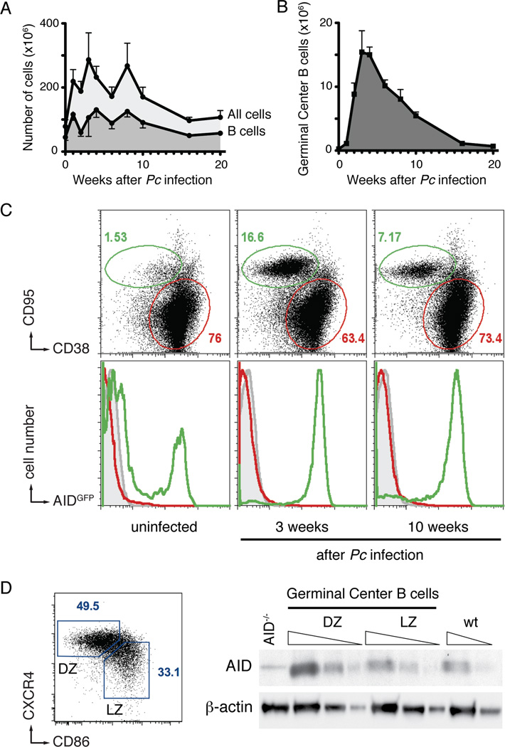
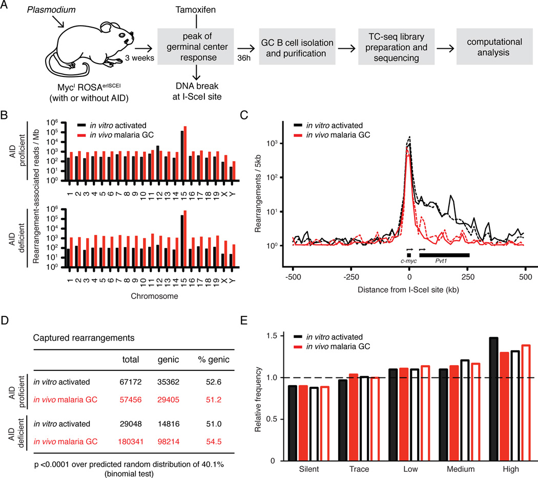
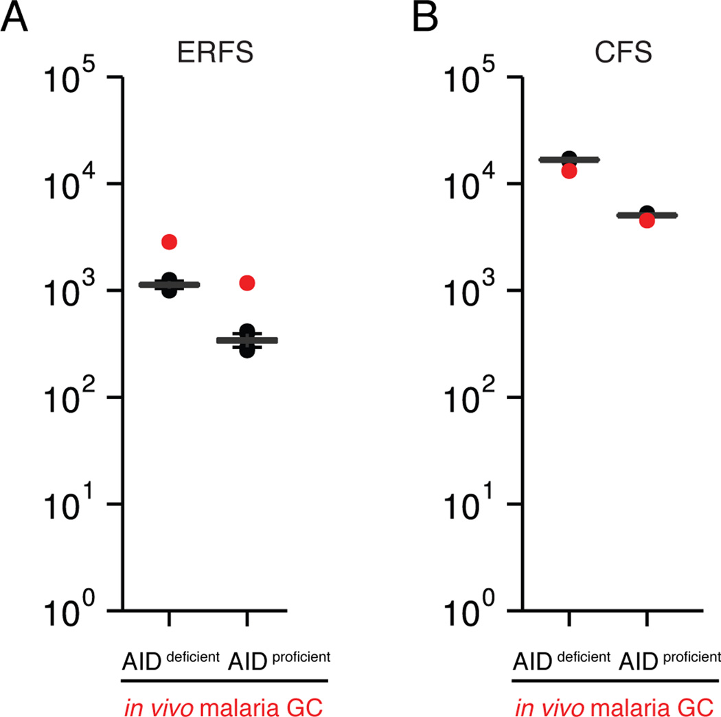
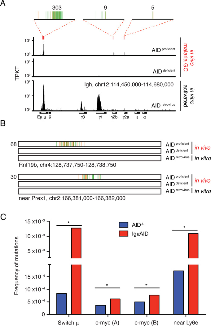
Chronic infection with Plasmodium falciparum was epidemiologically associated with endemic Burkitt's lymphoma, a mature B cell cancer characterized by chromosome translocation between the c-myc oncogene and Igh, over 50 years ago. Whether infection promotes B cell lymphoma, and if so by which mechanism, remains unknown. To investigate the relationship between parasitic disease and lymphomagenesis, we used Plasmodium chabaudi (Pc) to produce chronic malaria infection in mice. Pc induces prolonged expansion of germinal centers (GCs), unique compartments in which B cells undergo rapid clonal expansion and express activation-induced cytidine deaminase (AID), a DNA mutator. GC B cells elicited during Pc infection suffer widespread DNA damage, leading to chromosome translocations. Although infection does not change the overall rate, it modifies lymphomagenesis to favor mature B cell lymphomas that are AID dependent and show chromosome translocations. Thus, malaria infection favors mature B cell cancers by eliciting protracted AID expression in GC B cells. PAPERCLIP.


Figures

Figure 1
Figure 1. B cell responses to Plasmodium infection
A and B- Total spleen cellularity, total B cells (A), and germinal center B cells (B) following Pc infection. Mean with standard deviation is shown; at least five mice for each time point. C- AID expression is confined to germinal center B cells. Representative flow cytometry plots of splenocytes from Plasmodium infected AIDGFP mice. Top row shows the relative expansion of germinal center (green) over non-germinal center (red gate) B cells over time (gated on B220+). Bottom row shows the expression of AIDGFP in non-B cells (B220, grey), non-germinal center B cells (B220+ CD38+ CD95, red), and germinal center B cells (B220+CD38CD95+, green). At least three mice for each time point. D- AID protein in malaria GC B cells. Gating strategy (left) and semiquantitative Western Blot analysis (right) identifying AID in both light zone (LZ) and dark zone (DZ) cells sorted 3 weeks post inoculation. Triangles indicate threefold serial dilution. AID−/− and wild type (wt) control lanes are from in vitro activated B cells of the respective genotypes. Representative of two independent experiments. See also Figure S1.
Figure 2
Figure 2. Translocations in malaria GC B cells
A- Schematic diagram of the protocol to identify genomic rearrangements induced by Plasmodium in vivo. B- Chromosome distribution of rearrangement-associated reads captured by I-SceI breaks on chromosome 15. C- Profile of rearrangements around the I-SceI site. Dotted lines represent AID deficient samples. D- Proportion of genic rearrangements. E- Frequency of rearrangements at genes with various levels of transcription. Empty bars represent AID deficient samples. Dashed line represents the expected frequency based on random model. For all, rearrangements from malaria GC B cells are compared to cultured B cells (Klein et al., 2011). See also Figure S2.
Figure 3
Figure 3. AID-independent DNA damage in regions of DNA replication-associated fragility
A- Observed number of chromosome rearrangements within hotspots of viral integration at early replication fragile sites (ERFS, red dot), as compared to random Monte-Carlo simulation (p< 0.001 for both). B- Number of chromosome rearrangements within common fragile sites (CFS, red dot), as compared to random. See also Figure S3.
Figure 4
Figure 4. AID contributes to Plasmodium induced DNA damage
A- Translocations at the physiologic AID target Igh. Red rectangles indicate the location of AID hotspots, and 4kb regions at these hotspots are magnified on top, where vertical lines each represent a unique translocation event. Numbers on top indicate the total of translocations at each hotspot region. Rearrangements obtained in cultured B cells retrovirally expressing AID are shown for comparison (Klein et al., 2011). TPKT is the normalized number of translocations per kilobase per thousand translocations in the library. B- Examples of non-Igh hotspots of AID-dependent translocation induced by malaria. One kb region is shown, with each vertical line representing a unique translocation. Numbers on the left indicate the total of translocations. C- Mutational analysis of malaria GC B cells DNA by MutPE-seq. For c-myc, two adjacent regions in intron 1 were analyzed. P value is *<0.000001 for all (one-tailed Student’s T-test). See also Table S1 and Figure S4.
Figure 5
Figure 5. P53 suppresses and AID promotes Plasmodium induced lymphoma
A- Survival of Plasmodium infected mice. All mice are also CD19cre/+. B- Spleen histology of Plasmodium infected CD19cre/+p53lox/lox mice. L4 is AID proficient lymphoma and S2 is AID deficient benign B cell hyperplasia with marked extramedullary hematopoiesis. C- Lymphoma versus benign hyperplasia in Plasmodium infected mice. Lymphoid tissues were evaluated by histology, immunohistochemistry, and flow cytometry. “Benign hyperplasia” indicates mice with splenomegaly but normal B cell distribution, with B220+ cells confined to follicular areas. “Atypical hyperplasia or early neoplasia” denotes splenomegaly and B220+ cells expanding into the periarteriolar lymphoid sheats (PALS). “Lymphoma” defines abnormal lymphoid tissue architecture and/or dissemination to multiple organs. D- Extramedullary hematopoiesis in Plasmodium infected mice. Spleen sections were evaluated for the degree of extramedullary hematopoiesis. See also Figure S5 and Table S2.
Figure 6
Figure 6. Genomic rearrangements in Plasmodium induced lymphomas
A- Distribution of lymphoma phenotypes in Plasmodium-infected and control uninfected CD19cre/+p53lox/lox mice. B- Representative M-FISH images of metaphases from Plasmodium-induced CD19cre/+p53lox/lox lymphomas. Arrows point to chromosomes with detectable translocations. C- Circos diagram of the L23 genome. Red arches represent interchromosomal rearrangements, and green arches intrachromosomal ones. For genic rearrangements, the name of the gene is indicated. Asterisks indicate if the recombined site is a known AID target (red, (Hakim et al., 2012; Klein et al., 2011)), or within hotspots of viral integration at ERFS (black, M.J. and I.T.S., unpublished data). See also Figures S6, S7 and Tables S3, S4, and S5.

Comment in

References

    1. Achtman AH, Stephens R, Cadman ET, Harrison V, Langhorne J. Malaria-specific antibody responses and parasite persistence after infection of mice with Plasmodium chabaudi chabaudi. Parasite immunology. 2007;29:435–444. - PubMed
    1. Adams JM, Gerondakis S, Webb E, Corcoran LM, Cory S. Cellular myc oncogene is altered by chromosome translocation to an immunoglobulin locus in murine plasmacytomas and is rearranged similarly in human Burkitt lymphomas. Proc Natl Acad Sci U S A. 1983;80:1982–1986. - PMC - PubMed
    1. Adams JM, Harris AW, Pinkert CA, Corcoran LM, Alexander WS, Cory S, Palmiter RD, Brinster RL. The c-myc oncogene driven by immunoglobulin enhancers induces lymphoid malignancy in transgenic mice. Nature. 1985;318:533–538. - PubMed
    1. Barlow JH, Faryabi RB, Callen E, Wong N, Malhowski A, Chen HT, Gutierrez-Cruz G, Sun HW, McKinnon P, Wright G, et al. Identification of early replicating fragile sites that contribute to genome instability. Cell. 2013;152:620–632. - PMC - PubMed
    1. Bothmer A, Robbiani DF, Feldhahn N, Gazumyan A, Nussenzweig A, Nussenzweig MC. 53BP1 regulates DNA resection and the choice between classical and alternative end joining during class switch recombination. J Exp Med. 2010;207:855–865. - PMC - PubMed

Publication types

Substances