. 2015 Aug 13;162(4):836-48.
doi: 10.1016/j.cell.2015.07.036.

A Conserved Bicycle Model for Circadian Clock Control of Membrane Excitability

Affiliations

A Conserved Bicycle Model for Circadian Clock Control of Membrane Excitability

Matthieu Flourakis et al. Cell. .

Abstract

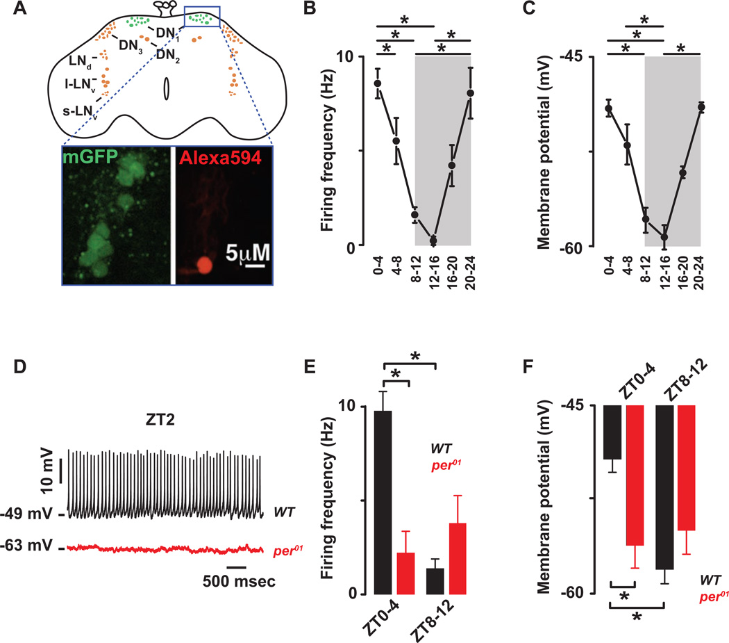
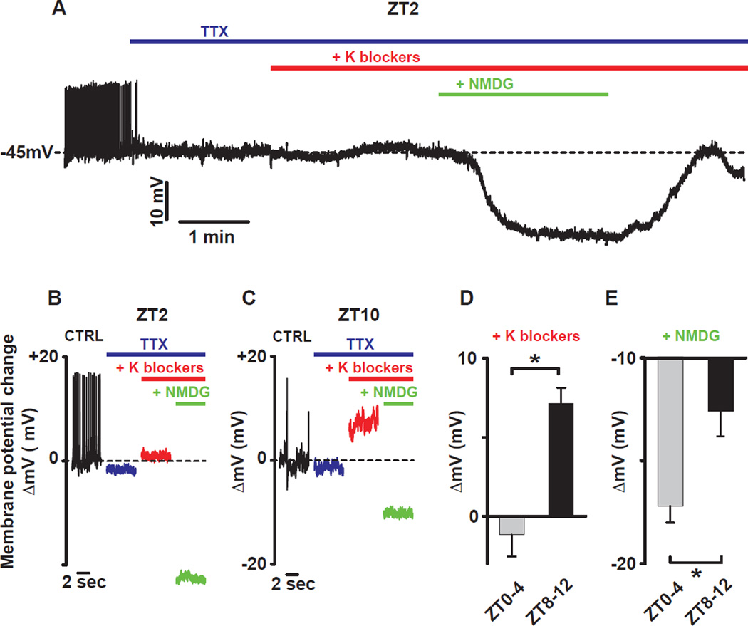
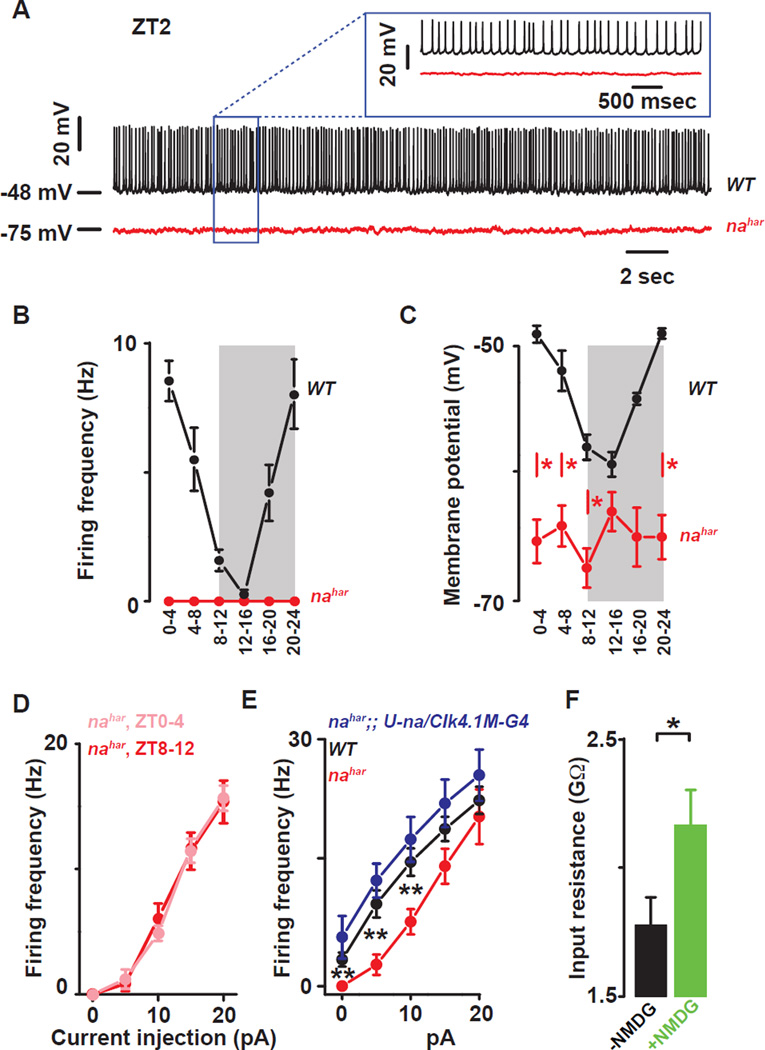
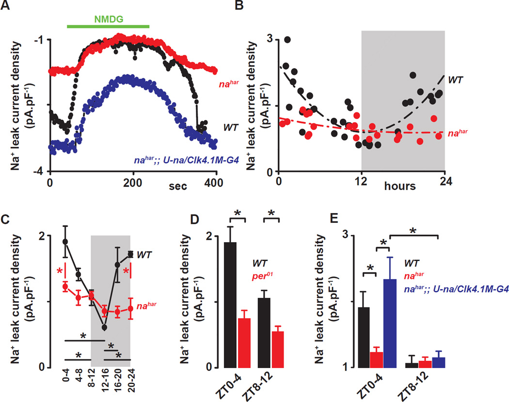
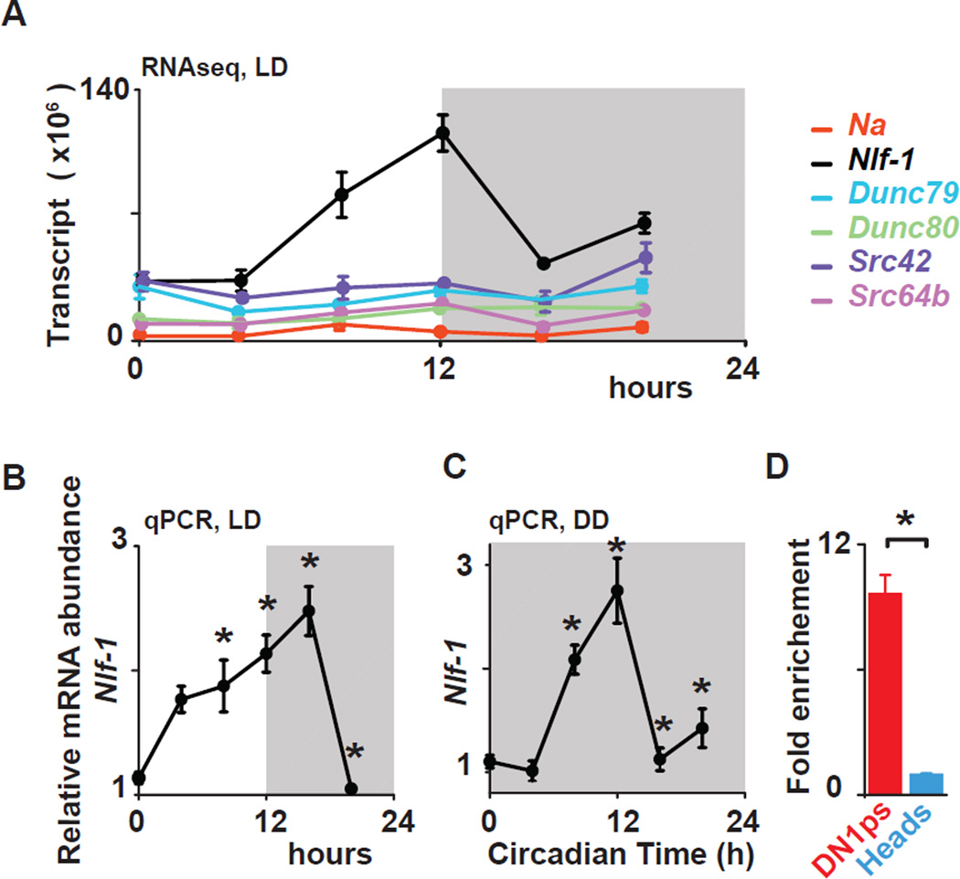
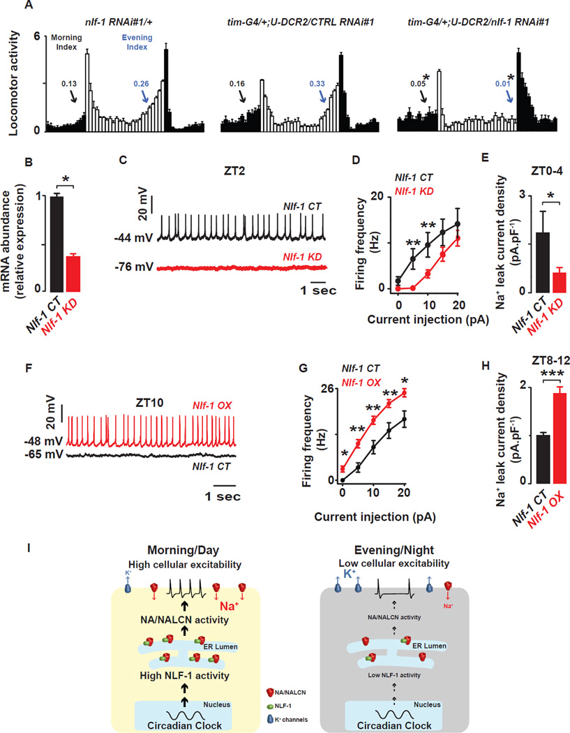
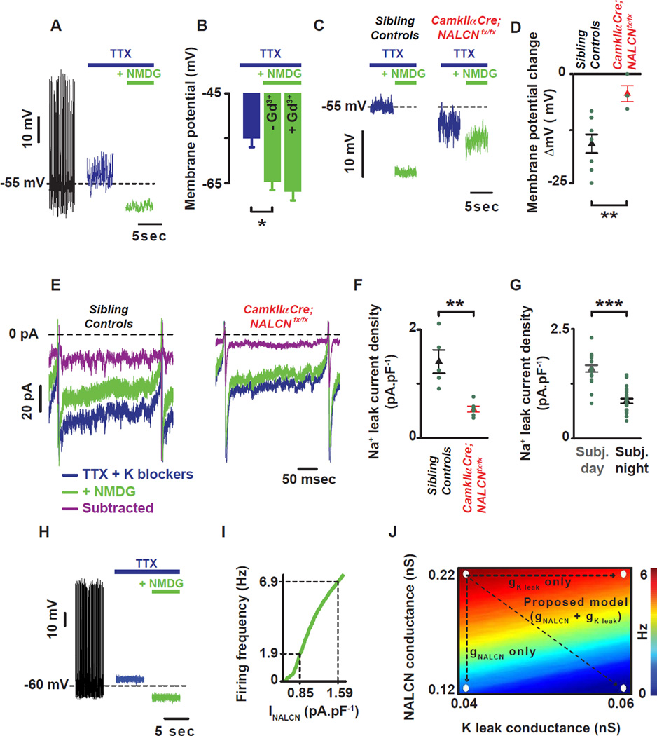
Circadian clocks regulate membrane excitability in master pacemaker neurons to control daily rhythms of sleep and wake. Here, we find that two distinctly timed electrical drives collaborate to impose rhythmicity on Drosophila clock neurons. In the morning, a voltage-independent sodium conductance via the NA/NALCN ion channel depolarizes these neurons. This current is driven by the rhythmic expression of NCA localization factor-1, linking the molecular clock to ion channel function. In the evening, basal potassium currents peak to silence clock neurons. Remarkably, daily antiphase cycles of sodium and potassium currents also drive mouse clock neuron rhythms. Thus, we reveal an evolutionarily ancient strategy for the neural mechanisms that govern daily sleep and wake.


Figures

Figure 1
Figure 1. The cellular excitability of the Drosophila DN1p circadian pacemaker neurons is clock controlled
(A) Schematic and image of the Drosophila brain indicating the location of the DN1ps and other clock neurons. Representative images of the GFP-expressing DN1ps in the intact Drosophila brain are shown below. The DN1ps were labeled by using the Clk4.1M–G4 driving the expression of U-CD8-GFP. Whole-cell access to GFP labeled neurons was confirmed following diffusion of Alexa Fluor 594 biocytin included in intracellular recording solution. All recorded WT neurons are plotted against time of day (in 4 hours bins) to show daily rhythms of firing frequency (B) and membrane potential (C). Grey areas represent the dark phase of the LD cycle. Asterisks indicate statistical significance (p<0.05) from a one-way ANOVA, Tukey’s post-hoc test. (D) Representative current clamp recordings at Zeitgeber Time-2 (ZT2) showing that the per01 DN1ps neurons (red) are hyperpolarized and silent compared to WT DN1p neurons (black). Histogram showing the decrease in firing frequency (E) and membrane potential (F) and lack of daily rhythm in per01 (red, 2.2±1.1Hz, −56±2 mV, n=15 at ZT0-4 and 3.9±1.5Hz, −55±1.9 mV, n=10 at ZT8–12, p>0.41) when compared to WT (black) DN1p neurons. Results are expressed as mean±SEM. Asterisks indicate statistical significance (p<0.05) from t-test performed in WT at ZT0-4 vs ZT8–12. See also Figures. S.1–2 and Tables S.1–2.
Figure 2
Figure 2. Time of day dependent effects of resting K and sodium leak conductance blockade on membrane potential in DN1p neurons
(A) Representative current clamp recording at ZT2 showing the effect of K and sodium conductance blockers on membrane potential. Bars indicate when drugs were applied (blue: TTX 10µM, red: TEA 10mM, 4-AP 5mM, CsCl 2mM and green: NMDG to replace the sodium from the extracellular solution). The effect of K blockers and sodium replacement on the membrane potential at different times of day are shown in (B) for ZT2 and (C) for ZT10. (D) Averaged changes of the membrane potential by K blockers (10mM TEA, 5mM 4-AP and 2mM CsCl): −1.2±1.4mV, n=5 between ZT0-4 and 7.1±1mV, n=5 between ZT8–12 and (E) sodium replacement with NMDG: −17.2±0.8mV, n=5 between ZT0-4 and − 12.6±1.2mV, n=5 between ZT8–12. Results are expressed as mean±SEM. Asterisks indicate statistical significance (p<0.05) from t-test.
Figure 3
Figure 3. The ion channel NARROW ABDOMEN controls Drosophila circadian pacemaker neuronal rhythms
(A) Representative current clamp recordings at ZT2 showing that the nahar DN1ps neurons (red) are hyperpolarized and silent compared to WT DN1p neurons (black). Statistical analysis comparing the firing frequency (B) and membrane potential (C) of the WT (black) and nahar (red) DN1p neurons. Red asterisks indicate statistical significance between WT and nahar neurons (p<0.05, from a one-way ANOVA, Tukey’s post-hoc test). (Data for WT neurons are also depicted in Fig 1B and 1C). (D) Depolarizing current injections confirm the lack of detectable rhythms in cellular excitability in the nahar neurons (light red: ZT0-4, dark red ZT8–12, p>0.35). (E) The decrease in cellular excitability can be restored by rescuing the expression of NA only in the DN1p in the mutant: WT (black), nahar (red) and nahar;; U-na/Clk4.1M–G4 (blue) DN1ps neurons. (F) Histograms showing that sodium substitution with NMDG induces an increase in the input resistance indicating that NA is open at rest (black and green columns are before and after NMDG substitution, respectively). Results are expressed as mean±SEM. Asterisks indicate statistical significance (t-test, p<0.05). See also Tables S.1–2.
Figure 4
Figure 4. The sodium leak current is under clock control in Drosophila circadian pacemaker neurons
(A) Representative time courses showing the sodium leak current (INA) recorded at −113mV from a ramp protocol in WT (black), nahar (red) and nahar;; U-na/Clk4.1M–G4 (blue) DN1p neurons. (B) All recorded WT neurons (black dots) and nahar neurons (red dots) are plotted against time of day for sodium leak current (INA). (C) Quantification and statistical analysis are shown. Grey areas represent the dark phase of the LD cycle. Red asterisks indicate statistical significance between WT and nahar neurons, and black asterisks indicate statistical significance between different time points in WT neurons (p<0.05) from a one-way ANOVA, Tukey’s post-hoc test. (D) Histograms showing the NA current in WT (black) and per01 (red) DN1ps recorded at different times of day ZT0-4 vs ZT8–12 (for per01, INA=0.7±0.2pA.pF−1, n=8 at ZT0-4 and 0.5±0.1pA.pF−1, n=7 at ZT8–12. Asterisks indicate statistical difference between WT and per01, p<0.05 from t-test). (E) Histograms showing the sodium leak current in WT (black), nahar (red) and nahar;; U-na/Clk4.1M–G4 (blue) DN1p neurons at different times of day (ZT0-4 vs ZT8–12) (for nahar;; U-na/Clk4.1M–G4, INA=2.3±0.3pA.pF−1, n=4 at ZT0-4 and 1.1±0.1pA.pF−1, n=4 at ZT8–12). Results are expressed as mean±SEM. Asterisks indicate statistical significance (p<0.05) from a t-test.
Figure 5
Figure 5. Nlf-1 is rhythmically expressed in DN1p neurons
(A) Nlf-1 mRNA shows rhythmic expression using RNA-seq data from FACS sorted DN1p neurons in LD (for isoform RB- shown in graph, BH corrected p=0.005). na, Unc79, Unc80, Src64B, and Src42a are not robustly cycling (graph shows isoforms with highest expression : BH= 0.28 for na-RF, 0.2 for Dunc79-RE, 0.85 for Dunc80-RE, 0.71 for Src42a–RA and 0.07 for Src64B–RJ). Nlf-1 cycles under LD (B) and during the first day of constant darkness (DD1) conditions (C) in DN1ps using qPCR. Based on two independent experiments, an asterisk indicates differences statistically significant one-way ANOVA, Tukey’s post-hoc test, LD ZT0 vs ZT12 p= 0.0011, ZT0 vs ZT16, p= 0.000142, ZT4 vs ZT16 p=0.029, ZT0 vs ZT8 p=0.022, ZT12 vs ZT20, p= 0.000441, ZT16 vs ZT20 p= 0.000136. DD1 CT0 vs CT8 p= 0.01081, CT0 vs CT12 p= 0.000142, CT12 vs CT16 p= 0.000145 and CT12 vs CT20 p= 0.000459. (D) Nlf-1 expression is enriched in the DN1ps vs whole head (t-test, p<0.02). Results are expressed as mean±SEM.
Figure 6
Figure 6. Nlf-1 is required for anticipatory behavior and NA current
(A) Nlf-1 RNAi expressing flies (tim-G4/+; U-Dcr2/Nlf-1 RNAi#1) show reduced morning anticipation (Morning Index) and evening anticipation (Evening Index) under LD conditions when compared to genetic controls (Nlf-1 RNAi#1/+ and tim-G4/+; U-Dcr2/CTRL RNAi#1), (t-test, p<0.05). (B) Nlf-1 expression is reduced in the DN1ps of Nlf-1 RNAi expressing flies (t-test, p<0.05) (C) Representative current clamp recordings at ZT2 showing that the Nlf-1 knockdown DN1p neurons (red) are hyperpolarized and silent compared to control DN1p neurons (black). (D) Depolarizing current injections confirm the decrease in cellular excitability in Nlf-1 knockdown neurons (red) vs control (black) (p<0.05). (E) Sodium leak current density is dramatically reduced in the Nlf-1 knockdown neurons (red) vs control neurons (black) (1.9±0.7pA.pF−1, n=4 in Nlf-1 CT and 0.6±0.2pA.pF−1, n=5 in Nlf-1 KD, measured at ZT0-4, p<0.05). (F) Representative current clamp recordings at ZT10 showing that the Nlf-1 overexpressing DN1p neurons (red) are depolarized and more active compared to control DN1p neurons (black). (G) Depolarizing current injections confirm the increase in cellular excitability in Nlf-1V5 overexpressing neurons (red) vs control (black) (p<0.05). (H) Sodium leak current density is also increased in the Nlf-1V5 overexpressing neurons (red) vs control neurons (black) (1±0.05pA.pF−1, n=4 in Nlf-1 CT and 1.9±0.1pA.pF−1, n=5 in Nlf-1 OX, measured at ZT8–12, p<0.05). Results are expressed as mean±SEM. Asterisks indicate statistical significance (p<0.05 from a t-test). A summary cartoon depicting the conserved bicycle model for controlling membrane excitability of circadian pacemaker neurons is shown in (I). In the morning/day, the molecular clock drives high NLF-1 activity increasing the sodium leak activity and K conductances are reduced thus increasing cellular excitability. In the evening/night, the sodium leak is decreased and, in parallel, K conductances are high, thus silencing the neurons. This dual regulation of the conductances responsible for the membrane properties is critical for driving high amplitude rhythmic oscillations of cellular excitability. See also Figures. S.3–5 and Tables S.2–4.
Figure 7
Figure 7. NALCN current is under clock control in mammalian SCN pacemaker neurons
(A) Representative current clamp recording showing the role of the TTX-resistant sodium leak (difference between green and blue) in setting the membrane potential of mammalian SCN neurons. (B) NMDG hyperpolarizes the cell with no additional effect in the presence of Gd3+. (C) NMDG-evoked hyperpolarization was reduced in a brain specific knockout of NALCN. (D) Quantification and statistical analysis of the NMDG-evoked hyperpolarization are shown: −15.9±2.0mV, n=9 in controls (black triangle) and −4.5±1.7mV, n=4 (red triangle). Asterisks indicate statistical significance (t-test, p=0.005). (E) Action potential clamp recordings showing the sodium leak flowing during the interspike interval in SCN neurons from sibling control (left) and CamkIIa-Cre;NALCNfx/fx animals (right). In the presence of TTX and K blockers (blue trace), the sodium leak current flowing during the interspike interval (INALCN) was reduced after sodium substitution with NMDG (green trace). The sodium leak current (INALCN = subtracted = purple trace) was revealed by subtracting the inward current in the presence of NMDG from the inward current present with TTX and K blockers. (F) INALCN was reduced in CamkIIa-Cre;NALCNfx/fx compared to sibling controls animals (0.5±0.1pA.pF−1, n=6 in CamkIIa-Cre;NALCNfx/fx (red triangle) and 1.4±0.2pA.pF−1, n=5 in sibling controls (black triangle)). Asterisks indicate statistical significance (t-test, p=0.002). (G) Circadian variation of INALCN: 1.6±0.1pA.pF−1, n=25 during the subjective day (grey columns) and 0.8±0.1pA.pF−1, n=23 during the subjective night (black columns). Asterisks indicate statistical significance (t-test, p<0.001). Green dots represent individual cells. (H) Simulations showing the role of TTX resistant sodium leak in setting the membrane potential using a mathematical model of SCN membrane excitability. Voltage traces from control simulation (gNa = 229 nS, gNALCN = 0.22 nS) and simulated application of TTX (gNa = 0 nS) and NMDG (gNALCN = 0 nS). (I) The model predicts the magnitude of change in firing rate as a function of magnitude of change in NALCN current density (gNALCN = 0.12 to 0.22 nS). A decrease of 0.74 pA.pF−1 in INALCN (observed between the subjective day and night (G)) leads to a 5 Hz decrease in firing rate. (J) Firing rate as a function of gNALCN and gKleak in a model SCN neuron. Arrows: decreasing gNALCN alone reduces firing rate from 7 Hz to 2 Hz, whereas increasing gKleak reduces firing rate from 7 Hz to 6 Hz. Concurrently decreasing gNALCN and increasing gKleak reduces firing rate from 7 Hz to 0.5 Hz. Results are expressed as mean±SEM. See also Figures. S.6–7.

References

    1. Abruzzi KC, Rodriguez J, Menet JS, Desrochers J, Zadina A, Luo W, Tkachev S, Rosbash M. Drosophila CLOCK target gene characterization: implications for circadian tissue-specific gene expression. Genes & development. 2011;25:2374–2386. - PMC - PubMed
    1. Allada R, Chung BY. Circadian organization of behavior and physiology in Drosophila. Annual review of physiology. 2010;72:605–624. - PMC - PubMed
    1. Cao G, Nitabach MN. Circadian control of membrane excitability in Drosophila melanogaster lateral ventral clock neurons. J Neurosci. 2008;28:6493–6501. - PMC - PubMed
    1. Casanova E, Fehsenfeld S, Mantamadiotis T, Lemberger T, Greiner E, Stewart AF, Schutz G. A CamKIIalpha iCre BAC allows brain-specific gene inactivation. Genesis. 2001;31:37–42. - PubMed
    1. Cavanaugh DJ, Geratowski JD, Wooltorton JR, Spaethling JM, Hector CE, Zheng X, Johnson EC, Eberwine JH, Sehgal A. Identification of a circadian output circuit for rest:activity rhythms in Drosophila. Cell. 2014;157:689–701. - PMC - PubMed

Publication types