. 2015 Oct;8(10):952-61.
doi: 10.1158/1940-6207.CAPR-15-0003. Epub 2015 Aug 14.

Colon Tumors with the Simultaneous Induction of Driver Mutations in APC, KRAS, and PIK3CA Still Progress through the Adenoma-to-carcinoma Sequence

Affiliations

Colon Tumors with the Simultaneous Induction of Driver Mutations in APC, KRAS, and PIK3CA Still Progress through the Adenoma-to-carcinoma Sequence

Jamie N Hadac et al. Cancer Prev Res (Phila). 2015 Oct.

Abstract

Human colorectal cancers often possess multiple mutations, including three to six driver mutations per tumor. The timing of when these mutations occur during tumor development and progression continues to be debated. More advanced lesions carry a greater number of driver mutations, indicating that colon tumors might progress from adenomas to carcinomas through the stepwise accumulation of mutations following tumor initiation. However, mutations that have been implicated in tumor progression have been identified in normal-appearing epithelial cells of the colon, leaving the possibility that these mutations might be present before the initiation of tumorigenesis. We utilized mouse models of colon cancer to investigate whether tumorigenesis still occurs through the adenoma-to-carcinoma sequence when multiple mutations are present at the time of tumor initiation. To create a model in which tumors could concomitantly possess mutations in Apc, Kras, and Pik3ca, we developed a novel minimally invasive technique to administer an adenovirus expressing Cre recombinase to a focal region of the colon. Here, we demonstrate that the presence of these additional driver mutations at the time of tumor initiation results in increased tumor multiplicity and an increased rate of progression to invasive adenocarcinomas. These cancers can even metastasize to retroperitoneal lymph nodes or the liver. However, despite having as many as three concomitant driver mutations at the time of initiation, these tumors still proceed through the adenoma-to-carcinoma sequence.


Conflict of interest statement

No conflicts of interest.

Figures

Figure 1
Figure 1
A minimally invasive technique for inoculation of the colon with Adeno-Cre initiates tumorigenesis in Apcfl/fl mice. We have developed a novel technique that can be utilized to sequester Adeno-Cre within the colon without laparotomy, as was required with prior methodologies. A, in this technique a slotted tube housing GelFoam allows for the treatment of the colon with trypsin. Abrasion of the colon is then performed prior to incubation with Adeno-cre-soaked GelFoam at the desired location. B, colonic tumors are visualized by endoscopy in 3–7 weeks following treatment with Adeno-Cre and monitored over time with serial endoscopy. C, interestingly, these tumors have varying growth patterns over 14 weeks of observation, despite a similar initiation. D, histological evaluation demonstrated that the majority (83%) of these lesions were adenomas with nuclear CTNNB1 consistent with activation of the WNT pathway. The area indicated by the rectangle is shown enlarged at the right; at the far right the same area in an adjacent slide stained for CTNNB1 is shown. Size bar = 1mm.
Figure 2
Figure 2
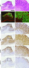
Multiple mutations were simultaneously expressed within the epithelium of the colon following a novel minimally invasive Adeno-Cre technique resulting in the development of colon tumors. To be certain that our mutations were being activated only in the epithelial cells of the colon with this technique we utilized mT/mG1 Apcfl/fl Pik3cap110* mice. Upon Cre recombination a gene encoding a red fluorescent marker in these mice is excised and a green fluorescent protein is expressed. A representative histological section is displayed demonstrating that Cre-mediated recombination has only occurred in the epithelial cells of these lesions. IHC confirmed CTNNB1 (β-catenin) localization to the nucleus and activation of the PI3K signaling cascade with intense staining for phosphorylated AKT (pAKT) and phosphorylated RPS6 (pRPS6), as expected. Activation of ERK1/2 was not observed in these tumors. The area indicated by each rectangle is shown enlarged at the right. Size bar = 1 mm.
Figure 3
Figure 3
Apcfl/fl KrasG12D/+(A2K1P0), Apcfl/fl Pik3cap110* (A2K0P1), and Apcfl/fl KrasG12D/+ Pik3cap110* (A2K1P)) mice were treated with Adeno-Cre at 40–50 days of age. A, incidence of tumors increases with the addition of KrasG12D/+ and/or Pik3cap110* to the mutation profile. (Due to the relatively small sample sizes and the similar tumor incidence rates among A2K1P0, A2K0P1 and A2K1P1 mice, the tumor incidence data from these strains was pooled (31/39) for comparison to A2K0P0 (18/31); p (one-sided) = 0.031, Barnard’s exact test). B, tumors were identified by endoscopy in the colon as early as 3–4 weeks after viral inoculation. Histological sectioning was performed on tumors shortly after identification on endoscopy revealing that these lesions were small adenomas without evidence of invasion into the muscularis mucosa. An adenoma from an Apcfl/fl Pik3cap110* mouse is shown here. Nuclear localization of CTNNB1 and phosphorylation of RPS6 were observed without phosphorylation of ERK1/2 indicating that recombination was occurring as expected. The area indicated by each rectangle is shown enlarged at the right. Size bar = 500µm.
Figure 4
Figure 4
The addition of Kras and Pik3ca mutations to the loss of APC has a modest effect on tumor proliferation. A, Apcfl/fl, (A2K0P0), Apcfl/fl Pik3cap110* (A2K0P1) and Apcfl/fl KrasG12D/+ Pik3cap110* (A2K1P1) mice were treated with Adeno-Cre and followed with serial endoscopy. B, change in percent lumen occlusion was used as a marker of tumor proliferation. The time required for tumors to occlude 75% of the lumen was determined. No difference was seen between groups. C, to confirm these results these tumors were sectioned and stained for Ki67. The Ki67 proliferation index was calculated for each tumor. D, no statistically significant increase in Ki67 staining was observed when additional mutations occurred concomitantly with loss of APC (p = 0.35, Kruskal-Wallis test). The area indicated by each rectangle is shown enlarged at the right. Size bar = 1mm.
Figure 5
Figure 5
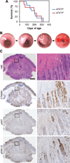
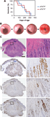
Tumors with multiple driver mutations present at the time of initiation progressed through the adenoma to carcinoma sequence. Apcfl/fl Pik3cap110* (A2K0P1) and Apcfl/fl KrasG12D/+ Pik3cap110* (A2K1P1) mice were treated with Adeno-Cre, as described, at 40–50 days of age and aged until moribund. A, no difference in survival between these two groups was noted. B, the resulting tumors were followed with serial endoscopy; they developed initially as polypoid lesions within the colon and progressed to occlude the majority of the lumen. These tumors could be followed as long as 12 months post viral inoculation and developed into "apple-core" lesions, as seen in the far right photo, similar to advanced colorectal tumors in humans. These mice became moribund due to anemia or intestinal obstruction. At necropsy, large colon tumors approached 1 cm in diameter. C, upon histological analysis, these lesions were invasive adenocarcinomas penetrating through muscularis propria and into the serosa in many instances. H&E staining of a large invasive cancer from an Apcfl/fl KrasG12D/+ Pik3cap110* (A2K1P1) mouse is shown. Nuclear localization of CTNNB1 and activation of AKT and ERK1/2 signaling was observed. The rea indicated by each rectangle shown enlarged at the right. Scale bar = 1 mm.
Figure 6
Figure 6
Metastatic colon adenocarcinoma was identified within Adeno-Cre treated mice. An Apcfl/fl KrasG12D/+ Pik3cap110* mouse became moribund after treatment with Adeno-Cre. A, 18F-FDG PET imaging demonstrated an avid para-arotic lymph node in addition to increased avidity of the primary tumor. Enlarged adenopathy was identified at necropsy. On histological sectioning, well-differentiated adenocarcinoma consistent with the colonic primary tumor was observed. Staining for CTNNB1 demonstrated nuclear localization consistent with loss of APC. B, in addition, metastatic adenocarcinoma in the liver was identified in a moribund Adeno-Cre treated Apcfl/fl Pik3cap110* mouse. A hepatic mass was observed grossly. H&E staining of histological sections demonstrated a well-differentiated adenocarcinoma arising within the liver. Staining demonstrated nuclear localization of CTNNB1 consistent with the colonic primary tumor. The area indicated by each rectangle in photos of stained sections is shown enlarged at the right. Size bars = 500µm.

References

    1. American Cancer Society. Cancer Facts & Figures 2014. Atlanta: American Cancer Society; 2014.
    1. Nowell PC. The colonal evolution of tumor cell populations. Science. 1976;194:23–28. - PubMed
    1. Fearon ER, Vogelstein B. A genetic model for colorectal tumorigenesis. Cell. 1990;61:759–767. - PubMed
    1. Goss KH, Groden J. Biology of the adenomatous polyposis coli tumor suppressor. J Clin Oncol. 2000;18:1967–1979. - PubMed
    1. Kinzler KW, Vogelstein B. Cancer-susceptibility genes. Gatekeepers and caretakers. Nature. 1997;386:761–763. - PubMed

Publication types

Substances