. 2015 Nov 1;24(21):6198-212.
doi: 10.1093/hmg/ddv336. Epub 2015 Aug 13.

Tau deposition drives neuropathological, inflammatory and behavioral abnormalities independently of neuronal loss in a novel mouse model

Affiliations

Tau deposition drives neuropathological, inflammatory and behavioral abnormalities independently of neuronal loss in a novel mouse model

Casey Cook et al. Hum Mol Genet. .

Abstract

Aberrant tau protein accumulation drives neurofibrillary tangle (NFT) formation in several neurodegenerative diseases. Currently, efforts to elucidate pathogenic mechanisms and assess the efficacy of therapeutic targets are limited by constraints of existing models of tauopathy. In order to generate a more versatile mouse model of tauopathy, somatic brain transgenesis was utilized to deliver adeno-associated virus serotype 1 (AAV1) encoding human mutant P301L-tau compared with GFP control. At 6 months of age, we observed widespread human tau expression with concomitant accumulation of hyperphosphorylated and abnormally folded proteinase K resistant tau. However, no overt neuronal loss was observed, though significant abnormalities were noted in the postsynaptic scaffolding protein PSD95. Neurofibrillary pathology was also detected with Gallyas silver stain and Thioflavin-S, and electron microscopy revealed the deposition of closely packed filaments. In addition to classic markers of tauopathy, significant neuroinflammation and extensive gliosis were detected in AAV1-Tau(P301L) mice. This model also recapitulates the behavioral phenotype characteristic of mouse models of tauopathy, including abnormalities in exploration, anxiety, and learning and memory. These findings indicate that biochemical and neuropathological hallmarks of tauopathies are accurately conserved and are independent of cell death in this novel AAV-based model of tauopathy, which offers exceptional versatility and speed in comparison with existing transgenic models. Therefore, we anticipate this approach will facilitate the identification and validation of genetic modifiers of disease, as well as accelerate preclinical assessment of potential therapeutic targets.


Figures

Figure 1.
Figure 1.
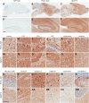
Widespread expression of human tau in AAV1-TauP301L model of tauopathy. Human tau-specific antibody (E1) reveals high level of expression throughout the brain and hippocampus in AAV1-TauP301L-injected (b and e) and rTg4510 mice (c and f), whereas AAV1-GFP-injected mice are negative (a and d). AAV1-TauP301L is also highly expressed throughout the hippocampal network (g–l), as well as other regions of the brain (s–x). The pattern of human tau expression in the hippocampal network (m–r) and other regions of the brain (y-dd) is shown in comparison with rTg4510 model. Scale bar in a–c equals 2 mm; scale bar in d–f equals 200 µm; scale bar in g–dd equals 100 µm.
Figure 2.
Figure 2.
Accumulation of hyperphosphorylated tau species in AAV-driven model of tauopathy. (a and b) Representative images depicting the deposition of PHF1- and CP13-positive tau species in the CA1 field of the hippocampus (a), as well as the cortex (b). (c) Mice injected with AAV1-TauP301L express 4-fold higher tau levels in the forebrain as assessed by Tau 5 (recognizing both mouse and human tau), leading to accumulation of tau that is hyperphosphorylated on multiple epitopes. (d) Quantification of PHF1 normalized to GAPDH (t = 3.46, P = 0.0018); (e) CP13 (t = 6.26, P < 0.0001); (f) 12E8 (t = 7, P < 0.0001); (g) Tau 5 (t = 7.45, P < 0.0001). Scale bar is equal to 100 µm.
Figure 3.
Figure 3.
Deposition of abnormally folded and protease-resistant tau. (a and b) Injection of AAV1-TauP301L leads to the accumulation of tau positive for MC1, a well-characterized marker of abnormally folded tau, as well as proteinase K-resistant tau that is positive for Ab39, a marker of mature NFTs. Representative images from the CA1 field of the hippocampus (a) and cortex (b). Scale bar is equal to 100 µm.
Figure 4.
Figure 4.
Thioflavin and Gallyas-positive tau deposition. (a and b) Expression of P301L-tau but not GFP leads to accumulation of insoluble tau species that are positive for Gallyas silver stain (a) and Thioflavin S (b) in CA1 and cortex. Scale bar is equal to 100 µm.
Figure 5.
Figure 5.
Tau filament formation in AAV1-TauP301L model of tauopathy. (a) Electron micrograph of a cortical neuron containing bundles of packed filaments that filled almost the entire cell body (N, nucleus). Arrow points to enlargement in (b) that shows the inclusion is not membrane-bound. (c) Further enlargement of (b) shows longitudinal, oblique and cross sections of filaments with diameters of 11–17 nm. Most of the filaments are straight. Some cross sections have a central lumen. Note the close proximity of filaments and mitochondria. (d) A filamentous aggregate excludes cytoplasmic organelles to its periphery. (e) Enlargement of the aggregate shows packed and loose filaments heavily labeled with E1 antibody that is specific for human tau. Scale bars are equal to 1 µm (a); 0.2 µm (b); 0.25 µm (d); 50 nm (c and e).
Figure 6.
Figure 6.
AAV1-TauP301L drives tau filament formation in neuronal processes. (a) A dentate granule cell (N) and two nearby neurites (arrows). (b and c) Enlargements of neurites in (a) show moderate labeling on less well-formed filaments (b) and heavy labeling of E1 on well-formed filaments (c). Arrows point to 18-nm gold particles. Arrowheads point to synapses. (d) A swollen, thinly myelinated neurite filled with E1-positive filaments. (e) Boxed area in (d) shows randomly orientated filaments. Arrowheads point to myelin sheathes. (f) A dense dystrophic neurite filled with E1 immunoreactivity. Arrowed area is enlarged in (g) showing many gold particles (arrows). Arrows point to gold particles. Scale bars are equal to 0.5 µm (a); 50 nm (b and c); 1 µm (d); 100 nm (e); 0.5 µm (f); 50 nm (g).
Figure 7.
Figure 7.
Tau pathology is associated with robust gliosis and inflammation. (a and b) Microgliosis and astrocytosis detected by Iba1 and GFAP, respectively, in the CA1 field of the hippocampus (a) and cortical regions (b). (cg) Inflammatory markers were evaluated by RT-qPCR. (c) Aif1 (t = 5.3, P < 0.0001), (d) Gfap (t = 2.5, P = 0.017), (e) Il1b (t = 3.15, P = 0.004), (f) Il6 (t = 3.17, P = 0.004) and (g) Tnfa (t = 2.9, P = 0.007) were all significantly elevated in AAV1-TauP301L mice compared with AAV1-GFP controls. Scale bar is equal to 100 µm *P < 0.05, **P < 0.01, ***P < 0.005, ****P < 0.0001.
Figure 8.
Figure 8.
Behavioral abnormalities in AAV1-TauP301L model of tauopathy. (ac) Behavior in the OFA revealed that AAV1-TauP301L mice exhibited an increase in total distance traveled (a; t = 3.1, P = 0.004) and time spent mobile (b; t = 3.9, P = 0.0006), and a decrease in the ratio of distance traveled in the center of the box compared with the total distance traveled (c; t = 3.6, P = 0.001). (d) Exploratory behavior was also evaluated in the EPM, which detected a significant increase in the time spent in open arms (t = 3.3, P = 0.0025). (e and f) Contextual fear conditioning was utilized to measure learning and memory. AAV1-TauP301L mice were significantly impaired in both contextual (e; t = 2.1, P = 0.04) and cued (f; t = 3, P = 0.005) versions of the task, indicative of hippocampal and amygdala-dependent learning and memory deficits. *P < 0.05, **P ≤ 0.005.
Figure 9.
Figure 9.
Aberrant proteolysis of PSD-95 in AAV1-TauP301L mice. (a) Accumulation of a 50-kDa PSD95 fragment was observed in mice injected with AAV1-TauP301L, whereas the levels of full-length PSD95 were unchanged. (b) Quantification of the 50-kDa fragment normalized to full-length PSD95 revealed a highly significant increase in AAV1-TauP301L mice (t = 7.47, P < 0.0001). (c) The 50-kDa/full-length PSD95 ratio is significantly correlated with the levels of human tau in animals injected with AAV1-TauP301L (r = 0.74, P = 0.0008). ***P < 0.0001.

References

    1. Hutton M., Lendon C.L., Rizzu P., Baker M., Froelich S., Houlden H., Pickering-Brown S., Chakraverty S., Isaacs A., Grover A. et al. (1998) Association of missense and 5′-splice-site mutations in tau with the inherited dementia FTDP-17. Nature, 393, 702–705. - PubMed
    1. Poorkaj P., Bird T.D., Wijsman E., Nemens E., Garruto R.M., Anderson L., Andreadis A., Wiederholt W.C., Raskind M., Schellenberg G.D. (1998) Tau is a candidate gene for chromosome 17 frontotemporal dementia. Ann. Neurol., 43, 815–825. - PubMed
    1. Spillantini M.G., Murrell J.R., Goedert M., Farlow M.R., Klug A., Ghetti B. (1998) Mutation in the tau gene in familial multiple system tauopathy with presenile dementia. Proc. Natl Acad. Sci. USA, 95, 7737–7741. - PMC - PubMed
    1. Kouri N., Carlomagno Y., Baker M., Liesinger A.M., Caselli R.J., Wszolek Z.K., Petrucelli L., Boeve B.F., Parisi J.E., Josephs K.A. et al. (2014) Novel mutation in MAPT exon 13 (p.N410H) causes corticobasal degeneration. Acta. Neuropathol., 127, 271–282. - PMC - PubMed
    1. Simon-Sanchez J., Schulte C., Bras J.M., Sharma M., Gibbs J.R., Berg D., Paisan-Ruiz C., Lichtner P., Scholz S.W., Hernandez D.G. et al. (2009) Genome-wide association study reveals genetic risk underlying Parkinson's disease. Nat. Genet., 41, 1308–1312. - PMC - PubMed

Publication types