

Rapid multi-orientation quantitative susceptibility mapping
- PMID: 26277773
- PMCID: PMC4691433
- DOI: 10.1016/j.neuroimage.2015.08.015
Rapid multi-orientation quantitative susceptibility mapping
Abstract
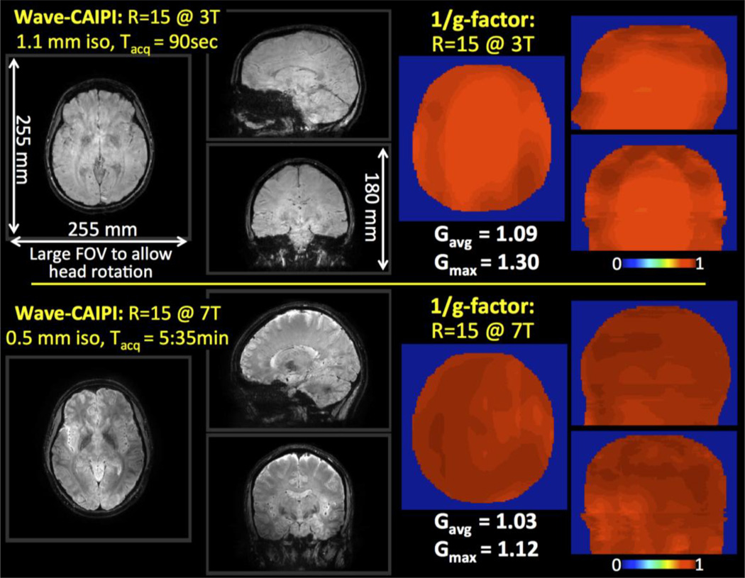
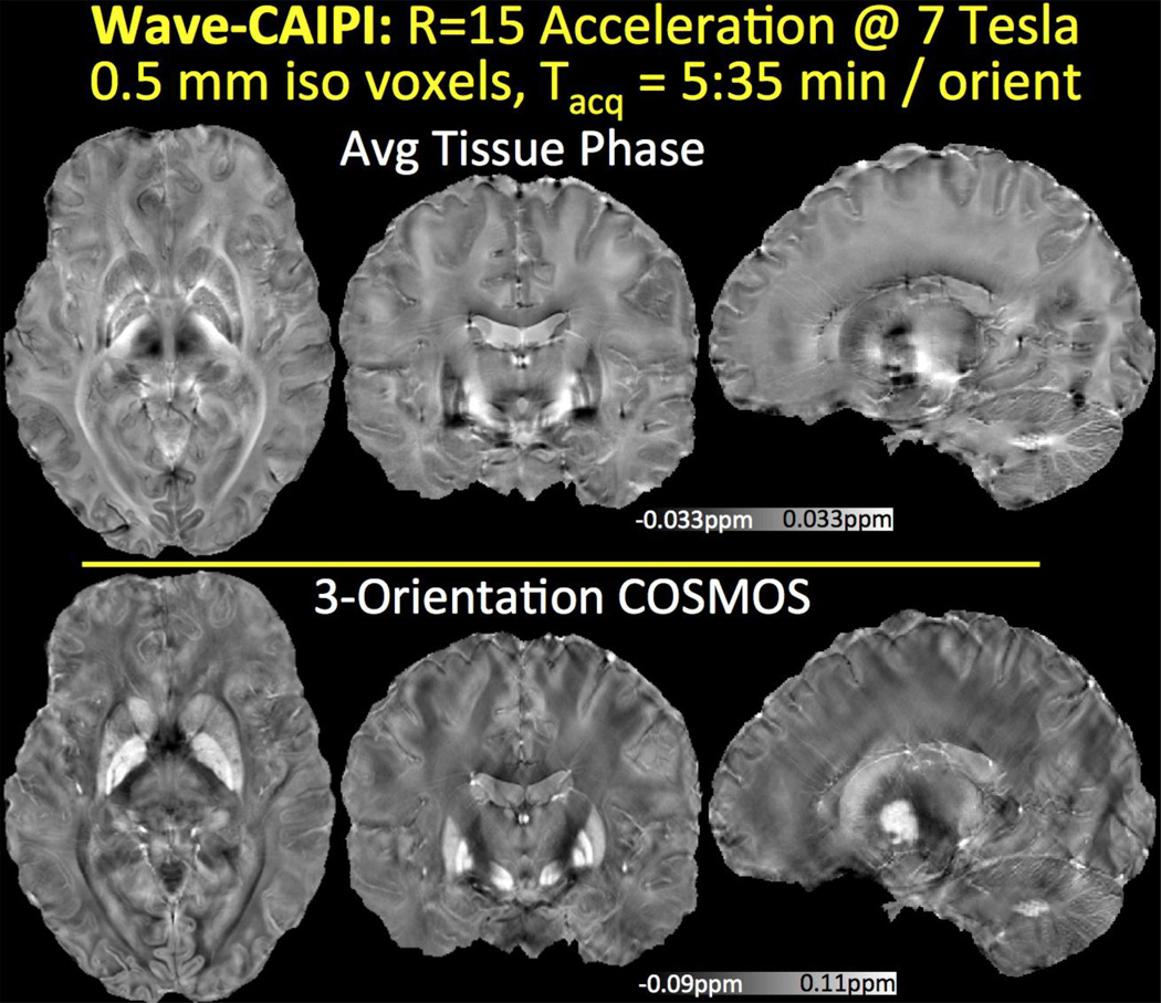
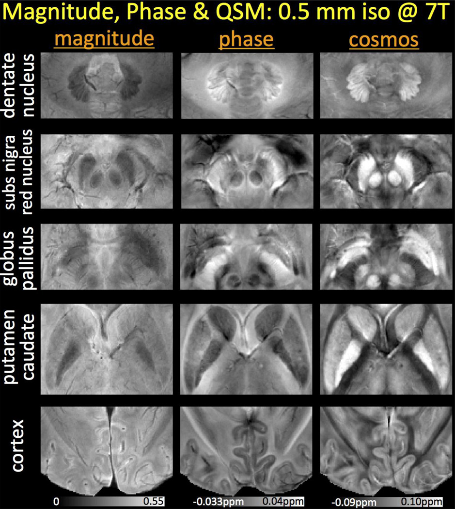
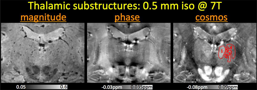
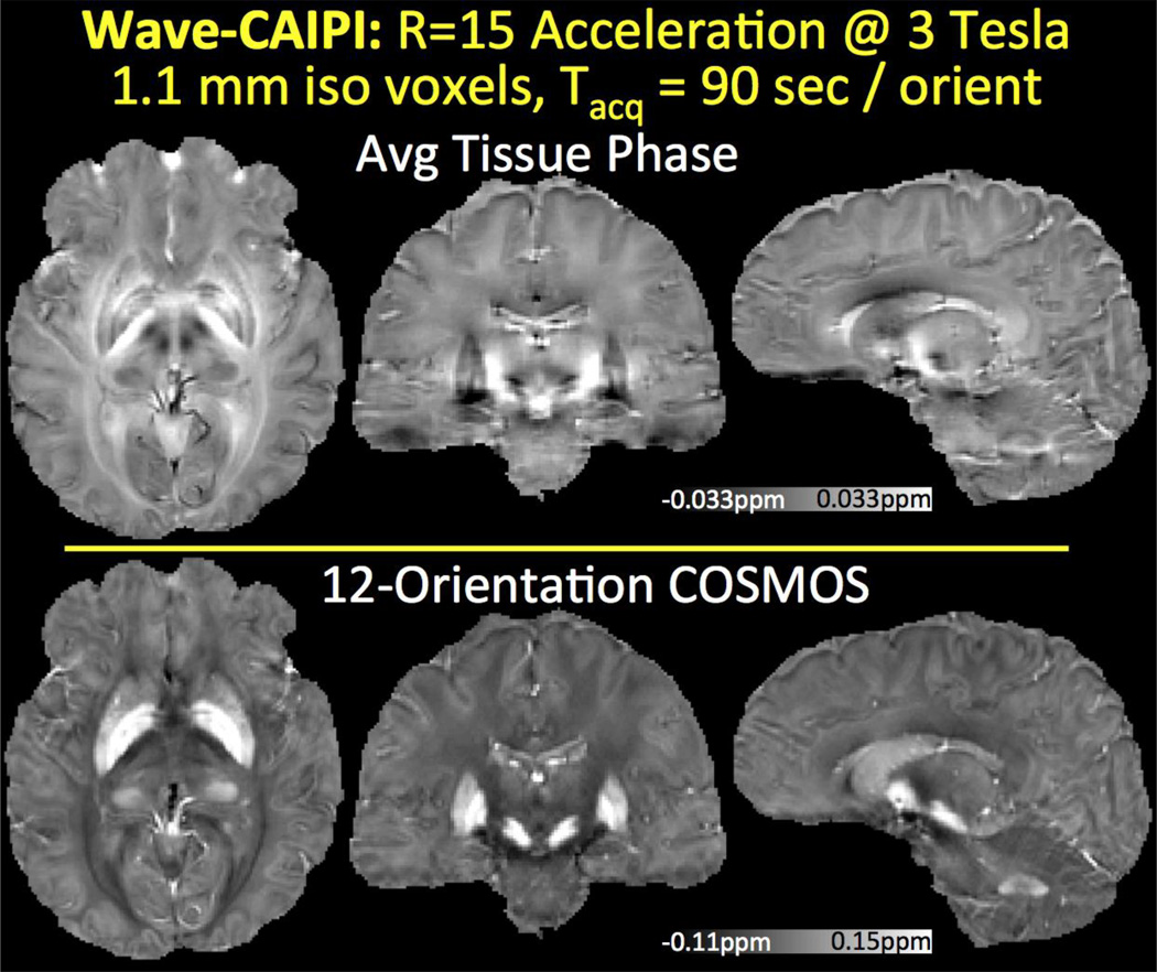
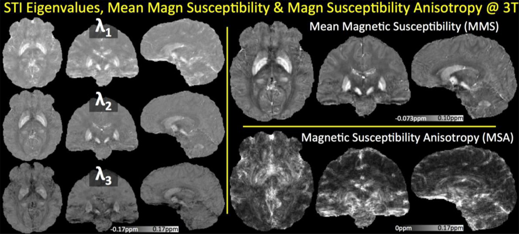
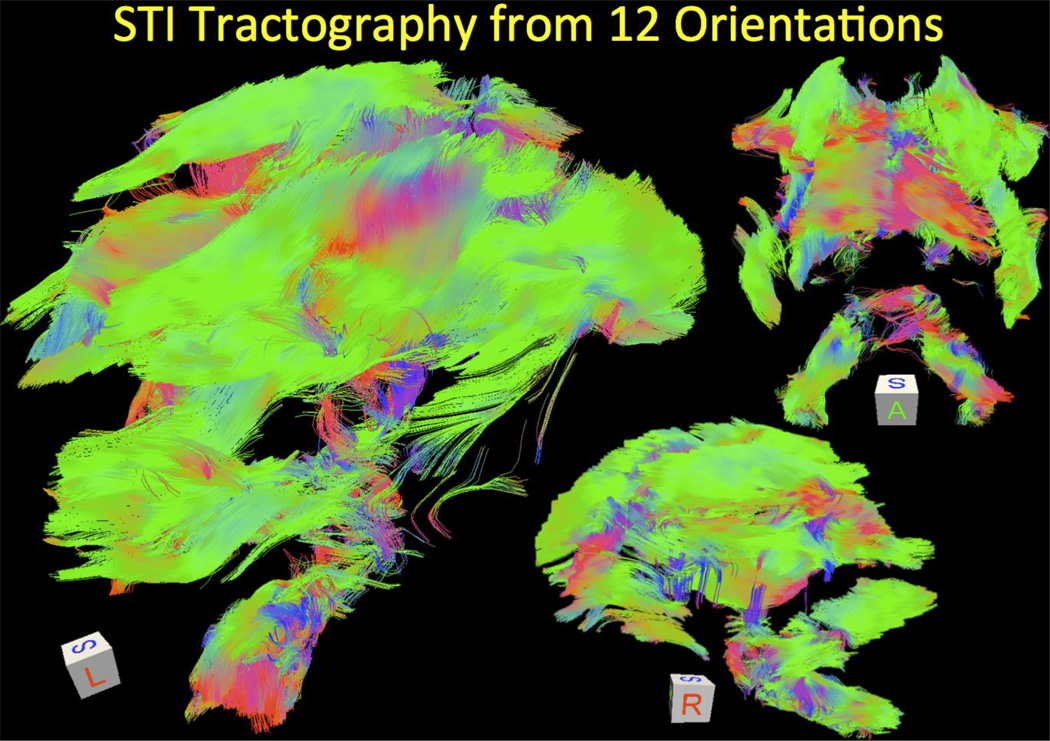
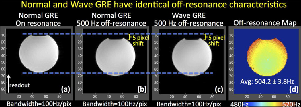
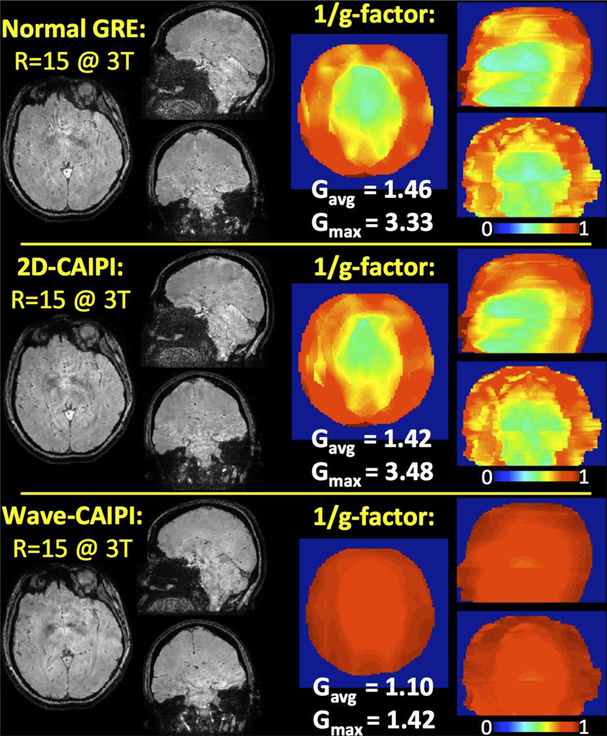
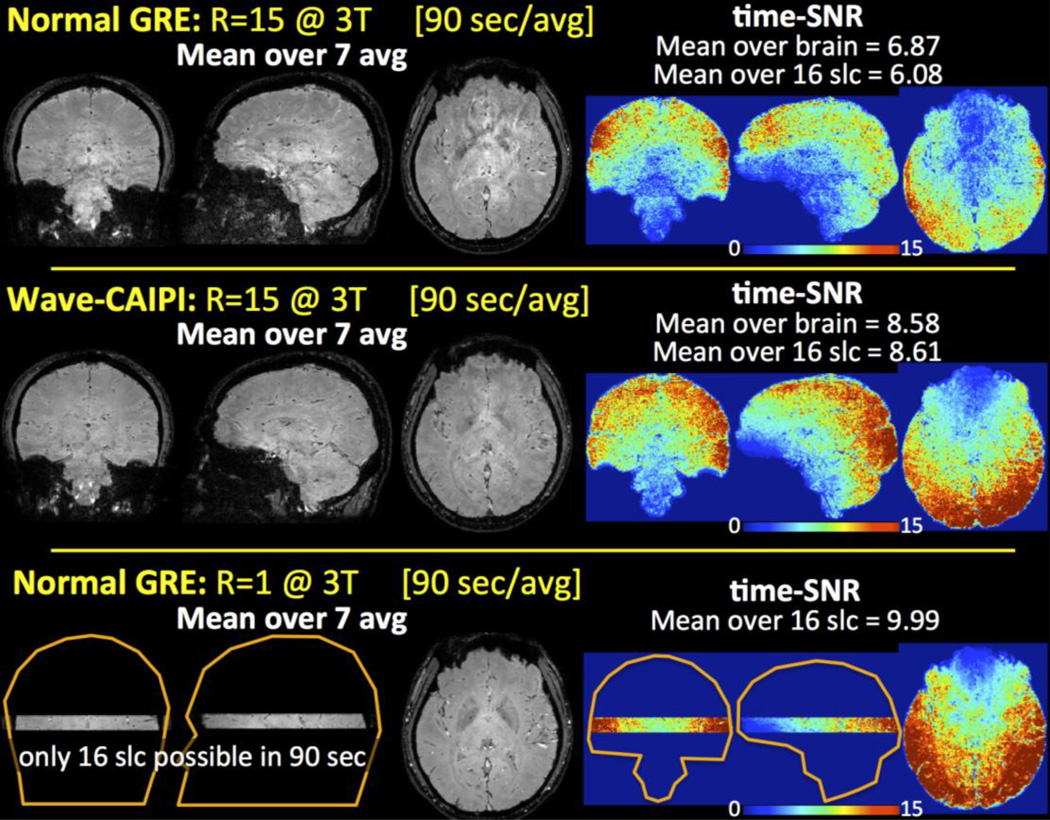
Three-dimensional gradient echo (GRE) is the main workhorse sequence used for susceptibility weighted imaging (SWI), quantitative susceptibility mapping (QSM), and susceptibility tensor imaging (STI). Achieving optimal phase signal-to-noise ratio requires late echo times, thus necessitating a long repetition time (TR). Combined with the large encoding burden of whole-brain coverage with high resolution, this leads to increased scan time. Further, the dipole kernel relating the tissue phase to the underlying susceptibility distribution undersamples the frequency content of the susceptibility map. Scans at multiple head orientations along with calculation of susceptibility through multi-orientation sampling (COSMOS) are one way to effectively mitigate this issue. Additionally, STI requires a minimum of 6 head orientations to solve for the independent tensor elements in each voxel. The requirements of high-resolution imaging with long TR at multiple orientations substantially lengthen the acquisition of COSMOS and STI. The goal of this work is to dramatically speed up susceptibility mapping at multiple head orientations. We demonstrate highly efficient acquisition using 3D-GRE with Wave-CAIPI and dramatically reduce the acquisition time of these protocols. Using R=15-fold acceleration with Wave-CAIPI permits acquisition per head orientation in 90s at 1.1mm isotropic resolution, and 5:35min at 0.5mm isotropic resolution. Since Wave-CAIPI fully harnesses the 3D spatial encoding capability of receive arrays, the maximum g-factor noise amplification remains below 1.30 at 3T and 1.12 at 7T. This allows a 30-min exam for STI with 12 orientations, thus paving the way to its clinical application.
Keywords: Parallel imaging; Phase imaging; Quantitative susceptibility mapping; Susceptibility tensor imaging; Wave-CAIPI.
Copyright © 2015 Elsevier Inc. All rights reserved.
Figures
References
-
- Breuer F, Blaimer M, Mueller MF, Seiberlich N, Heidemann RM, Griswold MA, Jakob PM. Controlled aliasing in volumetric parallel imaging (2D CAIPIRINHA) Magnetic Resonance in Medicine. 2006;55.3:549–556. - PubMed
-
- Conolly S, Nishimura D, Mackovski A, Glover G. Variable-rate selective excitation. Journal of Magnetic Resonance. 1988;78(3):440–458.
Publication types
MeSH terms
Grants and funding
- R01 MH096979/MH/NIMH NIH HHS/United States
- P41 EB015897/EB/NIBIB NIH HHS/United States
- U01 MH093765/MH/NIMH NIH HHS/United States
- 1U01MH093765/MH/NIMH NIH HHS/United States
- R01 NS079653/NS/NINDS NIH HHS/United States
- P41 RR014075/RR/NCRR NIH HHS/United States
- R00EB012107/EB/NIBIB NIH HHS/United States
- R01 EB017337/EB/NIBIB NIH HHS/United States
- R00 EB012107/EB/NIBIB NIH HHS/United States
- R21 HL122759/HL/NHLBI NIH HHS/United States
- R01NS079653/NS/NINDS NIH HHS/United States
- P41RR14075/RR/NCRR NIH HHS/United States
- R01MH096979/MH/NIMH NIH HHS/United States
- J 3480/FWF_/Austrian Science Fund FWF/Austria
- R01EB017337/EB/NIBIB NIH HHS/United States
- R21-HL122759/HL/NHLBI NIH HHS/United States
LinkOut - more resources
Full Text Sources
Other Literature Sources
