

Detection and Quantitation of Circulating Human Irisin by Tandem Mass Spectrometry
- PMID: 26278051
- PMCID: PMC4802359
- DOI: 10.1016/j.cmet.2015.08.001
Detection and Quantitation of Circulating Human Irisin by Tandem Mass Spectrometry
Abstract
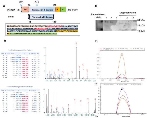
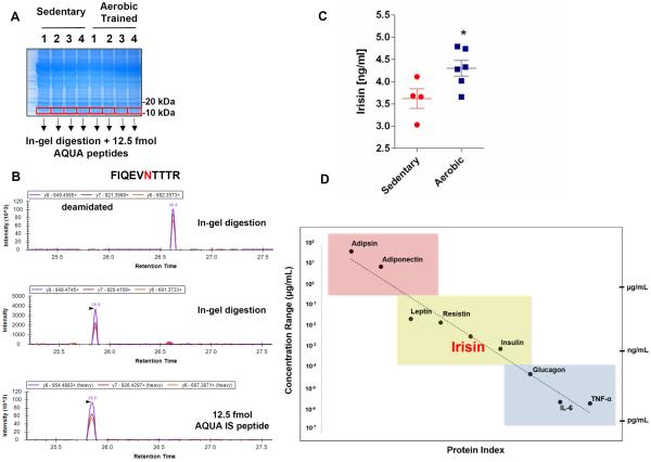
Exercise provides many health benefits, including improved metabolism, cardiovascular health, and cognition. We have shown previously that FNDC5, a type I transmembrane protein, and its circulating form, irisin, convey some of these benefits in mice. However, recent reports questioned the existence of circulating human irisin both because human FNDC5 has a non-canonical ATA translation start and because of claims that many human irisin antibodies used in commercial ELISA kits lack required specificity. In this paper we have identified and quantitated human irisin in plasma using mass spectrometry with control peptides enriched with heavy stable isotopes as internal standards. This precise state-of-the-art method shows that human irisin is mainly translated from its non-canonical start codon and circulates at ∼ 3.6 ng/ml in sedentary individuals; this level is increased to ∼ 4.3 ng/ml in individuals undergoing aerobic interval training. These data unequivocally demonstrate that human irisin exists, circulates, and is regulated by exercise.
Copyright © 2015 Elsevier Inc. All rights reserved.
Figures
Comment in
-
News: Circulating irisin confirmed by mass spectrometry.Nat Rev Endocrinol. 2015 Oct;11(10):568. doi: 10.1038/nrendo.2015.148. Epub 2015 Sep 1. Nat Rev Endocrinol. 2015. PMID: 26323660 No abstract available.
References
-
- Beausoleil SA, Villen J, Gerber SA, Rush J, Gygi SP. A probability-based approach for high-throughput protein phosphorylation analysis and site localization. Nat Biotechnol. 2006;24:1285–1292. - PubMed
-
- Chang KJ, Wang CC. Translation initiation from a naturally occurring non-AUG codon in Saccharomyces cerevisiae. The Journal of biological chemistry. 2004;279:13778–13785. - PubMed
-
- Daskalopoulou SS, Cooke AB, Gomez YH, Mutter AF, Filippaios A, Mesfum ET, Mantzoros CS. Plasma irisin levels progressively increase in response to increasing exercise workloads in young, healthy, active subjects. European journal of endocrinology / European Federation of Endocrine Societies. 2014;171:343–352. - PubMed
Publication types
MeSH terms
Substances
Grants and funding
- DK90861/DK/NIDDK NIH HHS/United States
- K99 NS087096/NS/NINDS NIH HHS/United States
- NS087096/NS/NINDS NIH HHS/United States
- UL1TR000135/TR/NCATS NIH HHS/United States
- RC4 DK090861/DK/NIDDK NIH HHS/United States
- R01 DK054477/DK/NIDDK NIH HHS/United States
- T32DK007352/DK/NIDDK NIH HHS/United States
- K01 DK098285/DK/NIDDK NIH HHS/United States
- DK31405/DK/NIDDK NIH HHS/United States
- UL1 TR000135/TR/NCATS NIH HHS/United States
- DK098285/DK/NIDDK NIH HHS/United States
- R01AG09531/AG/NIA NIH HHS/United States
- R37 DK031405/DK/NIDDK NIH HHS/United States
- R00 NS087096/NS/NINDS NIH HHS/United States
- T32 DK007352/DK/NIDDK NIH HHS/United States
- R01 DK031405/DK/NIDDK NIH HHS/United States
- R01 AG009531/AG/NIA NIH HHS/United States
- R01 GM067945/GM/NIGMS NIH HHS/United States
LinkOut - more resources
Full Text Sources
Other Literature Sources
Molecular Biology Databases
Research Materials