. 2015 Oct;36(10):1078-86.
doi: 10.1016/j.placenta.2015.08.003. Epub 2015 Aug 7.

Hypoplastic left heart syndrome is associated with structural and vascular placental abnormalities and leptin dysregulation

Affiliations

Hypoplastic left heart syndrome is associated with structural and vascular placental abnormalities and leptin dysregulation

Helen N Jones et al. Placenta. 2015 Oct.

Abstract

Introduction: Hypoplastic left heart syndrome (HLHS) is a severe cardiovascular malformation (CVM) associated with fetal growth abnormalities. Genetic and environmental factors have been identified that contribute to pathogenesis, but the role of the placenta is unknown. The purpose of this study was to systematically examine the placenta in HLHS with and without growth abnormalities.

Methods: HLHS term singleton births were identified from a larger cohort when placenta tissue was available. Clinical data were collected from maternal and neonatal medical records, including anthropometrics and placental pathology reports. Placental tissues from cases and controls were analyzed to assess parenchymal morphology, vascular architecture and leptin signaling.

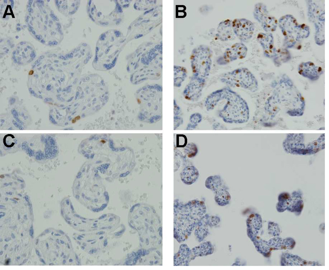
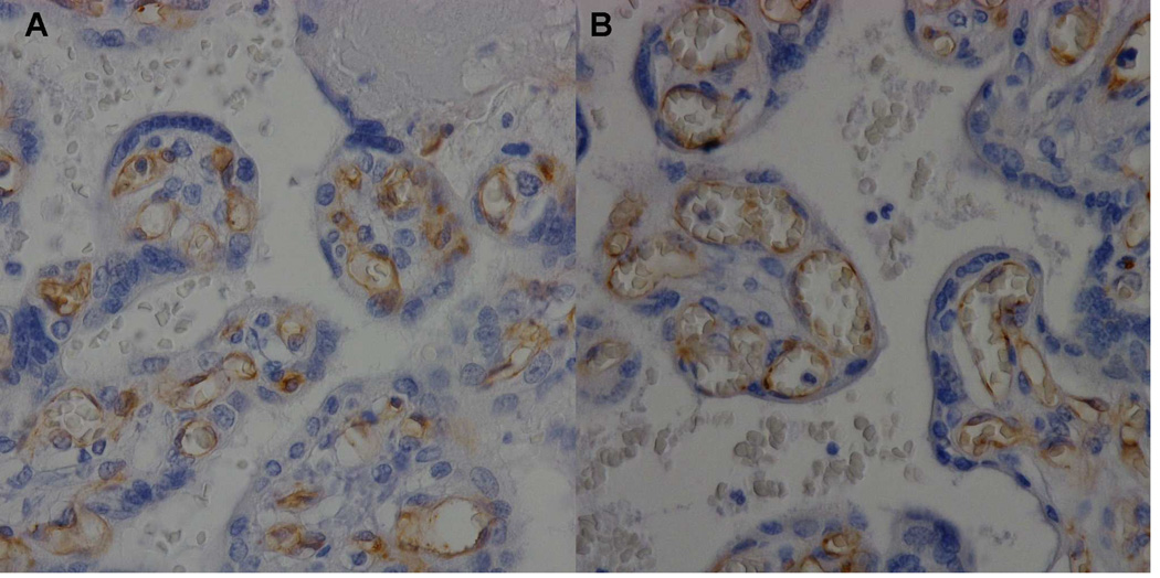
Results: HLHS cases (n = 16) and gestational age-matched controls (n = 18) were analyzed. Among cases, the average birth weight was 2993 g, including 31% that were small for gestational age. When compared with controls, gross pathology of HLHS cases demonstrated significantly reduced placental weight and increased fibrin deposition, while micropathology showed increased syncytial nuclear aggregates, decreased terminal villi, reduced vasculature and increased leptin expression in syncytiotrophoblast and endothelial cells.

Discussion: Placentas from pregnancies complicated by fetal HLHS are characterized by abnormal parenchymal morphology, suggesting immature structure may be due to vascular abnormalities. Increased leptin expression may indicate an attempt to compensate for these vascular abnormalities. Further investigation into the regulation of angiogenesis in the fetus and placenta may elucidate the causes of HLHS and associated growth abnormalities in some cases.

Keywords: Angiogenesis; Congenital heart disease; Vascular biology.


Figures

Figure 1
Figure 1. Placenta tissue in HLHS demonstrates morphologic abnormalities
Hematoxylin and Eoisin stain of representative placenta parenchyma of control (A) and HLHS cases (B–E), 40×. HLHS placenta (B) contains increased syncytial nuclear aggregates (arrows), increased fibrin deposits (outlined in black) and reduced fetal blood vessel density and area (yellow asterisks). In addition to subchorionic fibrin, as reported by gross pathology, microscopic fibrin is present within the parenchyma of the HLHS cases but not controls (outlined in black).
Figure 2
Figure 2. Proliferative activity is increased in placental tissue from HLHS cases
Ki67 expression in control (A, C) and HLHS cases (B, D). In control placenta, Ki67 staining (an indicator of proliferative activity) was sparse throughout the parenchyma, however, in HLHS cases, positively stained nuclei were present in syncytium, SNAs, and throughout the parenchyma.
Figure 3
Figure 3. Placenta tissue in HLHS is characterized by maladaptive angiogenesis
CD31 staining of representative placenta parenchyma in control (A) and HLHS cases (B), 40×. CD31 is limited to endothelial cells allowing analysis of vessel number, density and identification of vasculosyncytial membranes to aid villous maturation assessment (See Tables 3 and 4).
Figure 4
Figure 4. Placental weight is correlated with birthweight
Distribution of placental parameters vs. birthweight in control (A) and HLHS (B) cohorts. Placental weight is positively correlated with birthweight in both control (C, R square = 0.4951) and HLHS (D, R square = 0.3011) cohorts.
Figure 5
Figure 5. Placenta tissue in HLHS demonstrates leptin dysregulation
Leptin expression in control (A & C) and HLHS (B & D) placenta parenchyma. In control placenta leptin expression was localized in the syncytium (arrows). Leptin expression was increased in HLHS cases and localization expanded to the fetal endothelium and mesenchymal core of the villi as demonstrated by the brown staining.
Figure 6
Figure 6. Representative micrographs of FLK-1 expression in human placental parenchyma
Flk1 expression was localized to the fetal endothelium (arrowheads) in both control (A) and HLHS (B) with a reduction in Flk1 expression in the HLHS cases (B).
Figure 7
Figure 7. Model figure of the relationship between CVM, placental abnormalities and FGR
Multiple causative factors including genetics, environmental and nutrition may contribute to initial abnormal placental and/or cardiovascular formation and development. As gestation progresses, abnormal fetal cardiac output may impact the expansion of the peripheral fetal vasculature, including the placental villous vasculature, thought to branch in response to increased cardiac output as the fetus grows in utero. Consequently, associated growth abnormalities predispose the newborn with CVM to adverse outcomes. Placental responses to this hypoxemia and/or impaired villous tree expansion may include compensatory mechanisms to increase nutrient or oxygen transfer, such as increased leptin expression. Taken together, in some cases a combination of adverse factors may lead to a spectrum of placental abnormalities that contribute to FGR.

References

    1. Feinstein JA, Benson DW, Dubin AM, Cohen MS, Maxey DM, Mahle WT, et al. Hypoplastic left heart syndrome: current considerations and expectations. J Am Coll Cardiol. 2012;59(1 Suppl):S1–S42. Erratum in: J Am Coll Cardiol. 2012 59(5):544. - PMC - PubMed
    1. Gelb BD, Chung WK. Complex genetics and the etiology of human congenital heart disease. Cold Spring Harb Perspect Med. 2014;4(7):a013953. - PMC - PubMed
    1. Hinton RB, Martin LJ, Rame-Gowda S, Tabangin ME, Cripe LH, Benson DW. Hypoplastic left heart syndrome links to chromosomes 10q and 6q and is genetically related to biscuspid aortic valve. J Am Coll Cardiol. 2009;53:1065–1071. - PMC - PubMed
    1. Atz AM, Travison TG, Williams IA, Pearson GD, Laussen PC, Mahle WT, et al. Pediatric Heart Network Investigators. Prenatal diagnosis and risk factors for preoperative death in neonates with single right ventricle and systemic outflow obstruction: screening data from the Pediatric Heart Network Single Ventricle Reconstruction Trial(*) J Thorac Cardiovasc Surg. 2010;140:1245–1250. - PMC - PubMed
    1. Gaynor JW, Mahle WT, Cohen MI, Ittenbach RF, DeCampli WM, Steven JM, et al. Risk factors for mortality after the Norwood procedure. Eur J Cardiothorac Surg. 2002;22:82–89. - PubMed

Publication types