. 2015 Aug 20;22(8):1098-107.
doi: 10.1016/j.chembiol.2015.07.013. Epub 2015 Aug 13.

The Structure and Interactions of Periplasmic Domains of Crucial MmpL Membrane Proteins from Mycobacterium tuberculosis

Affiliations

The Structure and Interactions of Periplasmic Domains of Crucial MmpL Membrane Proteins from Mycobacterium tuberculosis

Nicholas Chim et al. Chem Biol. .

Abstract

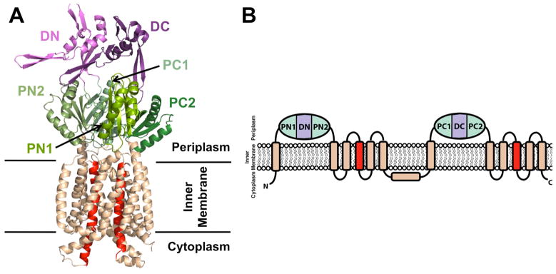
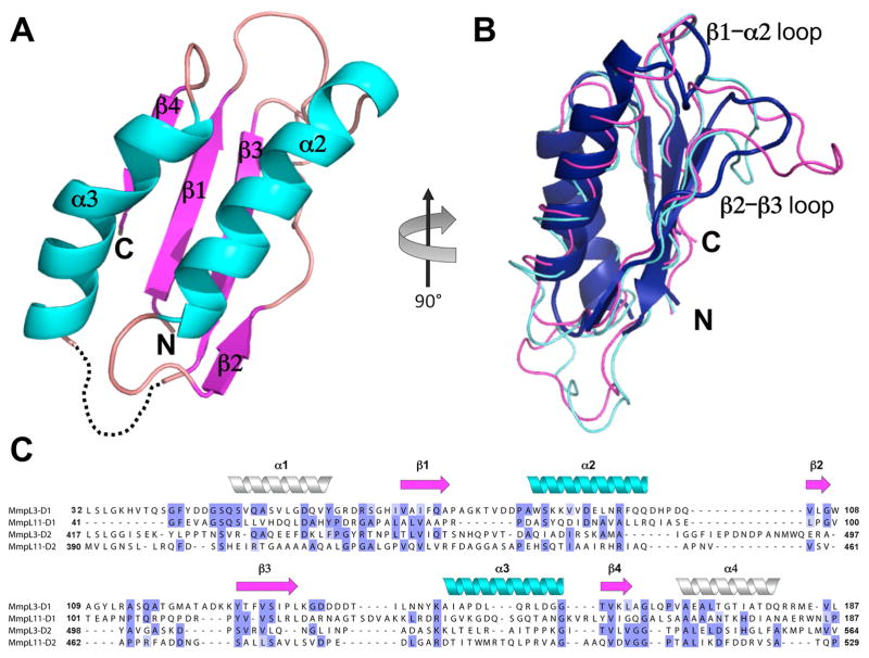
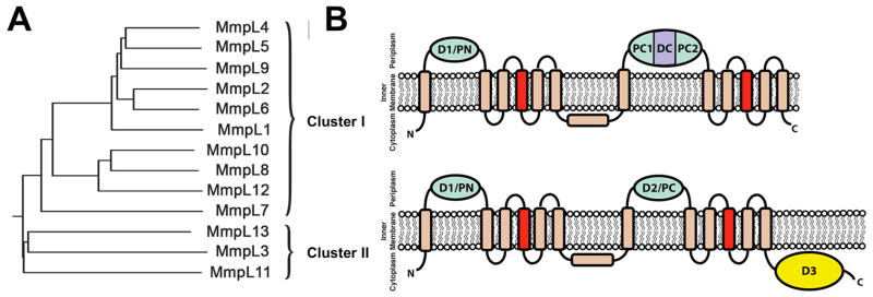
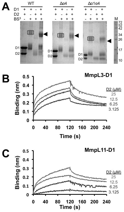
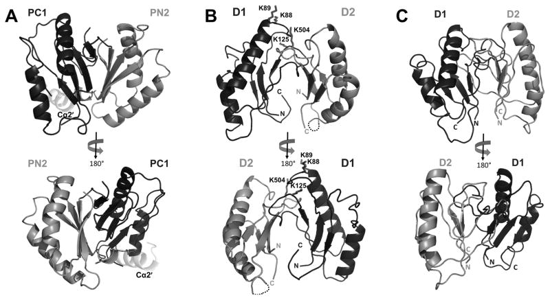
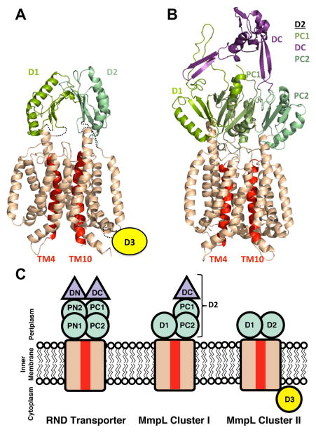
Mycobacterium tuberculosis mycobacterial membrane protein large (MmpL) proteins are important in substrate transport across the inner membrane. Here, we show that MmpL proteins are classified into two phylogenetic clusters, where MmpL cluster II contains three soluble domains (D1, D2, and D3) and has two full-length members, MmpL3 and MmpL11. Significantly, MmpL3 is currently the most druggable M. tuberculosis target. We have solved the 2.4-Å MmpL11-D2 crystal structure, revealing structural homology to periplasmic porter subdomains of RND (multidrug) transporters. The resulting predicted cluster II MmpL membrane topology has D1 and D2 residing, and possibly interacting, within the periplasm. Crosslinking and biolayer interferometry experiments confirm that cluster II D1 and D2 bind with weak affinities, and guided D1-D2 heterodimeric model assemblies. The predicted full-length MmpL3 and MmpL11 structural models reveal key substrate binding and transport residues, and may serve as templates to set the stage for in silico anti-tuberculosis drug development.

Keywords: MmpL; Mycobacterium tuberculosis; RND transporters; X-ray crystallography; biolayer interferometry; crosslinking; porter domain; tuberculosis.


Figures

Figure 1
Figure 1
(A) Cartoon representation of monomeric CusA (PDB code: 4DNT) showing the docking, porter, and transmembrane subdomains. The N- and C-terminal docking (DN and DC) and porter (PN1, PN2, PC1, and PC2) subdomains are colored shades of purple and green, respectively, while the transmembrane subdomain is colored wheat, except for the central transmembrane helices, TM4 and TM10, which are colored red. (B) RND transporter membrane topology with two periplasmic domains, each containing two porter subdomains and one docking subdomain. An additional extracytosplasmic α-helix between TM6 and TM7 is located near the cytoplasmic membrane surface and runs almost parallel to it. Subdomain color designation is as in (A).
Figure 2
Figure 2
(A) Cartoon representation of MmpL11-D2 structure with missing residues 479 – 489 depicted as dashed lines. α-helices, β-strands, and loops are colored cyan, magenta, and wheat, respectively (B) MmpL11-D2 colored blue, is rotated 90° clockwise from (A) and structurally aligned with RND PC1 porter subdomains from ZneA (PDB code: 4K0E) and MexB (PDB code: 2V50), colored magenta and cyan, respectively. (C) MmpL11 and MmpL3 Cluster II D1 and D2 domain sequence alignment based on secondary structural prediction and MmpL11-D2 structure. Cylinders (α-helices) and arrows (β-sheets) colors correspond to the secondary structural elements in (A). The predicted α-helices (α1 and α4) are shown as white cylinders.
Figure 3
Figure 3
(A) Phylogenetic tree of MmpL proteins reveals two distinct clusters where the (B) predicted membrane topologies of MmpL Clusters I and II proteins are based on RND transporters. The predicted porter domains (N-terminal D1 (PN) and Cluster I C-terminal D2 (PC1 and PC2) and Cluster II C-terminal D2 (PC)) are colored green and the predicted Cluster I C-terminal docking domain (DC) is colored purple. D3 is colored yellow while the transmembrane subdomain is colored wheat, except for the central transmembrane helices, TM4 and TM10, which are colored red. The predicted additional extra-cytoplasmic α-helix located between TM6 and TM7 is shown almost parallel to the cytoplasmic membrane surface, as observed in RND transporter structures (Fig. 1B).
Figure 4
Figure 4
MmpL3 and MmpL11 D1 and D2 domains interact. (A) SDS-PAGE of MmpL3 D1 and D2 domains and their respective truncated constructs (Δα4 and Δα1α4) in the presence of BS3, suggesting that α1 helix is essential for heterodomain interaction. In all instances, the MmpL3-D1 homodimer (38.3 kDa for WT) is boxed whereas arrowheads identify the MmpL3-D1-D2 heterodimer (34.9 kDa for WT). Notably, for the Δα1α4 constructs, the heterodimer is absent. Biolayer interferometry experiments to assess interactions between (B) MmpL3 D1 (biotinylated) and D2 domains, and (C) MmpL11 D1 (biotinylated) and D2 domains. All reactions were performed at 25 °C in 20 mM sodium phosphate pH 7.4 and 150 mM NaCl. Immobilized biotinylated D1 domains were exposed to different concentrations (25 – 3.125 μM) of D2 domains, where interaction (association and dissociation) is assessed by a wavelength shift (nm).
Figure 5
Figure 5
(A) Cartoon representation of CusA PC1 and PN2 heterodimer (PDB code: 4DNT). PC1 and PN2 are colored black and dark grey, respectively, while PC1 Cα2′ is colored light grey. Cartoon representation of the heterodimer models of (B) MmpL11-D1-D2, depicting crosslinked lysine residues as sticks and (C) MmpL3-D1-D2. MmpL3/11 D1 and D2 are colored black and dark grey, respectively.
Figure 6
Figure 6
Phyre2 models of (A) MmpL11 with additional restraints from the crosslinking results and (B) MmpL4. The transmembrane domains are colored wheat, except for TM4 and TM10, which are colored red. The different periplasmic porter subdomains are in shades of green and the proposed MmpL4 docking domain is colored purple. The Cluster II MmpL (MmpL11) cytoplasmic D3 domain is signified by a yellow circle. (C) A cartoon representing the domain architecture of RND transporters, MmpL Cluster I and II proteins. Subdomain color designations are as in (A) and (B).

References

    1. Adams PD, Afonine PV, Bunkoczi G, Chen VB, Davis IW, Echols N, Headd JJ, Hung LW, Kapral GJ, Grosse-Kunstleve RW, et al. PHENIX: a comprehensive Python-based system for macromolecular structure solution. Acta Crystallogr D Biol Crystallogr. 2010;66:213–221. - PMC - PubMed
    1. Battye TG, Kontogiannis L, Johnson O, Powell HR, Leslie AG. iMOSFLM: a new graphical interface for diffraction-image processing with MOSFLM. Acta Crystallogr D Biol Crystallogr. 2011;67:271–281. - PMC - PubMed
    1. Bolla JR, Su CC, Do SV, Radhakrishnan A, Kumar N, Long F, Chou TH, Delmar JA, Lei HT, Rajashankar KR, et al. Crystal structure of the Neisseria gonorrhoeae MtrD inner membrane multidrug efflux pump. PLoS One. 2014;9:e97903. - PMC - PubMed
    1. Carel C, Nukdee K, Cantaloube S, Bonne M, Diagne CT, Laval F, Daffe M, Zerbib D. Mycobacterium tuberculosis proteins involved in mycolic acid synthesis and transport localize dynamically to the old growing pole and septum. PLoS One. 2014;9:e97148. - PMC - PubMed
    1. Chen VB, Arendall WB, 3rd, Headd JJ, Keedy DA, Immormino RM, Kapral GJ, Murray LW, Richardson JS, Richardson DC. MolProbity: all-atom structure validation for macromolecular crystallography. Acta Crystallogr D Biol Crystallogr. 2010;66:12–21. - PMC - PubMed

Publication types

MeSH terms

Substances

LinkOut - more resources