

Clinical and pathological features of intraductal papillary neoplasm of the biliary tract and gallbladder
- PMID: 26278323
- PMCID: PMC4557656
- DOI: 10.1111/hpb.12460
Clinical and pathological features of intraductal papillary neoplasm of the biliary tract and gallbladder
Abstract
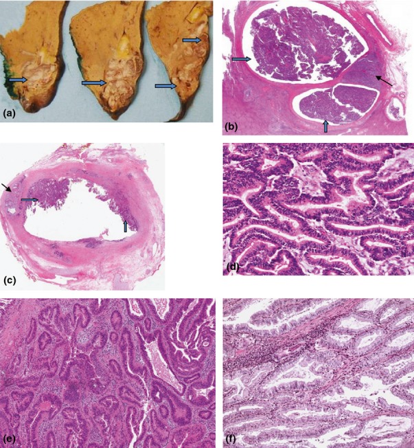
Background: Intraductal papillary neoplasms of the biliary tract (IPNB) and intracholecystic papillary neoplasms (ICPN) are rare tumours characterized by intraluminal papillary growth that can be associated with invasive carcinoma. Their natural history remains poorly understood. This study examines clinicopathological features and outcomes.
Methods: Patients who underwent surgery for IPNB/ICPN (2008-2014) were identified. Descriptive statistics and survival data were generated.
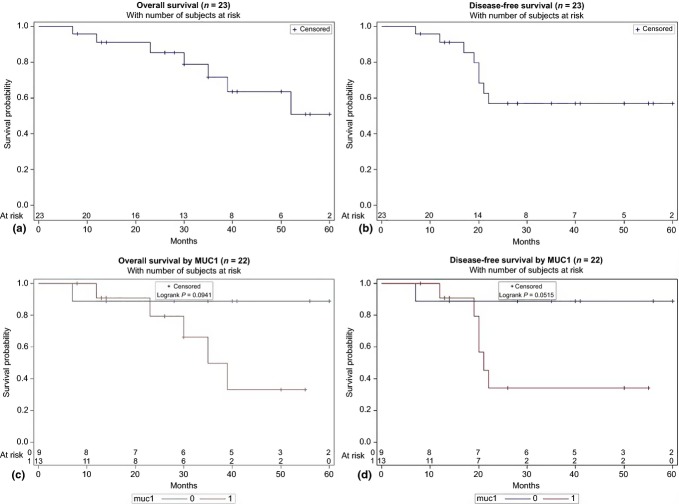
Results: Of 23 patients with IPNB/ICPN, 10 were male, and the mean age was 68 years. The most common presentations were abdominal pain (n = 10) and jaundice (n = 9). Tumour locations were: intrahepatic (n = 5), hilar (n = 3), the extrahepatic bile duct (n = 8) and the gallbladder (n = 7). Invasive cancer was found in 20/23 patients. Epithelial subtypes included pancreatobiliary (n = 15), intestinal (n = 7) and gastric (n = 1). The median follow-up was 30 months. The 5-year overall (OS) and disease-free survivals (DFS) were 51% and 57%, respectively. Decreased OS (P = 0.09) and DFS (P = 0.05) were seen in patients with tumours expressing MUC1 on immunohistochemistry (IHC).
Conclusion: IPNB/ICPN are rare precursor lesions that can affect the entire biliary epithelium. At pathology, the majority of patients have invasive carcinoma, thus warranting a radical resection. Patients with tumours expressing MUC1 appear to have worse OS and DFSs.
© 2015 International Hepato-Pancreato-Biliary Association.
Figures
References
-
- Zen Y, Aishima S, Ajioka Y, Haratake J, Kage M, Kondo F, et al. Proposal of histological criteria for intraepithelial atypical/proliferative biliary epithelial lesions of the bile duct in hepatolithiasis with respect to cholangiocarcinoma: preliminary report based on interobserver agreement. Pathol Int. 2005;55:180–188. - PubMed
-
- Zen Y, Adsay NV, Bardadin K, Colombari R, Ferrell L, Haga H, et al. Biliary intraepithelial neoplasia: an international interobserver agreement study and proposal for diagnostic criteria. Mod Pathol. 2007;20:701–709. - PubMed
-
- Zen Y, Sasaki M, Fujii T, Chen TC, Chen MF, Yeh TS, et al. Different expression patterns of mucin core proteins and cytokeratins during intrahepatic cholangiocarcinogenesis from biliary intraepithelial neoplasia and intraductal papillary neoplasm of the bile duct—an immunohistochemical study of 110 cases of hepatolithiasis. J Hepatol. 2006;44:350–358. - PubMed
-
- Aishima S, Kubo Y, Tanaka Y, Oda Y. Histological features of precancerous and early cancerous lesions of biliary tract carcinoma. J Hepatobiliary Pancreat Sci. 2014;21:448–452. - PubMed
MeSH terms
Substances
LinkOut - more resources
Full Text Sources
Other Literature Sources
Medical
Research Materials
Miscellaneous
