Comparative Study
. 2015 Dec;149(7):1742-51.
doi: 10.1053/j.gastro.2015.08.005. Epub 2015 Aug 14.

The Functional Lumen Imaging Probe Detects Esophageal Contractility Not Observed With Manometry in Patients With Achalasia

Affiliations
Comparative Study

The Functional Lumen Imaging Probe Detects Esophageal Contractility Not Observed With Manometry in Patients With Achalasia

Dustin A Carlson et al. Gastroenterology. 2015 Dec.

Abstract

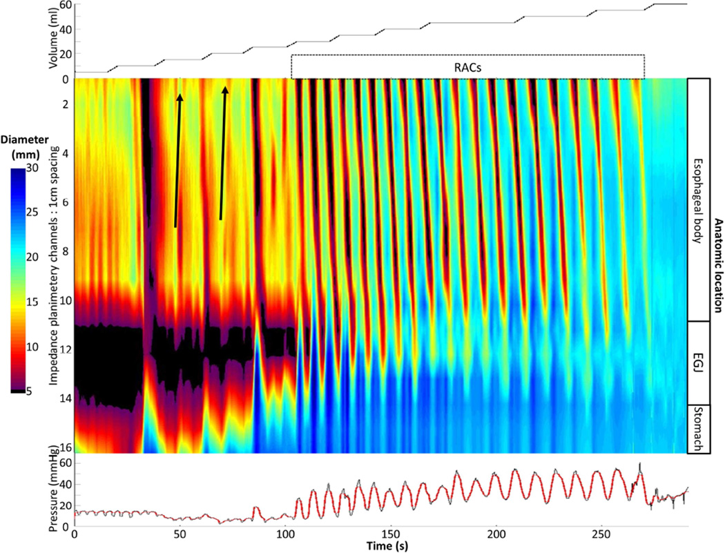
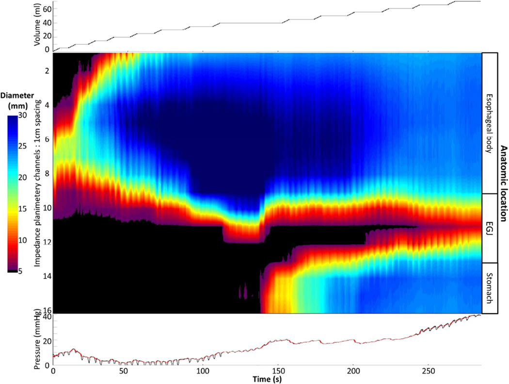
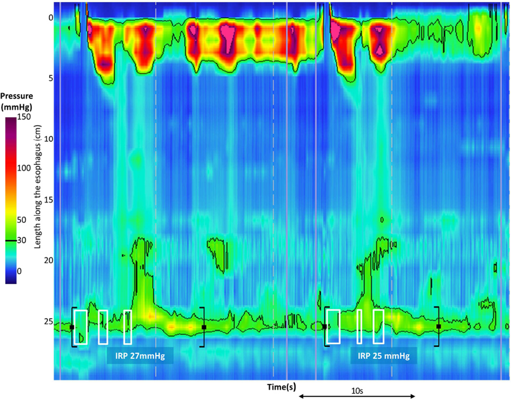
Background & aims: The functional lumen imaging probe (FLIP) could improve the characterization of achalasia subtypes by detecting nonocclusive esophageal contractions not observed with standard manometry. We aimed to evaluate esophageal contractions during volumetric distention in patients with achalasia using FLIP topography.

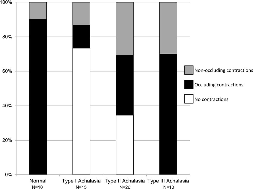
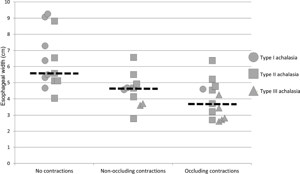
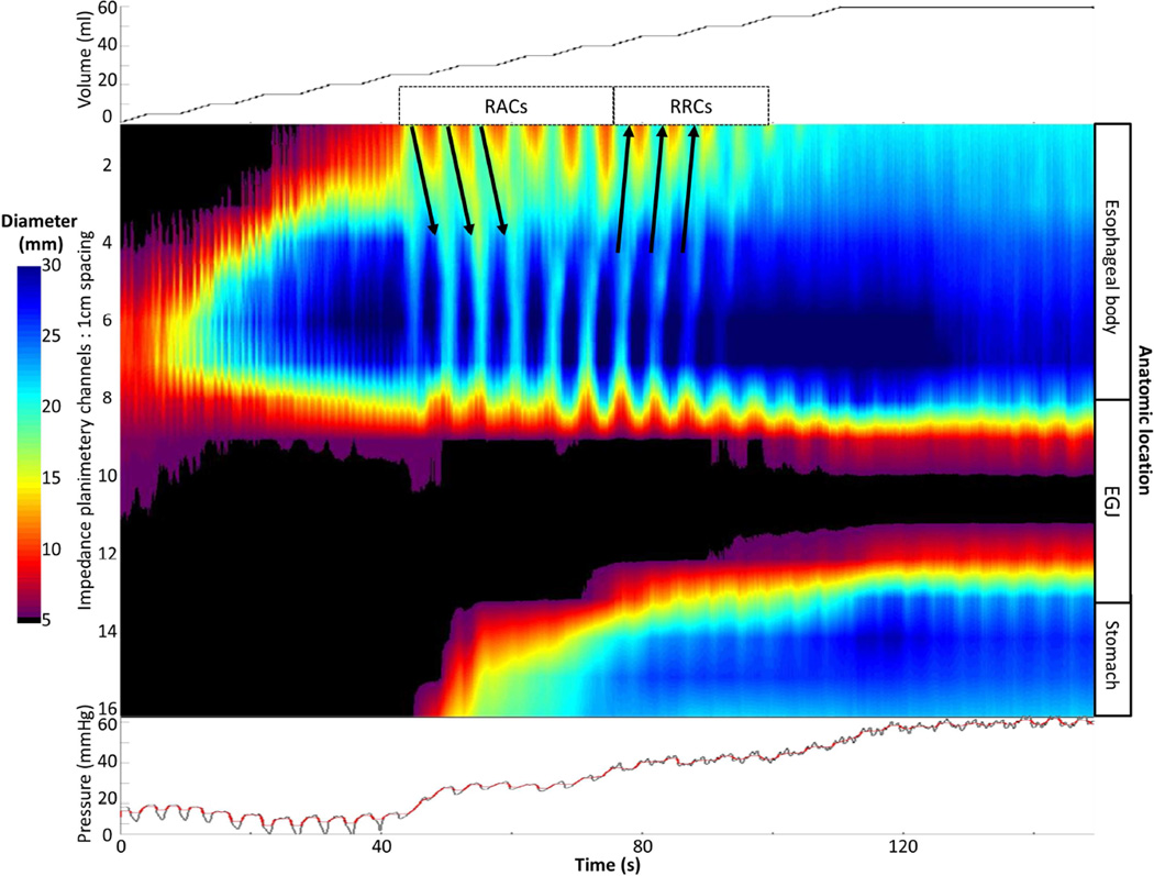
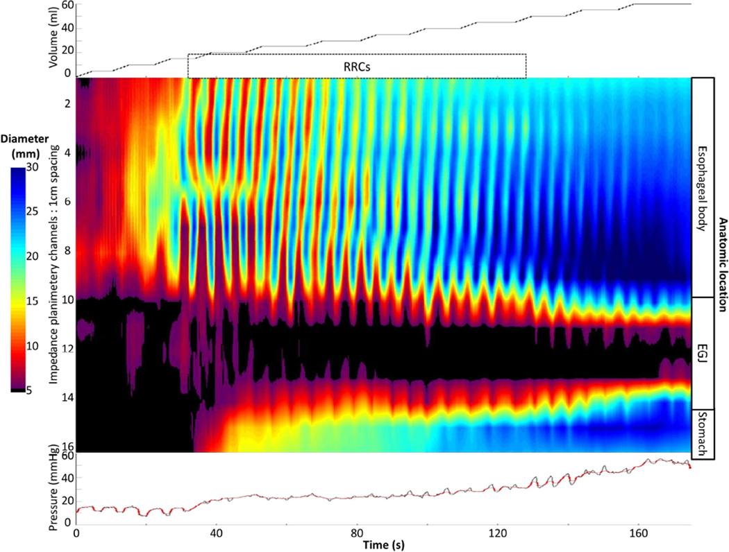
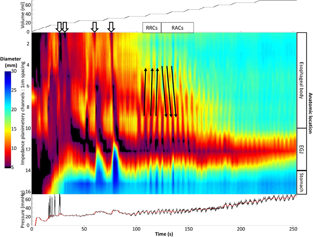
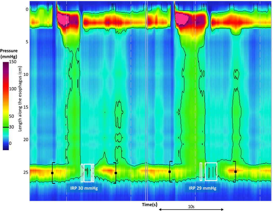
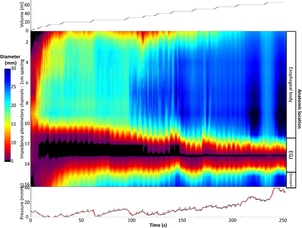
Methods: Fifty-one treatment-naive patients with achalasia, defined and subclassified by high-resolution esophageal pressure topography, and 10 asymptomatic individuals (controls) were evaluated with the FLIP during endoscopy. During stepwise distension, simultaneous intrabag pressures and 16 channels of cross-sectional areas were measured; data were exported to software that generated FLIP topography plots. Esophageal contractility was identified by noting periods of reduced luminal diameter. Esophageal contractions were characterized further by propagation direction, repetitiveness, and based on whether they were occluding or nonoccluding.

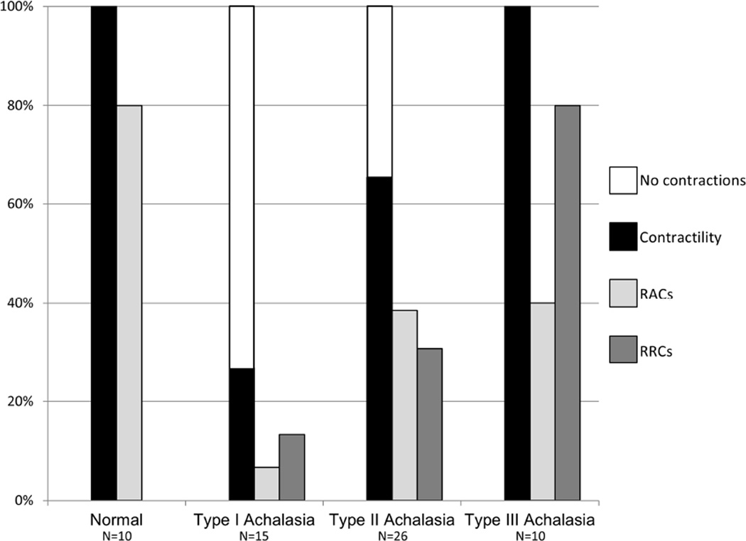
Results: Esophageal contractility was detected in all 10 controls: 8 of 10 had repetitive antegrade contractions and 9 of 10 had occluding contractions. Contractility was detected in 27% (4 of 15) of patients with type I achalasia and in 65% (18 of 26, including 9 with occluding contractions) of patients with type II achalasia. Contractility was detected in all 10 patients with type III achalasia; 8 of these patients had a pattern of contractility that was not observed in controls (repetitive retrograde contractions).

Conclusions: Esophageal contractility not observed with manometry can be detected in patients with achalasia using FLIP topography. The presence and patterns of contractility detected with FLIP topography may represent variations in pathophysiology, such as mechanisms of panesophageal pressurization in patients with type II achalasia. These findings could have implications for additional subclassification to supplement prediction of the achalasia disease course.

Keywords: EndoFLIP; Esophagus; Motility; Peristalsis.


Figures

Figure 1
Figure 1. FLIP topography of a normal control
Data output (Top: distension volume, thus representing the distension protocol; Middle: Topography; Bottom: Intra-bag pressure) generated by the customized MATLAB program over the course of the study protocol from a single control subject are displayed. Two retrograde contractions (noted with black arrows) are present in a period of contractility prior to the onset of repetitive, antegrade, contractions (RACs). Figure used with permission from the Esophageal Center at Northwestern.
Figure 2
Figure 2. Non-occlusive contractions detected with FLIP
Patients were considered to have occlusive contractions if any observed contraction on FLIP topography achieved a luminal diameter ≤ 6 mm and non-occlusive if the minimal diameter reached was > 6mm. Frequencies varied between normal controls and achalasia (p = 0.005) and among achalasia subtypes (p = 0.004: type I vs type II, p = 0.057; type I vs type III, p = 0.001; type II vs type III, p = 0.022).
Figure 3
Figure 3. Association of esophageal width, achalasia subtype, and presence of occluding and non-occluding contractions
Each patient that had an esophagram available for esophageal width measurement is represented according to their achalasia subtype. Median values within each contraction group are represented by the dashed horizontal lines.
Figure 4
Figure 4. Frequencies of contractility patterns observed with FLIP topography among normal controls and achalasia subtypes
Other than no contractions and contractility, which were mutually exclusive, a subject may have demonstrated more than one pattern. RACs - Repetitive, antegrade, contractions. RRCs - Repetitive, retrograde, contractions (RRCs).
Figure 5
Figure 5. Patterns of esophageal contractility detected with FLIP topography in achalasia
A). An example of absent contractility on FLIP topography is depicted in a patient with type I achalasia. B) In this example of FLIP topography in a type II patient, propagating contractions, both repetitive antegrade contractions (RACs; downward pointing black arrows)) and repetitive, retrograde contractions (RRCs; upward pointing black arrows) were observed. The narrowest luminal diameter achieved during contractions in this patient was 9.7 thus indicating non-occlusive contractions. C) On FLIP topography in this type III patient, RRCs, the most common pattern observed among type III achalasia patients, are observed. In each of the FLIP topographies (A – C) the narrow diameters at the esophagogastric junction (EGJ) throughout the distension protocol can be noted: The EGJ-distensibility index at in each of these patients was < 1.5 mm2/mmHg at 50 and 60-ml distension volumes. Figure used with permission from the Esophageal Center at Northwestern.
Figure 6
Figure 6. Potential for supplementary outcome prediction based on FLIP topography assessment
Two swallows from two patients’ high-resolution manometries that met criteria for type I (A) and type II (C) achalasia are displayed. On FLIP topography, the type I patient (B) demonstrated contractility in the esophageal body, including both repetitive, antegrade contractions (RACs) and repetitive, retrograde contractions (RRCs); lumen occluding contractions were actually detected on FLIP topography (white arrows). Six months after 30-mm pneumatic dilation, the patient was asymptomatic. The FLIP topography for the type II patient (D) demonstrated absent contractility. After per-oral endoscopic myotomy and pneumatic dilation to 30 then 35- mm, the patient continued to have dysphagia on a daily basis. Figure used with permission from the Esophageal Center at Northwestern.

References

    1. Vaezi MF, Pandolfino JE, Vela MF. ACG clinical guideline: diagnosis and management of achalasia. The American journal of gastroenterology. 2013;108(8):1238–1249. quiz 50. - PubMed
    1. Pandolfino JE, Kwiatek MA, Nealis T, et al. Achalasia: a new clinically relevant classification by high-resolution manometry. Gastroenterology. 2008;135(5):1526–1533. - PMC - PubMed
    1. Kahrilas PJ, Bredenoord AJ, Fox M, et al. The Chicago Classification of esophageal motility disorders, v3.0. Neurogastroenterology and motility : the official journal of the European Gastrointestinal Motility Society. 2015;27(2):160–174. - PMC - PubMed
    1. Salvador R, Costantini M, Zaninotto G, et al. The preoperative manometric pattern predicts the outcome of surgical treatment for esophageal achalasia. Journal of gastrointestinal surgery : official journal of the Society for Surgery of the Alimentary Tract. 2010;14(11):1635–1645. - PubMed
    1. Pratap N, Kalapala R, Darisetty S, et al. Achalasia cardia subtyping by high-resolution manometry predicts the therapeutic outcome of pneumatic balloon dilatation. Journal of neurogastroenterology and motility. 2011;17(1):48–53. - PMC - PubMed

Publication types

MeSH terms

LinkOut - more resources