. 2015 Nov;192(2):204-8.
doi: 10.1016/j.jsb.2015.08.006. Epub 2015 Aug 13.

Automatic estimation and correction of anisotropic magnification distortion in electron microscopes

Affiliations

Automatic estimation and correction of anisotropic magnification distortion in electron microscopes

Timothy Grant et al. J Struct Biol. 2015 Nov.

Abstract

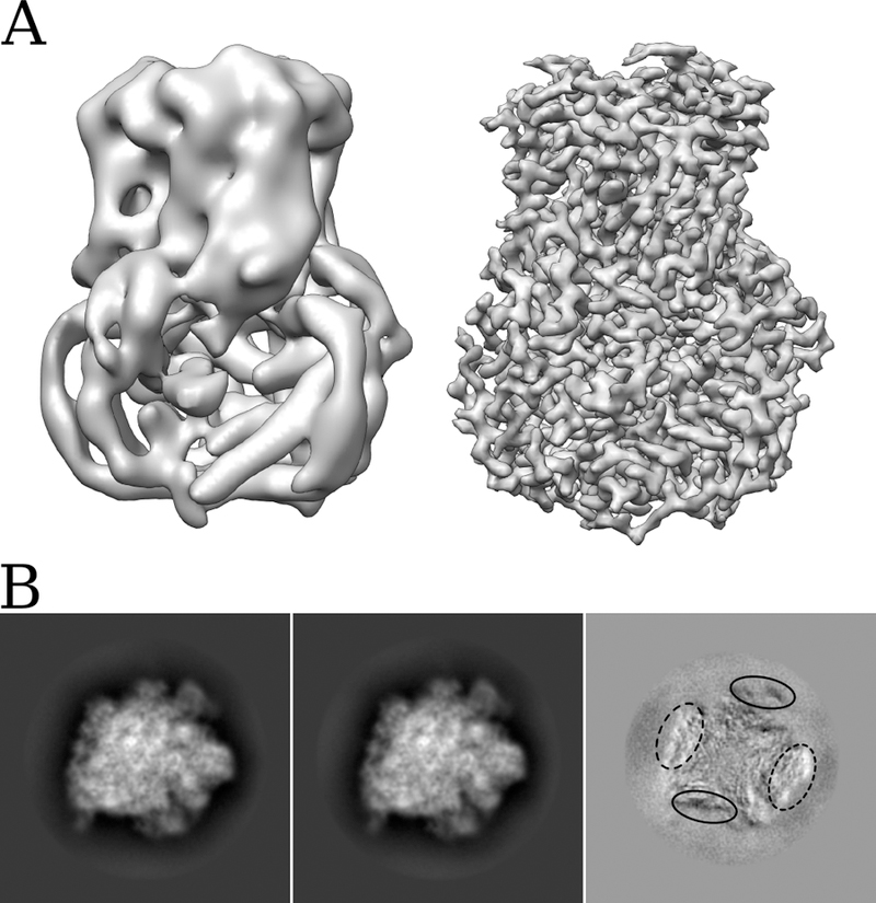
We demonstrate a significant anisotropic magnification distortion, found on an FEI Titan Krios microscope and affecting magnifications commonly used for data acquisition on a Gatan K2 Summit detector. We describe a program (mag_distortion_estimate) to automatically estimate anisotropic magnification distortion from a set of images of a standard gold shadowed diffraction grating. We also describe a program (mag_distortion_correct) to correct for the estimated distortion in collected images. We demonstrate that the distortion present on the Titan Krios microscope limits the resolution of a set of rotavirus VP6 images to ∼7 Å, which increases to ∼3 Å following estimation and correction of the distortion. We also use a 70S ribosome sample to demonstrate that in addition to affecting resolution, magnification distortion can also interfere with the classification of heterogeneous data.

Keywords: Electron cryo-microscopy; Image correction; Magnification anisotropy; Resolution.


Figures

Figure 1
Figure 1
(A) An anisotropic magnification distortion can be described with 3 parameters (in this case, two scale factors which describe the scaling along the major and minor axes and α, which describes the orientation of the major axis). (B) Sum of the amplitude spectra from 10 images of a polycrystalline gold covered diffraction grating. The images were taken at a nominal magnification of 22,500 on an FEI Titan Krios. The ~2.4 Å and ~2 Å gold rings are visible in the image and are slightly elliptical suggesting anisotropic magnification. (C) Top – half of the image shown in B, with the gold rings masked out and a path tracing the ~2.4 Å gold ring. Bottom – half of the rotational average of the image shown in B, also with the gold rings masked out and a path tracing the ~2.4 Å gold ring. The dashed white box illustrates the area, which is shown zoomed and overlaid in panel D. (D) Overlay of the section of the paths traced in C surrounded by the dashed white box. The path from the original image is different compared with the rotationally averaged version, indicating anisotropic magnification. In this instance the difference is ~1%, which combined with a ~1% difference in the orthogonal direction indicates a ~2% anisotropic magnification distortion at this magnification.
Figure 2
Figure 2
(A) Comparison of reconstructions of rotavirus VP6 obtained before estimation and correction of anisotropic magnification distortion (left), and after correction (right). The resolution improves from ~7 Å to ~3 Å. (B) Projections of two classes (first two images) obtained after 3D classification of a 70S ribosome dataset before estimation and correction of anisotropic magnification, and the result of subtracting image 2 from image 1 (third image). Superimposition of the two projections clearly demonstrates that the main difference is a stretching in one direction, coupled with a shrinking in the orthogonal direction. The difference image also demonstrates the edges of the molecule are most strongly affected by the distortion.

References

    1. Bartesaghi A, Merk A, Banerjee S, Matthies D, Wu X, Milne JLS, Subramaniam S, 2015. 2.2 Å resolution cryo-EM structure of β-galactosidase in complex with a cell-permeant inhibitor. Science 348, 1147–1151. doi:10.1126/science.aab1576 - DOI - PMC - PubMed
    1. Brilot AF, Chen JZ, Cheng A, Pan J, Harrison SC, Potter CS, Carragher B, Henderson R, Grigorieff N, 2012. Beam-induced motion of vitrified specimen on holey carbon film. J. Struct. Biol 177, 630–7. doi:10.1016/j.jsb.2012.02.003 - DOI - PMC - PubMed
    1. Campbell MG, Cheng A, Brilot AF, Moeller A, Lyumkis D, Veesler D, Pan J, Harrison SC, Potter CS, Carragher B, Grigorieff N, 2012. Movies of ice-embedded particles enhance resolution in electron cryo-microscopy. Structure 20, 1823–8. doi:10.1016/j.str.2012.08.026 - DOI - PMC - PubMed
    1. Campbell MG, Veesler D, Cheng A, Potter CS, Carragher B, 2015. 2.8 Å resolution reconstruction of the Thermoplasma acidophilum 20S proteasome using cryo-electron microscopy. Elife 4, e06380. doi:10.7554/eLife.06380 - DOI - PMC - PubMed
    1. Faruqi AR, Henderson R, 2007. Electronic detectors for electron microscopy. Curr Opin Struct Biol 17, 549–555. doi:10.1016/j.sbi.2007.08.014 - DOI - PubMed

Publication types

MeSH terms

LinkOut - more resources