. 2015 Aug 27;162(5):1003-15.
doi: 10.1016/j.cell.2015.07.042. Epub 2015 Aug 13.

PAF1, a Molecular Regulator of Promoter-Proximal Pausing by RNA Polymerase II

Affiliations

PAF1, a Molecular Regulator of Promoter-Proximal Pausing by RNA Polymerase II

Fei Xavier Chen et al. Cell. .

Abstract

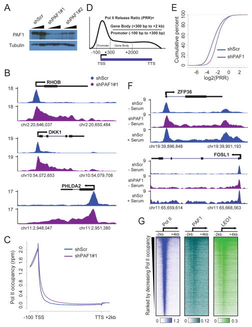
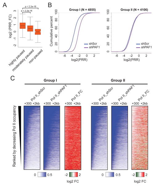
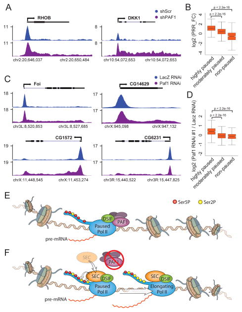
The control of promoter-proximal pausing and the release of RNA polymerase II (Pol II) is a widely used mechanism for regulating gene expression in metazoans, especially for genes that respond to environmental and developmental cues. Here, we identify that Pol-II-associated factor 1 (PAF1) possesses an evolutionarily conserved function in metazoans in the regulation of promoter-proximal pausing. Reduction in PAF1 levels leads to an increased release of paused Pol II into gene bodies at thousands of genes. PAF1 depletion results in increased nascent and mature transcripts and increased levels of phosphorylation of Pol II's C-terminal domain on serine 2 (Ser2P). These changes can be explained by the recruitment of the Ser2P kinase super elongation complex (SEC) effecting increased release of paused Pol II into productive elongation, thus establishing PAF1 as a regulator of promoter-proximal pausing by Pol II.


Figures

Figure 1
Figure 1. Depletion of PAF1 results in a redistribution of Pol II from promoters to gene bodies
(A) Cells transduced with shScr or two independent shRNAs targeting PAF1 (shPAF1#1 and shPAF1#2) were subjected to western blotting with antibodies against PAF1 and β-Tubulin. (B) Representative genome browser track examples of total Pol II ChIP-seq in HCT116 cells transduced with shScr or shPAF1#1 for the indicated genes. The x-axis indicates the chromosome position, and the y-axis represents normalized read density in reads per million (rpm). (C) Metagene analysis showing total Pol II occupancy measured by ChIP-seq in cells transduced with shScr or shPAF1#1. The 9,333 genes included for analysis meet the criteria of having Pol II (rpm > 1) at promoters in the control condition, are longer than 2 kb, and do not have nearby genes within 1 kb to avoid the inclusion of reads for Pol II from nearby genes. (D) Schematic representation of the modified traveling ratio or body/promoter ratio used to calculate the Pol II Release Ratio (herein called the PRR). The promoter is defined as the region covering 100 bp upstream to 300 bp downstream of the TSS; The gene body is defined as the region from 300 bp to 2 kb downstream of the TSS. (E) The empirical cumulative distribution function (ECDF) plot of the PRR distribution in cells transduced with shScr or shPAF1. (F) Track examples of total Pol II ChIP-seq in cells transduced with shScr or shPAF1 in starved (− serum) or serum-induced (+ serum) conditions for the indicated genes. (G) Heatmaps of Pol II, PAF1 and LEO1 occupancy around the TSS region. Rows are sorted by decreasing Pol II occupancy in the −2 kb to +4 kb region. Color-scaled intensities are in units of rpm.
Figure 2
Figure 2. PAF1 depletion has a greater effect on genes that are highly paused
(A) Box plot analysis of PRR fold change (PRR_FC) after PAF1 depletion for genes with different degrees of pausing as measured by pausing index (the ratio of Pol II occupancy at promoters to occupancy over gene bodies). The 9,333 genes were divided into three groups: highly paused (pausing index >4; n=4184), moderately paused (1 < pausing index < 4; n=4743), and non-paused (pausing index < 1; n=406). PRR values were then plotted in pausing index groups and p-values were then calculated with a two-sided t test. (B) ECDF plots of PRR for more affected (group I) and less affected (group II) genes in cells transduced with shScr or shPAF1. Group I constitutes 4,855 genes with a more than 1.5 increase of PRR_FC (log2 (PRR_FC) > 0.585). Group II constitutes 4,106 genes with a PRR_FC less than 1.5 in either direction (0.585 > log2 (PRR_FC) > −0.585). (C) Heatmaps of Pol II occupancy within the gene body regions, 300 bp to 2 kb downstream of the TSS, in cells transduced with shScr or shPAF1. Rows are sorted by decreasing Pol II occupancy in the shScr condition. Color-scaled intensities are in units of rpm. For the heatmaps of fold changes (Pol II_FC), the color bars depict log2 values.
Figure 3
Figure 3. Pause-release following PAF1 depletion represented by GRO-seq
(A) Genome browser tracks of GRO-seq in cells transduced with shScr or shPAF1 for the same set of genes shown in Figure 1B. Positive and negative values on the y-axis represent normalized reads (rpm) mapping to the positive and negative strands, respectively. (B) Metagene analysis showing Pol II occupancy as measured by GRO-seq in cells transduced with shScr or shPAF1. (C) Box plots of log2 PRR values of GRO-seq signal in cells transduced with shScr or shPAF1. (D) Heatmaps of GRO-seq signal around TSS regions in cells transduced with shScr or shPAF1 for group I and group II genes. Rows are sorted by decreasing Pol II occupancy in the −2 kb to +4 kb region in the shScr condition. Color-scaled intensities are in units of rpm.
Figure 4
Figure 4. PAF1 depletion leads to deregulation of gene expression
(A–B) Genome browser tracks of nascent RNA-seq (A) and total RNA-seq (B) in cells transduced with shScr or shPAF1 for the same set of genes shown in Figure 1B. Positive and negative values on the y-axis represent normalized reads (rpm) mapping to the positive and negative strands, respectively. (C–E) Box plots showing the log2 fold change of RNA levels from GRO-seq (C), nascent RNA-seq (D) and total RNA-seq (E) following the knockdown of PAF1 in group I and group II. (F) Hierarchical clustering demonstrating similarities in the log2 fold changes of RNA levels as determined by GRO-seq, nascent RNA-seq and total RNA-seq following PAF1 depletion.
Figure 5
Figure 5. Changes in CTD phosphorylation, but not H2B monoubiquitination, are associated with the release of Pol II into gene bodies in PAF1-depleted cells
(A) Cells transduced with shScr, shPAF1, or two independent shRNAs targeting BRE1A (shBRE1A#1 and shBRE1A#2) were subjected to Western blotting with antibodies against H2Bub, BRE1A, PAF1 and β-Tubulin. (B) ECDF plots of the PRR distributions in cells transduced with shScr or shBRE1A#1 for group I and group II. (C) Western blotting with antibodies against total Pol II (N20), Ser2P (3E10) and β-Tubulin for cells transduced with shScr or shPAF1. (D) Box plots showing the ratio of Ser2P and total Pol II at promoters for group I and group II genes. (E) Heatmaps of Ser2P levels around TSS regions in cells transduced with shScr or shPAF1 for all genes. Rows are sorted by decreasing Pol II occupancy in the –2 kb to +4 kb region in the shScr condition. Color-scaled intensities are in units of rpm. For the heatmaps of fold changes (Ser2P_FC), the color bars depict log2 values. (F) Metagene analysis showing average Ser2P occupancy as measured by ChIP-seq in cells transduced with shScr or shPAF1 for group I and group II genes.
Figure 6
Figure 6. PAF1 regulates CTD phosphorylation of elongating Pol II through recruitment of SEC
(A) Western blotting with antibodies against SEC subunits (CDK9, AFF4 and ELL2) and β-Tubulin for cells transduced with shScr or shPAF1. (B) Track examples of AFF4, ELL2 and CDK9 occupancy in cells transduced with shScr or shPAF1 for the same set of genes shown in Figure 1B. (C–E) Heatmaps of AFF4 (C), ELL2 (D) and CDK9 (E) levels around TSS regions in cells transduced with shScr or shPAF1 for all genes. Rows are sorted by decreasing Pol II occupancy in the −2 kb to +4 kb region in the shScr condition. Color-scaled intensities are in units of rpm. (F–H) Metagene analyses showing the occupancies of AFF4 (F), ELL2 (G) and CDK9 (H) as measured by ChIP-seq in cells transduced with shScr or shPAF1.
Figure 7
Figure 7. The role of PAF1 in regulating Pol II pausing in metazoans
(A) Track examples of total Pol II ChIP-seq in MCF7 cells transduced with shScr or shPAF1 for the indicated genes. (B) Box plot analysis of PRR fold change (PRR_FC) after PAF1 depletion for genes with different degrees of pausing as measured by pausing index (the ratio of Pol II occupancy at promoters to occupancy over gene bodies) in MCF7 cells. (C) Track examples of total Pol II ChIP-seq in Drosophila S2 cells with non-targeting (LacZ) or Paf1 RNAi for the indicated genes. (D) Box plot analysis of PRR fold change (PRR_FC) after PAF1 depletion by dsRNA#1 targeting Paf1 for genes with different degrees of pausing as measured by pausing index (the ratio of the Pol II occupancy at promoters to the occupancy over gene bodies). Genes that have Pol II (rpm > 1) at promoters in the control condition, are longer than 1 kb, and do not have nearby genes within 200 bp were selected and then divided into three groups: highly paused (pausing index >10; n=1003), moderately paused (2 < pausing index < 10; n=3089), and non-paused (pausing index < 2; n=625). PRR values were then plotted in pausing index groups and p-values were then calculated with a two-sided t test. (E) In metazoans, Pol II is frequently found to be pausing near the promoter and is phosphorylated at serine 5 of the CTD (Ser5P, red circles). The release into gene bodies is restrained by PAF1, a component of the PAF complex, which is recruited to direct interactions with Pol II and DSIF. (F) Upon depletion of PAF1, the CTD kinase SEC is recruited and subsequently phosphorylates Pol II on serine 2 (Ser2P, yellow circles), which facilitates release of paused Pol II into productive elongation.

References

    1. Bentley DL, Groudine M. A block to elongation is largely responsible for decreased transcription of c-myc in differentiated HL60 cells. Nature. 1986;321:702–706. - PubMed
    1. Boehm AK, Saunders A, Werner J, Lis JT. Transcription factor and polymerase recruitment, modification, and movement on dhsp70 in vivo in the minutes following heat shock. Molecular and cellular biology. 2003;23:7628–7637. - PMC - PubMed
    1. Chen F, Gao X, Shilatifard A. Stably paused genes revealed through inhibition of transcription initiation by the TFIIH inhibitor triptolide. Genes Dev. 2015;29:39–47. - PMC - PubMed
    1. Chen Y, Yamaguchi Y, Tsugeno Y, Yamamoto J, Yamada T, Nakamura M, Hisatake K, Handa H. DSIF, the Paf1 complex, and Tat-SF1 have nonredundant, cooperative roles in RNA polymerase II elongation. Genes Dev. 2009;23:2765–2777. - PMC - PubMed
    1. Cheng B, Li T, Rahl PB, Adamson TE, Loudas NB, Guo J, Varzavand K, Cooper JJ, Hu X, Gnatt A, et al. Functional association of Gdown1 with RNA polymerase II poised on human genes. Molecular cell. 2012;45:38–50. - PMC - PubMed

Publication types

Associated data