. 2015 Aug 27;162(5):1090-100.
doi: 10.1016/j.cell.2015.07.043. Epub 2015 Aug 13.

Rational Design of an Epstein-Barr Virus Vaccine Targeting the Receptor-Binding Site

Affiliations

Rational Design of an Epstein-Barr Virus Vaccine Targeting the Receptor-Binding Site

Masaru Kanekiyo et al. Cell. .

Abstract

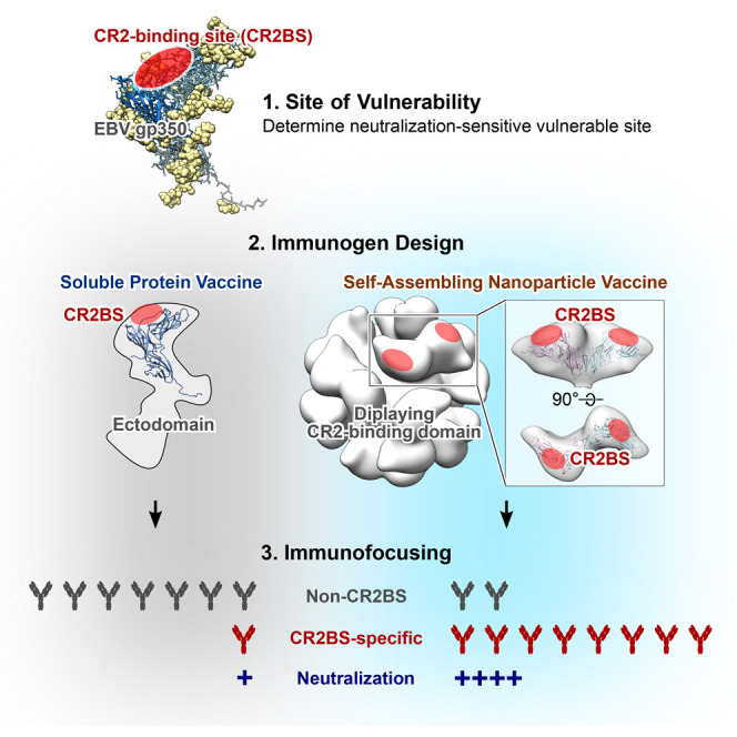
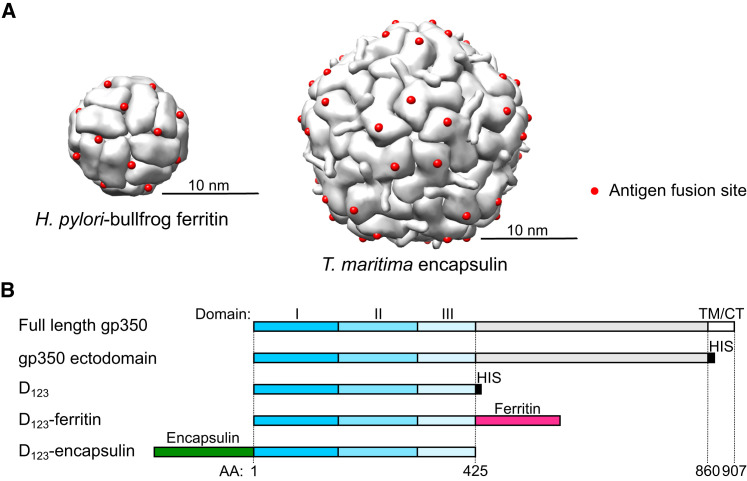
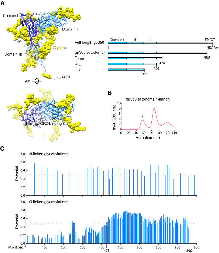
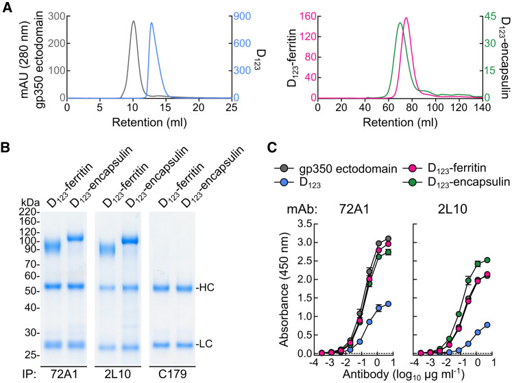
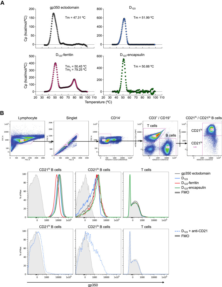
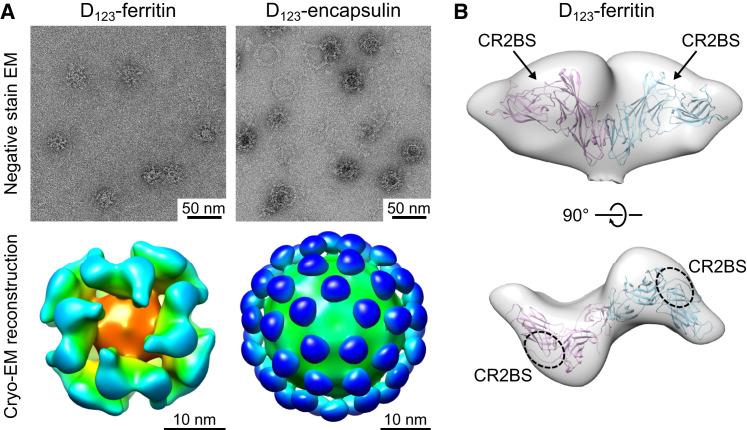
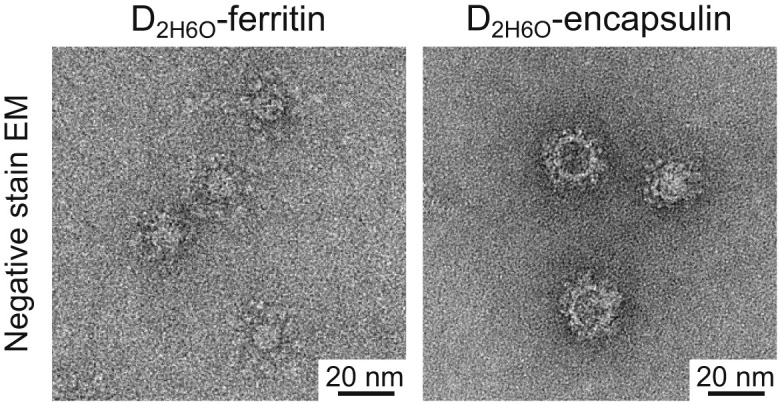
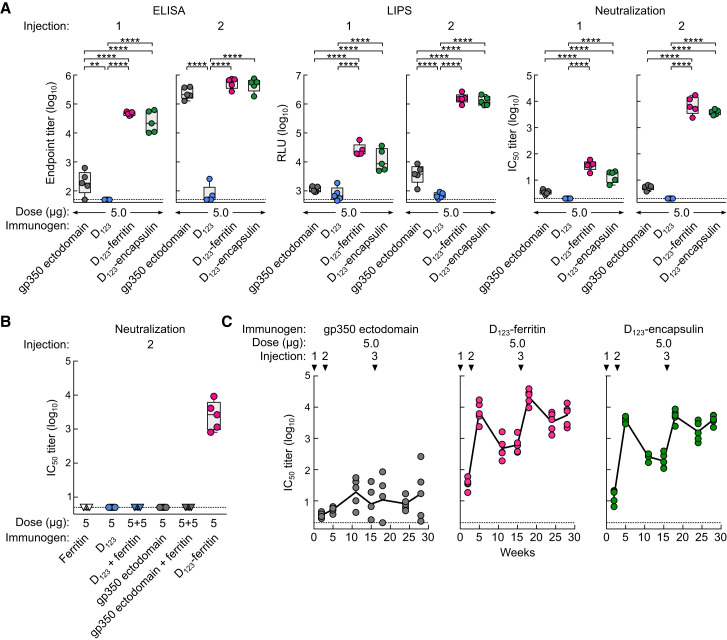
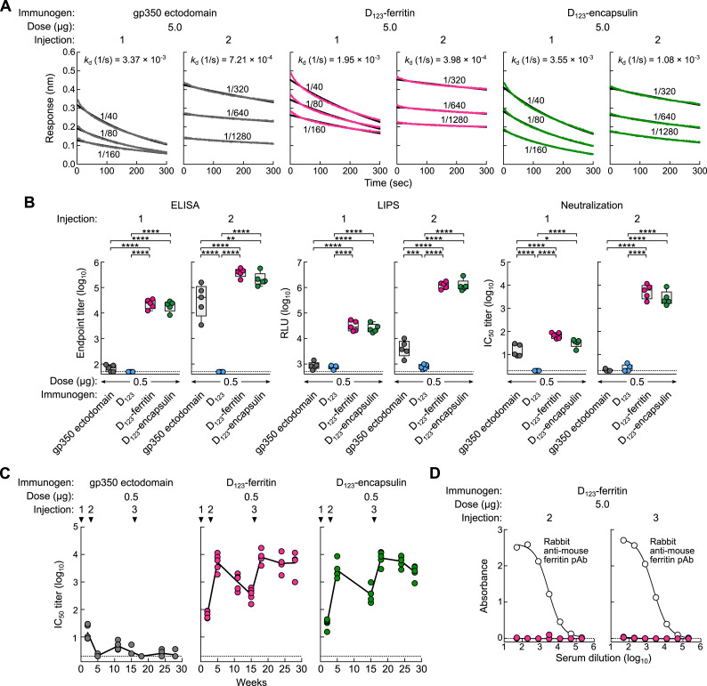
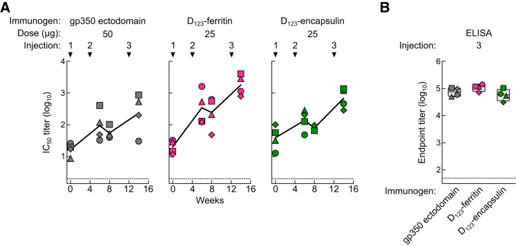
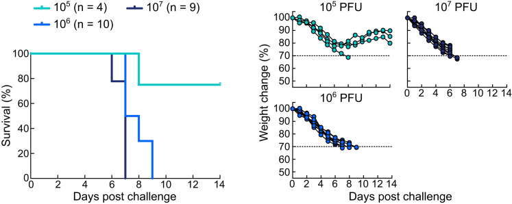
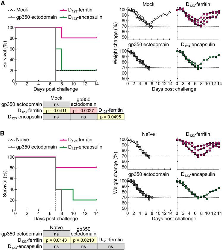
Epstein-Barr virus (EBV) represents a major global health problem. Though it is associated with infectious mononucleosis and ∼200,000 cancers annually worldwide, a vaccine is not available. The major target of immunity is EBV glycoprotein 350/220 (gp350) that mediates attachment to B cells through complement receptor 2 (CR2/CD21). Here, we created self-assembling nanoparticles that displayed different domains of gp350 in a symmetric array. By focusing presentation of the CR2-binding domain on nanoparticles, potent neutralizing antibodies were elicited in mice and non-human primates. The structurally designed nanoparticle vaccine increased neutralization 10- to 100-fold compared to soluble gp350 by targeting a functionally conserved site of vulnerability, improving vaccine-induced protection in a mouse model. This rational approach to EBV vaccine design elicited potent neutralizing antibody responses by arrayed presentation of a conserved viral entry domain, a strategy that can be applied to other viruses.


Figures

None
Graphical abstract
Figure 1
Figure 1
Molecular Design of gp350-Based Nanoparticles (A) Assembled hybrid ferritin model (left) and encapsulin (right). Engineered insertion sites for exogenous antigens are indicated as red spheres. (B) Schematic representation of full-length gp350, its truncated variants, and gp350-based nanoparticles. Domains I, II, and III of gp350 are color coded in dark blue, sky blue, and pale blue, respectively. Structurally undefined region and transmembrane (TM)/cytoplasmic tail (CT) are colored in gray and white, respectively. Hexa-histidine tag (HIS), ferritin, and encapsulin are colored in black, red, and green, respectively. Amino acid (AA) position relative to full-length gp350 is indicated at the bottom. See also Figure S1.
Figure S1
Figure S1
Design of Truncated gp350 Variants, Related to Figure 1 (A) Crystal structure of gp350 receptor-binding domain (Szakonyi et al., 2006) (PDB: 2H6O) (left). Structure of amino terminal portion of gp350 (residues 4-443) is shown. Structurally defined domains I, II and III are colored in blue, sky blue and pale blue, respectively. Attached N-linked glycans are shown in yellow spheres. Inferred receptor-binding site (CR2BS) is located between domains I and II and is indicated by a dotted circle. Schematic representation of truncated gp350 constructs (right). Amino acid (AA) position of the last residue of each design is denoted. Domains I, II and III are colored as shown in the left panel. (B) Size exclusion chromatography profile of gp350 ectodomain-ferritin purified by GNA lectin resin. Arrow indicates the predicted size of assembled particle. (C) Predicted N- and O-linked glycosylation sites in gp350. N-linked (top) and O-linked (bottom) glycosylation sites in the full-length gp350 (UniProtKB: P03200, spanning residues 1-907) were predicted using NetNGlyc 1.0 and NetOGlyc 3.1 programs (CBS Prediction Servers at http://www.cbs.dtu.dk/services), respectively. Glycosylation potential scores above the dotted line (0.5) were considered as positive.
Figure 2
Figure 2
Biochemical and Antigenic Characterization of gp350-Based Nanoparticles (A) Size exclusion chromatography profiles of soluble gp350 ectodomain, D123, and D123 nanoparticles. After affinity purification with Ni-NTA (soluble gp350 proteins) or GNA lectin (D123-ferritin and D123-encapsulin), proteins were separated by size exclusion chromatography using a Superdex 200 10/300 (soluble gp350 proteins) (left) or a Sephacryl S-500 16/60 (D123 nanoparticles) column (right). (B and C) Binding of anti-gp350 mAbs to purified proteins was assessed by immunoprecipitation (B) and ELISA (C). HC and LC indicate heavy and light chains of antibody, respectively. Each symbol represents mean ± SD. See also Figure S2.
Figure S2
Figure S2
Thermostability and CR2-Binding Properties of Soluble gp350s and gp350-Based Nanoparticles, Related to Figure 2 (A) Thermostability of gp350 ectodomain, D123, D123-ferritin and D123-encapsulin was measured by differential scanning calorimetry (DSC) using a Microcal VP-capillary DSC apparatus (GE Healthcare). Purified proteins were diluted to 0.5 mg ml−1 in PBS and subjected to DSC measurement (30-100°C, 1°C min−1). (B) Binding of gp350-based immunogens to human B cells. Human peripheral blood mononuclear cells were stained with the following monoclonal antibodies: CD3 Cy7-APC, IgG BV421, IgM-Cy5.5-peridinin chlorophyll protein (PerCP), CR2 (CD21) Cy5PE (BD Biosciences), CD14 BV510, CD56 BV510 (Biolegend), CD27-QD605 (Life Technologies), CD19 ECD (Beckman Coulter), CD38 Ax680 (In house: OKT10). Cell viability was assessed using Aqua Blue amine-reactive dye (Life Technologies). Approximately 0.1 ug of EBV gp350 conjugated protein was used for each sample. The gp350 ectodomain, D123, D123-ferritin and D123-encapsulin were biotinylated through primary amine group prior to conjugated with streptavidin-PE. Cells were analyzed on a LSR-II flow cytometer (BD Biosciences) and the data were processed in FlowJo software version 9.8.2 (FlowJo). Cell gating strategy is shown (top). Binding profile of gp350-based immunogens was represented as a histogram for each cell subset (middle). Binding profile of the sample pre-incubated with an anti-CR2 antibody (BD Biosciences, Clone 1048) (bottom).
Figure 3
Figure 3
Structural Characterization of gp350-Based Nanoparticles (A) Negative-stain transmission EM images (top) and cryo-EM reconstruction model (bottom) of D123 nanoparticles. (B) Fit model of gp350 D123 protrusion on ferritin nanoparticle. PDB entry 2H6O of gp350 (residues 4–425) was superimposed into cryo-EM density map. Inferred CR2BS is indicated. EM density maps of D123-ferritin and D123-encapsulin were deposited into EMDB under accessions EMDB-3025 and EMDB-6341, respectively. See also Figure S3.
Figure S3
Figure S3
Electron Microscopic Analysis of gp350 D2H6O Nanoparticles, Related to Figure 3 Negative-stain EM images of gp350 D2H6O-ferritin and D2H6O-encapsulin. The D2H6O-nanoparticles were prepared and purified as described for the D123-nanoparticles. The observed protrusions of gp350 D2H6O are slightly larger than that of D123, accounting for the slightly larger overall size of the nanoparticles.
Figure 4
Figure 4
Immunogenicity of Soluble gp350 Monomer and gp350-Based Nanoparticles (A) Groups of BALB/c mice (n = 5) were immunized with 5.0 μg of soluble gp350 ectodomain, D123, D123-ferritin, or D123-encapsulin in adjuvant at weeks 0 and 3. Immune sera were collected 2 weeks after the first (1) and the second (2) immunizations and analyzed by gp350 ELISA, LIPS, and virus neutralization assays. Endpoint binding titer (left), LIPS relative light units (RLU) (middle), and neutralization IC50 titer (right) were determined. ∗∗p < 0.01; ∗∗∗p < 0.001; ∗∗∗∗p < 0.0001. (B) Neutralization titers from mice 2 weeks after the second immunization with ferritin alone, ferritin mixed with gp350, or D123-ferritin. The data are shown as box-and-whiskers plots (box indicates lower and upper quartiles with line at median, and whiskers span minimum and maximum data points) with individual data points. (C) Kinetics of serum neutralization titers after immunization with gp350 ectodomain or gp350-based nanoparticles. Groups of BALB/c mice (n = 5) were immunized with 5.0 μg of gp350 ectodomain, D123-ferritin, or D123-encapsulin in adjuvant at weeks 0, 3, and 16. Immune sera were collected periodically after immunization, and serum neutralization IC50 titers were determined and plotted. Each dot represents an individual mouse. The horizontal dotted line represents the detection limit of the assay. See also Figure S4.
Figure S4
Figure S4
Affinity Maturation, Low-Dose Immunogenicity, and Anti-Autologous Ferritin Antibody after Immunization with Soluble gp350s and gp350-Based Nanoparticles, Related to Figure 4 (A) Avidity of anti-gp350 antibody after immunization with gp350-based immunogens. Serum antibody binding to gp350 was measured after the first (1) and second (2) immunizations with 5.0 μg of gp350 ectodomain, D123-ferritin or D123-encapsulin with adjuvant by biolayer interferometry (fortéBio Octet HTX, Pall). Dilutions of pooled sera (n = 5) are indicated. Antibody dissociation rates (off-rate, kd) were calculated by Octet analysis software version 8.0 using 1:2 bivalent analyte model. (B) Groups of BALB/c mice (n = 5) were immunized intramuscularly with 0.5 μg of soluble gp350 ectodomain, D123, D123-ferritin or D123-encapsulin mixed with adjuvant at weeks 0 and 3. Immune sera were collected 2 weeks after the first (1) and the second (2) immunizations and analyzed by gp350 ELISA, gp350 LIPS, and virus neutralization assays. Endpoint binding titer (left panels), LIPS relative light units (RLU) (middle panels), and IC50 neutralization titer (right panels) were determined and plotted. The data are shown as box-and-whiskers plots (box indicates lower and upper quartiles with bar at median and whiskers spanning minimum and maximum data points) with individual data points. p < 0.05; ∗∗p < 0.01; ∗∗∗p < 0.001; ∗∗∗∗p < 0.0001. The horizontal dotted line represents the detection limit of the assay. (C) Kinetics of serum neutralization titers after immunization with gp350 ectodomain or gp350-based nanoparticles. Groups of BALB/c mice (n = 5) were immunized intramuscularly with 0.5 μg of gp350 ectodomain, D123-ferritin or D123-encapsulin mixed with adjuvant at weeks 0, 3 and 16. Immune sera were collected periodically after immunization and serum neutralization IC50 titers were determined and plotted. Each dot represents an individual mouse. The horizontal dotted line represents the detection limit of the assay. (D) Antibody response to murine ferritin. Serum antibody response to autologous murine ferritin was measured after the second (2) and third (3) immunization with 5.0 μg of gp350 D123-ferritin with adjuvant by ELISA. Rabbit anti-mouse ferritin polyclonal antibody (pAb) was used as positive control in the assay. The horizontal dotted line represents the detection limit of the assay.
Figure 5
Figure 5
Immunogenicity of gp350-Based Nanoparticles in Cynomolgus Macaques (A) Neutralization titers in monkeys immunized with 50 μg of gp350 ectodomain, 25 μg of gp350 D123-ferritin, or D123-encapsulin in adjuvant at weeks 0, 4, and 12. Plasma was collected prior to immunization and periodically after immunization. (B) Plasma gp350 binding antibody titer in immunized monkeys after three immunizations. Each symbol represents an individual monkey. The horizontal dotted line represents the detection limit of the assay. The data are shown as box-and-whiskers plot with individual data points.
Figure S5
Figure S5
Titration of Recombinant Vaccinia Virus Expressing gp350 in Mice, Related to Figure 6 Groups of BALB/c mice were challenged intranasally with recombinant vaccinia virus expressing gp350 (rVV-gp350) at 105, 106 or 107 PFU. Kaplan-Meier survival curve (left) and body weight change (right) are plotted.
Figure 6
Figure 6
Protective Immunity against an Experimental Infection with Recombinant Vaccinia Virus Expressing EBV gp350 in Mice (A and B) Kaplan-Meier survival curve and body weight change after recombinant gp350-vaccinia virus challenge. Mice (n = 5) were immunized with 5.0 μg of gp350 ectodomain, gp350 D123-ferritin, or D123-encapsulin at weeks 0, 3, and 16 and were challenged at 2 months after the final immunization (A) or immunized three times with 0.5 μg of vaccines and challenged at 10 months after the final immunization (B). Summary of p values between groups (generated by log-rank Mantel-Cox test) is shown in each panel (ns, not significant). Mock, irrelevant nanoparticle-immunized mice (n = 5); control, age-matched naive mice (n = 5). See also Figure S5.
Figure S6
Figure S6
SPR-Based Antibody Cross-Competition Assay, Related to Figure 7 A sensor chip immobilized with purified soluble gp350 ectodomain through amine coupling reaction was used. Either irrelevant mAb (C179) or competing mAb (72A1 or 2L10) was first injected onto the gp350-chip to saturate the mAb’s binding site and then the immune serum was injected onto the mAb-saturated gp350-chip to measure residual binding. In the case of irrelevant mAb-saturated gp350-chip, antibodies in the immune serum can bind to any gp350 surface thus measuring maximum binding (left). In the case of competing mAb-saturated gp350-chip, serum antibodies can only bind to gp350 surface not occupied by the competing mAb thus measuring serum gp350 binding excluding sites blocked by the competing mAb (right).
Figure 7
Figure 7
Detection of CR2BS-Directed Antibodies in Immune Sera (A) SPR-based cross-competition assay of immune sera with mAbs. Binding of immune sera (after the second immunization) to the mAb-saturated gp350 was measured by Biacore. Each curve represents an individual serum. All data were normalized with C179-saturated curves and are shown as fraction response. Cross-competition of immune sera by 72A1 (left) and 2L10 (right) are shown for different immunization groups. (B) Specificity of gp350-binding antibodies in immune sera. Percent inhibition of serum antibodies to bind gp350 by 72A1 or 2L10 was calculated. (C) Generation of EBV gp350 CR2BS mutants. The gp350 domains I and II are shown in blue and sky blue, respectively. Attached glycans are shown as yellow spheres. Residues 162 and 208 are shown in red, and inferred CR2BS is indicated (PDB: 2H6O) (left). Binding of gp350 WT and CR2BS mutants to mAbs 72A1 and 2L10 was measured by ELISA (right). Each symbol represents mean ± SD. (D) Serum neutralization titers determined in the absence (closed circle) or presence of gp350 WT (open circle) and glyc162/208 (open square) proteins. (E) Preferential elicitation of antibody response to CR2BS in mice and monkeys. Serum antibody response to either gp350 WT or glyc162/208 was measured after the second immunization with indicated immunogens. Endpoint ELISA antibody titers for both gp350 WT and glyc162/208 were determined, and ratios of those titers were calculated (WT ⁄ glyc162/208). The data in (B), (D), and (E) are shown as box-and-whiskers plots with individual data points. p < 0.05; ∗∗p < 0.01; ∗∗∗p < 0.001; ∗∗∗∗p < 0.0001. See also Figure S6.

References

    1. Bachmann M.F., Zinkernagel R.M. Neutralizing antiviral B cell responses. Annu. Rev. Immunol. 1997;15:235–270. - PubMed
    1. Bachmann M.F., Kalinke U., Althage A., Freer G., Burkhart C., Roost H., Aguet M., Hengartner H., Zinkernagel R.M. The role of antibody concentration and avidity in antiviral protection. Science. 1997;276:2024–2027. - PubMed
    1. Baker M.L., Zhang J., Ludtke S.J., Chiu W. Cryo-EM of macromolecular assemblies at near-atomic resolution. Nat. Protoc. 2010;5:1697–1708. - PMC - PubMed
    1. Blasco R., Moss B. Selection of recombinant vaccinia viruses on the basis of plaque formation. Gene. 1995;158:157–162. - PubMed
    1. Chen Z., Earl P., Americo J., Damon I., Smith S.K., Yu F., Sebrell A., Emerson S., Cohen G., Eisenberg R.J. Characterization of chimpanzee/human monoclonal antibodies to vaccinia virus A33 glycoprotein and its variola virus homolog in vitro and in a vaccinia virus mouse protection model. J. Virol. 2007;81:8989–8995. - PMC - PubMed

Publication types

MeSH terms