. 2015 Aug 25;12(8):1252-60.
doi: 10.1016/j.celrep.2015.07.039. Epub 2015 Aug 13.

Intramolecular C2 Domain-Mediated Autoinhibition of Protein Kinase C βII

Affiliations

Intramolecular C2 Domain-Mediated Autoinhibition of Protein Kinase C βII

Corina E Antal et al. Cell Rep. .

Abstract

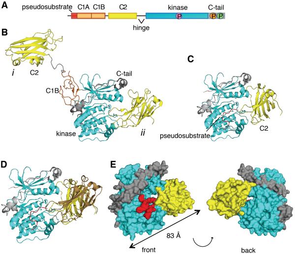
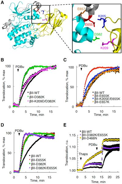
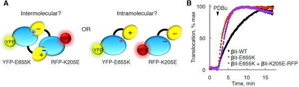
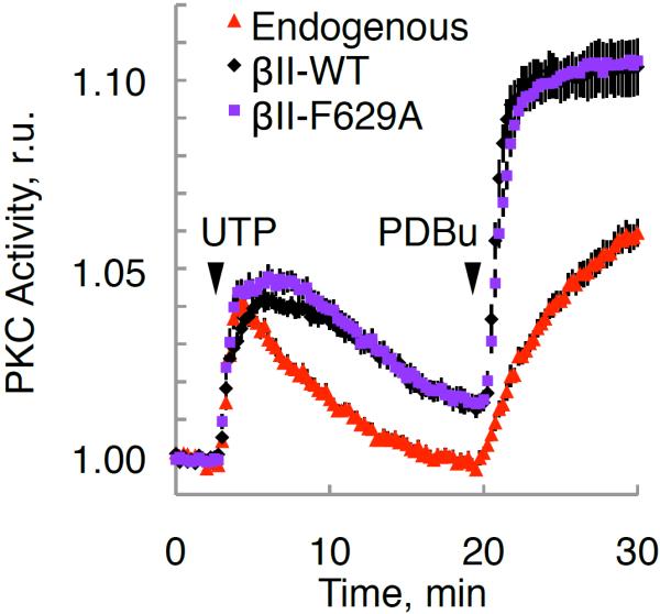
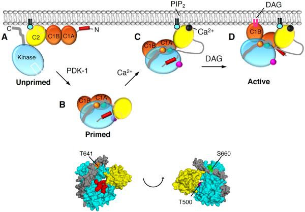
The signaling output of protein kinase C (PKC) is exquisitely controlled, with its disruption resulting in pathophysiologies. Identifying the structural basis for autoinhibition is central to developing effective therapies for cancer, where PKC activity needs to be enhanced, or neurodegenerative diseases, where PKC activity should be inhibited. Here, we reinterpret a previously reported crystal structure of PKCβII and use docking and functional analysis to propose an alternative structure that is consistent with previous literature on PKC regulation. Mutagenesis of predicted contact residues establishes that the Ca(2+)-sensing C2 domain interacts intramolecularly with the kinase domain and the carboxyl-terminal tail, locking PKC in an inactive conformation. Ca(2+)-dependent bridging of the C2 domain to membranes provides the first step in activating PKC via conformational selection. Although the placement of the C1 domains remains to be determined, elucidation of the structural basis for autoinhibition of PKCβII unveils a unique direction for therapeutically targeting PKC.


Figures

Figure 1
Figure 1. The C2 Domain of PKCβII Interacts with the Kinase Domain and C-Terminal Tail
(A) Schematic of the primary structure of PKCβII showing domain composition, priming phosphorylation sites (activation loop in pink, turn motif in orange, and hydrophobic motif in green), and the proteolytically-labile hinge that separates the regulatory and catalytic moieties. (B) Crystal structure of PKCβII (PDBID:3PFQ) showing the original interpretation of the location of the C2 domain that traces polypeptide from the C1B to the C2 (mode i) and the alternative interpretation in which the C2 domain binds the kinase domain by an intramolecular interaction (mode ii). Both modes are present in the crystal packing. (C) C2 domain (yellow) docked onto a complex of the PKCβII kinase domain (cyan) and the modeled pseudosubstrate (red). (D) Complex of the PKCβII kinase domain (cyan) and the mode ii C2 domain (yellow) from the crystal packing with the modeled pseudosubstrate (red), superimposed with the docked C2 domain (brown). (E) Structure of the kinase domain:C2 domain:pseudosubstrate complex showing the opening between the kinase domain (cyan) and the C2 domain (yellow), through which the pseudosubstrate:C1A linker (red) can be accommodated. See also Figure S2.
Figure 2
Figure 2. Mutational Analysis Corroborates a C2:Kinase Domain Interface
(A) Crystal structure of PKCβII (PDBID:3PFQ) showing predicted ion pairs between the kinase domain (cyan) or the C-terminal tail (gray) and the C2 domain (yellow). (B–D) Normalized FRET ratio changes (mean ± SEM) representing PDBu- (200 nM) induced PKC translocation in COS7 cells co-expressing YFP-tagged PKCβII WT or mutants and plasma membrane-targeted CFP. (E) FRET-ratio changes (mean ± SEM) representing thapsigargin- (5μM) followed by PDBu- (200 nM) induced PKC translocation in COS7 cells co-expressing YFP-tagged PKCβII WT or mutant and plasma membrane-targeted CFP. See also Figure S1.
Figure 3
Figure 3. The PKCβII Kinase Domain Binds the C2 Domain through an Intramolecular Interaction
(A) Charge reversal of the ion pair partner in the C2 domain (K205E) would rescue the fast translocation kinetics of the E655K C-terminal tail mutation in the case of an intermolecular (left) but not intramolecular (right) C2:kinase interaction. (B) Normalized FRET ratio changes (mean ± SEM) representing PDBu- (200 nM) induced PKC translocation in COS7 cells co-expressing plasma membrane-targeted CFP and either YFP-PKCβII-WT, YFP-PKCβII-E655K, or both YFP-PKCβII-E655K and RFP-PKCβII-K205E.
Figure 4
Figure 4. Mutation of Phe629 Does Not Affect PKC Activation
Normalized FRET ratio changes (mean ± SEM) showing agonist-dependent PKC activity of the indicated RFP-tagged PKCβII constructs or RFP control (endogenous) in COS7 cells co-expressing CKAR.
Figure 5
Figure 5. Model of PKCβII Activation
(A) Unprimed PKCβII is in a membrane-associated, open conformation in which its C1A, C1B, and C2 domains are fully exposed and the pseudosubstrate and C-terminal tail are unmasked. (B) Upon priming phosphorylation at its activation loop (T500, magenta) by PDK-1, followed by autophosphorylation at the turn motif (T641, orange) and the hydrophobic motif (S660, green), PKCβII matures into a closed conformation in which the C2 domain interfaces with the kinase domain and traps the pseudosubstrate into the substrate-binding site, both C1 domains become masked, and the primed enzyme localizes to the cytosol. (C) In response to agonists that promote PIP2 hydrolysis, Ca2+ binds cytosolic PKCβII via a low affinity interaction such that upon the next diffusion-controlled membrane encounter, the Ca2+-bound C2 domain is retained at the plasma membrane via Ca2+-bridging to anionic lipids and binding to PIP2. (D) Pre-targeted PKC binds the membrane-embedded ligand, DAG, predominantly via the C1B domain, resulting in release of the pseudosubstrate from the substrate-binding cavity, thereby activating PKC. Only one of the C1 domains binds DAG in the membrane at a time.

References

    1. Antal CE, Hudson AM, Kang E, Zanca C, Wirth C, Stephenson NL, Trotter EW, Gallegos LL, Miller CJ, Furnari FB, et al. Cancer-Associated Protein Kinase C Mutations Reveal Kinase's Role as Tumor Suppressor. Cell. 2015;160:489–502. - PMC - PubMed
    1. Antal CE, Violin JD, Kunkel MT, Skovso S, Newton AC. Intramolecular conformational changes optimize protein kinase C signaling. Chemistry & biology. 2014;21:459–469. - PMC - PubMed
    1. Banci L, Cavallaro G, Kheifets V, Mochly-Rosen D. Molecular dynamics characterization of the C2 domain of protein kinase Cbeta. The Journal of biological chemistry. 2002;277:12988–12997. - PubMed
    1. Belin RJ, Sumandea MP, Allen EJ, Schoenfelt K, Wang H, Solaro RJ, de Tombe PP. Augmented protein kinase C-alpha-induced myofilament protein phosphorylation contributes to myofilament dysfunction in experimental congestive heart failure. Circulation research. 2007;101:195–204. - PubMed
    1. Bernado P. Effect of interdomain dynamics on the structure determination of modular proteins by small-angle scattering. Eur Biophys J. 2010;39:769–780. - PubMed

Publication types