. 2014 Apr-Jun;2(2):e16.
doi: 10.15190/d.2014.8.

PLK1 is a binding partner and a negative regulator of FOXO3 tumor suppressor

Affiliations

PLK1 is a binding partner and a negative regulator of FOXO3 tumor suppressor

Octavian Bucur et al. Discoveries (Craiova). 2014 Apr-Jun.

Abstract

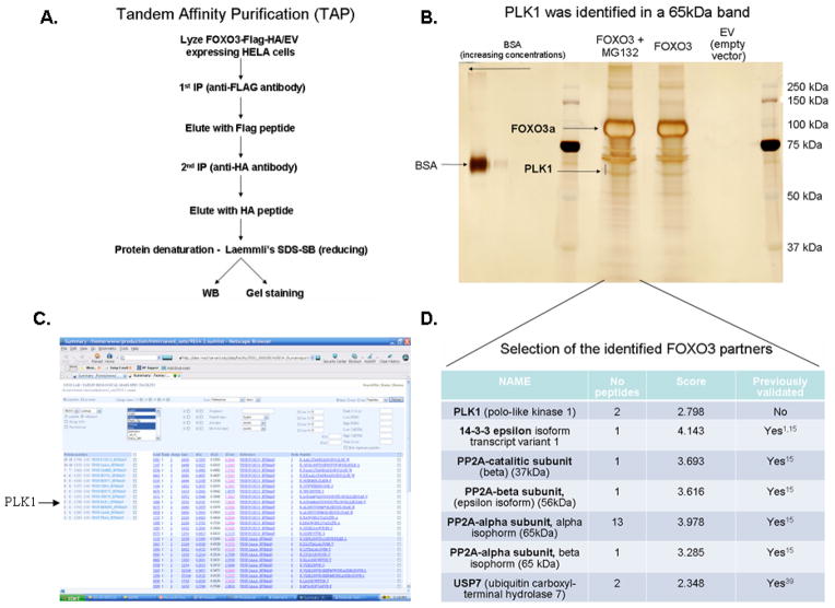
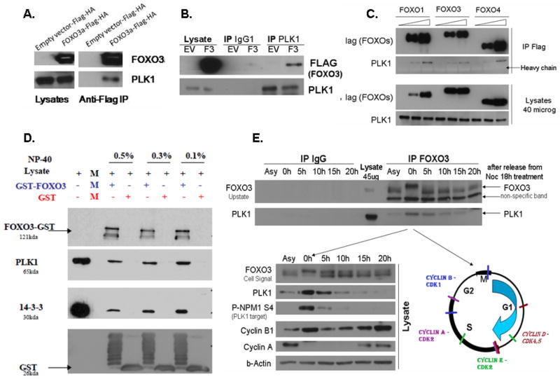
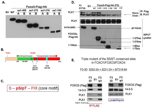
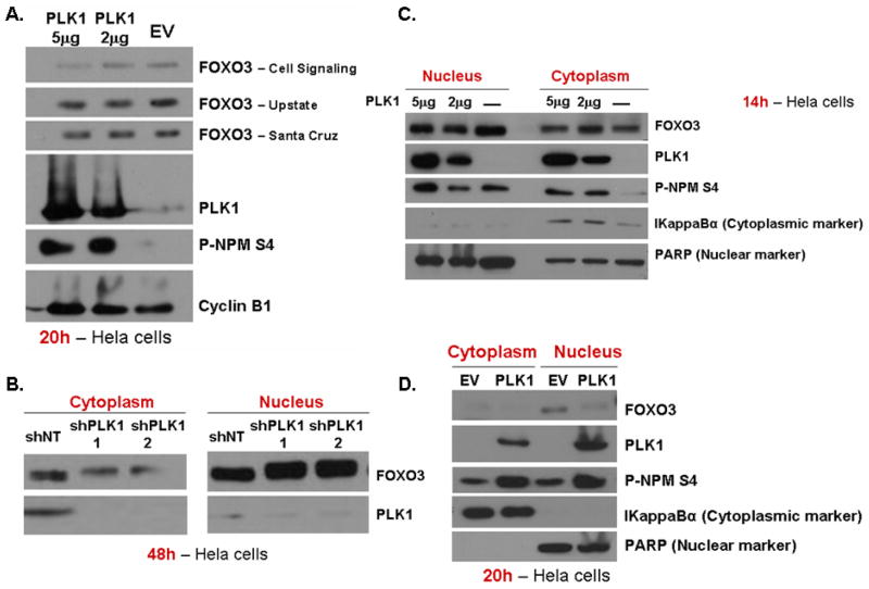
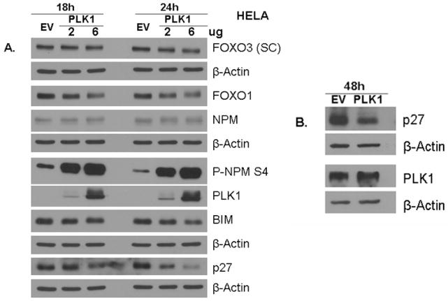
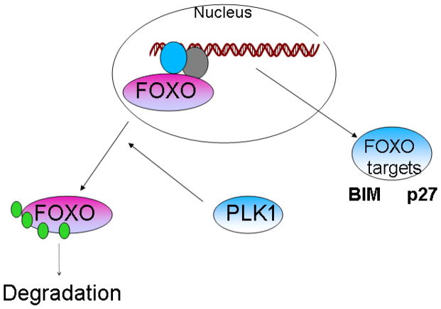
FOXO family members (FOXOs: FOXO1, FOXO3, FOXO4 and FOXO6) are important transcription factors and tumor suppressors controlling cell homeostasis and cell fate. They are characterized by an extraordinary functional diversity, being involved in regulation of cell cycle, proliferation, apoptosis, DNA damage response, oxidative detoxification, cell differentiation and stem cell maintenance, cell metabolism, angiogenesis, cardiac and other organ's development, aging, and other critical cellular processes. FOXOs are tightly regulated by reversible phosphorylation, ubiquitination, acetylation and methylation. Interestingly, the known kinases phosphorylate only a small percentage of the known or predicted FOXOs phosphorylation sites, suggesting that additional kinases that phosphorylate and control FOXOs activity exist. In order to identify novel regulators of FOXO3, we have employed a proteomics screening strategy. Using HeLa cancer cell line and a Tandem Affinity Purification followed by Mass Spectrometry analysis, we identified several proteins as binding partners of FOXO3. Noteworthy, Polo Like Kinase 1 (PLK1) proto-oncogene was one of the identified FOXO3 binding partners. PLK1 plays a critical role during cell cycle (G2-M transition and all phases of mitosis) and in maintenance of genomic stability. Our experimental results presented in this manuscript demonstrate that FOXO3 and PLK1 exist in a molecular complex through most of the phases of the cell cycle, with a higher occurrence in the G2-M cell cycle phases. PLK1 induces translocation of FOXO3 from the nucleus to the cytoplasm and suppresses FOXO3 activity, measured by the decrease in the pro-apoptotic Bim protein levels and in the cell cycle inhibitor protein p27. Furthermore, PLK1 can directly phosphorylate FOXO3 in an in vitro kinase assay. These results present the discovery of PLK1 proto-oncogene as a binding partner and a negative regulator of FOXO3 tumor suppressor.

Keywords: FOXO tumor suppressors; FOXO1; FOXO3 binding partners; PI3K-Akt pathway; Polo Like Kinase 1; apoptosis; cell cycle; proto-oncogene; transcription factors.


Conflict of interest statement

Conflict of Interest:

The authors do not declare any conflict of interest.

Figures

Figure 1
Figure 1. Novel FOXO3 binding partners discovered by using a TAP-MS strategy
A. Tandem Affinity Purification (TAP) followed by protein identification by Mass Spectrometry (MS) method was employed to identify novel FOXO3 binding partners. Lysates from HeLa cells transfected with either FOXO3-Flag-HA or EV-Flag-HA were immunoprecipitated with an anti-FLAG antibody, followed by a second purification with an anti-HA antibody. Denatured proteins (reducing Laemmli’s SDS-SB) were separated by SDS-PAGE. B. Proteins were stained with Silver stain. The bands corresponding to FOXO3 and PLK1 proteins identified by MS are presented. BSA (Bovine Serum Albumin) was used as a marker for protein quantity. C. Proteins identified by MS in the 60–65 kDa band presented in (B). PLK1 was one of the identified proteins (the arrow indicates PLK1). D. Most important/promising proteins identified after analyzing multiple bands by MS, covering 25–200 kDa gel range, are presented. The “Score” represents a probability Score determine with Sequest. EV – empty vector; MG132 was used to enrich for potential E3 ligases.
Figure 2
Figure 2. Screening validation: PLK1 kinase forms a complex with FOXO3 and other FOXO family members (FOXO1 & FOXO4)
A. HeLa cells were transfected with either pcDNA3-EV-FLAG-HA (Control) or pcDNA3-FOXO3-FLAG-HA and exogenous FOXO3 was immunoprecipitated by using an anti-FLAG antibody. The results show that exogenous FOXO3 is co-immunoprecipitated with endogenous PLK1 in HeLa cells. B. HeLa cells were transfected with either pcDNA3-EV-FLAG-HA (Control) or pcDNA3-FOXO3-FLAG-HA and endogenous PLK1 was immunoprecipitated by using an anti-PLK1 antibody. The results show that endogenous PLK1 is co-immunoprecipitated with exogenous FOXO3 in HeLa cells (revers immunoprecipitation). C. HeLa cells were transfected with either pcDNA3-EV-FLAG (Control), pcDNA3-FOXO1-FLAG, pcDNA3-FOXO3-FLAG and pcDNA3-FOXO4-FLAG. Exogenous FOXO1/FOXO3/FOXO4 was immunoprecipitated by using an anti-FLAG antibody. The results demonstrate that all three FOXO family members can form a complex with endogenous PLK1; D. Purified GST-FOXO3 protein was incubated with lysates from PLK1-FLAG transfected HeLa cells. GST pull-down reveals that GST-FOXO3 forms a complex with PLK1 (detected with an anti-FLAG antibody). GST is used as a negative control, while 14-3-3, a known scaffold protein of FOXO3, is used as a positive control. Results obtained with several different concentrations of NP40 detergent are shown. E. Asynchronous and synchronized proliferating HeLa cells in different phases of the cell cycle were used to evaluate the endogenous-endogenous interaction between FOXO3 and PLK1. Cells were synchronized with Nocodazole and released. Cells collected at 0, 5, 10, 15 and 20h after Nocodazole release were evaluated for cell cycle phases by analyzing the Cyclins expression (B1, A), which are particularly expressed in specific phases of the cell cycle (see right panel of the lower figure). PLK1 is also expressed at highest levels in mitosis and can itself be used as an indicator of cell cycle phase. Immunoprecipitated endogenous FOXO3 co-immunoprecipitates with endogenous PLK1. While the FOXO3-PLK1 complex can be seen during all phases of the cells cycle and in asynchronous cells, most PLK1 is bound to FOXO3 during the M/G2-M phases of the cell cycle. This is probably due to the fact that PLK1 has the highest expression during these phases of the cell cycle; PLK1 activity is determined by measuring the phosphorylation of NPM at Ser4 (site phosphorylated by PLK1).
Figure 3
Figure 3. PLK1 binds FOXO3 at specific sites
A. Testing of the FOXO3 C-terminal deletion mutants – size dependent SDS-PAGE migration - HeLa cells were transfected with either pcDNA3-EV (Control) or pcDNA3-FOXO3 deletion mutants and detected with an anti-FLAG antibody. Constructs were validated by checking both the correlation between their size and migration on SDS-PAGE, and sequencing of each individual construct. Two different clones were selected after the mutagenesis process and named A and B. Clone A was further selected for the following experiments; B. Schematic structure of FOXO3 protein underscoring its major functional domains (reproduced with permission). C. PLK1 PBD conserved core consensus sequence as determined by Lewis C. Cantley & Michael B. Yaffe, (“S” is Serine, “pS” is phosphorylated Serine, “pT” is phosphorylated Threonine and “P” is Proline); D. Finding PLK1 binding region in FOXO3. HeLa cells were transfected with either pcDNA3-EV (Control) or pcDNA3-FOXO3 deletion mutants and exogenous FOXO mutants were co-immunoprecipitated (using an anti-FLAG antibody) with endogenous PLK1, in HeLa cells. Results show that the region between the amino acids (aa) 219–270 of FOXO3 is important for PLK1 binding; E. A mutant of the three SS/ST conserved sites between FOXO family members significantly suppresses the binding of PLK1 to FOXO3.
Figure 4
Figure 4. PLK1 regulates FOXO3 nuclear localization and total levels
A. HeLa cells were transfected with either EV-FLAG or increasing concentrations (2ug, 5ug) of PLK1-FLAG. FOXO3 total levels are downregulated by PLK1 overexpression after 20h from transfection. Cyclin B1 is used as an internal loading control. B. HeLa cells were transfected with either two different shRNA for PLK1 or a non-targeting shRNA (shNT), for 48h. PLK1 knockdown induces FOXO3 relocalization into the nucleus. Nuclei and cytoplasm were isolated and FOXO3 levels were evaluated in each fraction. C. & D. HeLa cells were transfected with either EV-FLAG or PLK1-FLAG. PLK1 expression induces a decrease of the nuclear FOXO3 fraction at both 14h (C.) and 20h (D.), suggesting a decrease in FOXO3 activity. PARP is used as a nuclear marker, while IkappaBα is employed as a cytoplasmic marker; PLK1 activity is determined by measuring the phosphorylation of NPM at Ser4 (site known to be phosphorylated by PLK1);
Figure 5
Figure 5. PLK1 downregulates FOXO’s targets Bim and p27
A. HeLa cells were transfected with either EV-FLAG or increasing concentrations (2μg, 6μg) of PLK1-FLAG and the expression of FOXO1, FOXO3, BIM, p27, PLK1, P-NPM1 Ser4 & NPM1 was monitored by western blot. PLK1 induces a decrease in BIM (24h) and p27 (18h and 24h), well known FOXO targets; B. HeLa cells were transfected with EV-FLAG or PLK1-FLAG. Expression of p27 was evaluated. The results show that PLK1 decreases p27 levels; β-Actin was used as a loading control; PLK1 activity is determined by measuring the phosphorylation of NPM at Ser4 (site known to be phosphorylated by PLK1); SC = anti-FOXO3 antibody from Santa Cruz Biotechnology;
Figure 6
Figure 6
Model of how PLK1 controls FOXO3 localization, levels and activity

Similar articles

Cited by

References

    1. Calnan DR, Brunet A. The FoxO code. Oncogene. 2008 Apr 7;27(16):2276–88. doi: 10.1038/onc.2008.21;. - DOI - PubMed
    1. Dumitrascu GR, Bucur O. Critical physiological and pathologic al functions of Forkhead Box O tumor suppressors. Discoveries. 2013 Oct-Dec;1(1):e5. doi: 10.15190/d.2013.5. - DOI - PMC - PubMed
    1. Bucur O, Stancu AL, Khosravi-Far R, Almasan A. Analysis of apoptosis methods recently used in Cancer Research and Cell Death & Disease publications. Cell Death Dis. 2012;3:e263. doi: 10.1038/cddis.2012.2. - DOI - PMC - PubMed
    1. Parody JP, Ceballos MP, Quiroga AD, Frances DE, Carnovale CE, Pisani GB, et al. FoxO3a modulation and promotion of apoptosis by interferon-α2b in rat preneoplastic liver. Liver Int. 2013 doi: 10.1111/liv.12421. - DOI - PubMed
    1. Bucur O, Ray S, Bucur MC, Almasan A. APO2 ligand/tumor necrosis factor-related apoptosis-inducing ligand in prostate cancer therapy. Front Biosci. 2006 May 1;11:1549–68. doi: 10.2741/1903. - DOI - PubMed

LinkOut - more resources