. 2015 Jul 13;1(7):481-493.
doi: 10.1021/acsbiomaterials.5b00181.

High and low molecular weight hyaluronic acid differentially influence macrophage activation

Affiliations

High and low molecular weight hyaluronic acid differentially influence macrophage activation

Jamie E Rayahin et al. ACS Biomater Sci Eng. .

Abstract

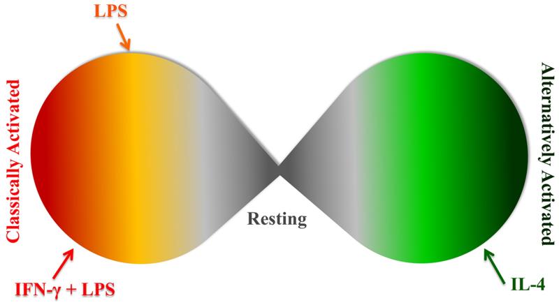
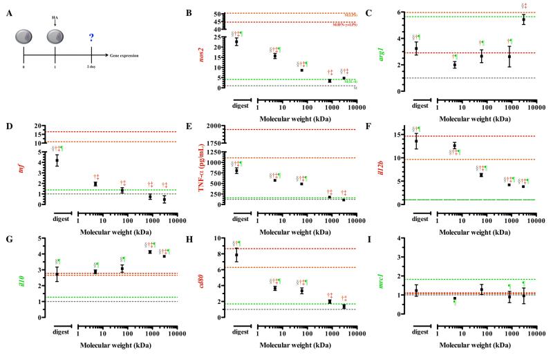
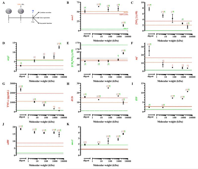
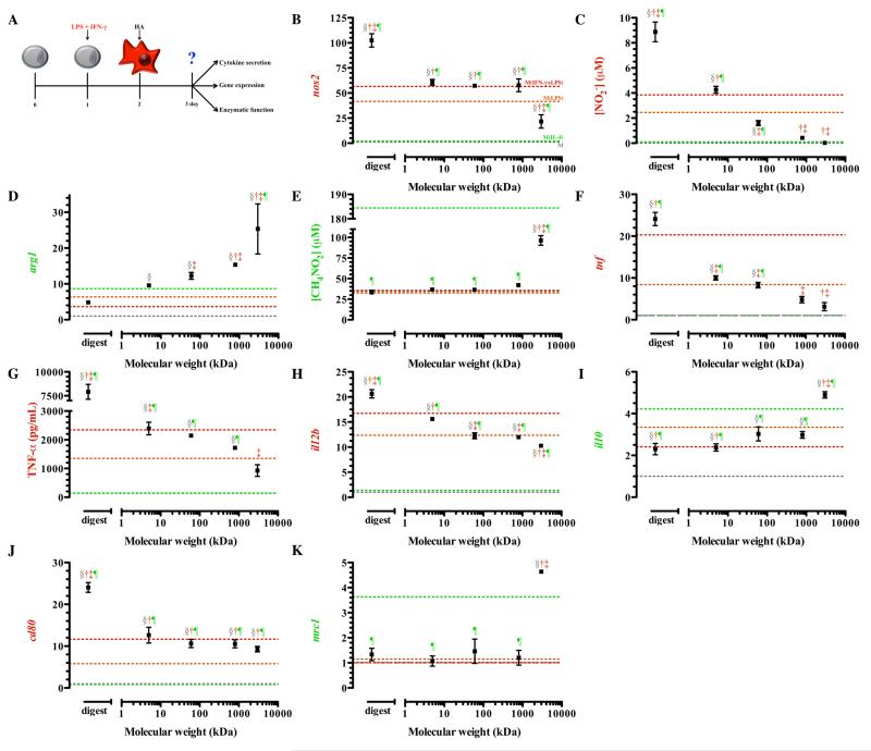
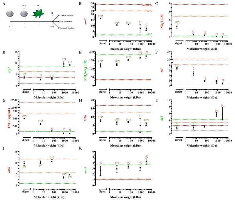
Macrophages exhibit phenotypic diversity permitting wide-ranging roles in maintaining physiologic homeostasis. Hyaluronic acid, a major glycosaminoglycan of the extracellular matrix, has been shown to have differential signaling based on its molecular weight. With this in mind, the main objective of this study was to elucidate the role of hyaluronic acid molecular weight on macrophage activation and reprogramming. Changes in macrophage activation were assessed by activation state selective marker measurement, specifically quantitative real time polymerase chain reaction, and cytokine enzyme-linked immunoassays, after macrophage treatment with differing molecular weights of hyaluronic acid under four conditions: the resting state, concurrent with classical activation, and following inflammation involving either classically or alternatively activated macrophages. Regardless of initial polarization state, low molecular weight hyaluronic acid induced a classically activated-like state, confirmed by up-regulation of pro-inflammatory genes, including nos2, tnf, il12b, and cd80, and enhanced secretion of nitric oxide and TNF-α. High molecular weight hyaluronic acid promoted an alternatively activated-like state, confirmed by up regulation of pro-resolving gene transcription, including arg1, il10, and mrc1, and enhanced arginase activity. Overall, our observations suggest that macrophages undergo phenotypic changes dependent on molecular weight of hyaluronan that correspond to either (1) pro-inflammatory response for low molecular weight HA or (2) pro-resolving response for high molecular weight HA. These observations bring significant further understanding of the influence of extracellular matrix polymers, hyaluronic acid in particular, on regulating the inflammatory response of macrophages. This knowledge can be used to guide the design of HA-containing biomaterials to better utilize the natural response to HAs.

Keywords: alternatively activated; classically activated; hyaluronic acid; macrophage; molecular weight; polarization.


Figures

Figure 1
Figure 1. Macrophage activation is a continuum
Classically and alternatively activated macrophages are two extremes of a continuous spectrum. Many stimuli have the ability to polarize macrophages and even stimuli that give generally same phenotype (LPS or IFN-γ with LPS) do not result in the same activated state.
Figure 2
Figure 2. Gene expression of resting macrophages in response to hyaluronic acid
Resting macrophages were treated with 1μM HAs for 24 hours before phenotypic assessment was made. The schematic depicts the time course of the experiments (A). Low molecular weight HAs induced transcription of nos2 (B), decreased arg1 transcription (C), and enhanced tnf transcription (D) and TNF-a (E). High molecular weight HAs stimulated minimal il12b (F) transcription and enhanced il10 (G) transcription. Low molecular weight HAs enhanced transcription of cd80 (H) but did not change mrc1 (I) transcription in resting macrophages. Points represent mean plus or minus (±) standard deviation of three independent experiments. The symbols indicate significant difference (p-value less than 0.05) from the reference states, which are represented by corresponding colored dotted lines: resting macrophages (§; M), classically activated macrophages, stimulated with LPS (†; M(LPS)) or LPS and IFN-γ (‡; M(IFN-γ+LPS)), or alternative activated macrophages, stimulated with IL-4 (¶; M(IL-4)).
Figure 3
Figure 3. Gene expression, cytokine secretion, and enzymatic function of classically activated macrophages (M(LPS) treated simultaneously with HAs
After plating, macrophages were simultaneously treated with LPS and HAs. The schematic depicts the time course of the experiments (A). Expression of nos2 (B) and nitrite production (C) was elevated in groups treated with low molecular weight HAs. Treatment of macrophages with LPS and high molecular weight HAs enhanced arg1 (D) transcription and production of urea (E). Expression (F) and secretion (G) of tumor necrosis factor alpha was lowest in groups treated with high molecular weight HAs. Expression of il12b (H) and cd80 (J) was elevated in groups treated with low molecular weight HAs. Transcription of il10 (I) and mrc1 (K) steadily increased with increasing molecular weight. Points represent mean plus or minus (±) standard deviation of at least three independent experiments. The symbols indicate significant difference (p-value less than 0.05) from the reference states, which are represented by corresponding colored dotted lines: resting macrophages (§; M), classically activated macrophages, stimulated with LPS (†; M(LPS)) or LPS and IFN-γ (‡; M(IFN-γ+LPS)), or alternative activated macrophages, stimulated with IL-4 (¶; M(IL-4)).
Figure 4
Figure 4. Gene expression, cytokine secretion, and enzymatic function of classically activated macrophages (M(IFN-γ+LPS )) treated with HAs
After plating, macrophages were classically activated with interferon-γ and LPS for 24 hours, after which media was replaced with media containing HAs for treatment (A). Expression of nos2 (B) and production of nitrite (C) was elevated in groups treated with low molecular weight HAs, whereas arg1 (D) and urea production (E) were elevated in groups treated with high molecular weight HAs. (B). Expression (F) and secretion (G) of tumor necrosis factor alpha was significantly reduced in groups treated with high molecular weight HAs. Expression of il12b (H) was elevated in groups treated with lower molecular weight HAs. High molecular weight HAs potentiated il10 expression (I), but did not change cd80 (J) or mrc1 (K) expression. Points represent mean plus or minus (±) standard deviation of at least three independent experiments. The symbols indicate significant difference (p-value less than 0.05) from the reference states, which are represented by corresponding colored dotted lines: resting macrophages (§; M), classically activated macrophages, stimulated with LPS (†; M(LPS)) or LPS and IFN-γ (‡; M(IFN-γ+LPS)), or alternative activated macrophages, stimulated with IL-4 (¶; M(IL-4)).
Figure 5
Figure 5. Gene expression, cytokine secretion, and enzymatic function of alternatively activated macrophages (M(IL-4)) treated with HAs
After plating, macrophages were alternatively activated with IL-4 for 24 hours, after which they were washed and media was replaced with media containing HAs for treatment (A). Expression of nos2 was elevated in all treatment groups (B), but nitrite was only elevated in groups treated with the HA digest (C). Transcription of arg1 was enhanced by high molecular weight HAs (D). Treatment with high molecular weight HAs maintained production of urea (E). Expression (F) and secretion (G) of tumor necrosis factor alpha was significantly elevated in groups treated with low molecular weight HAs. Elevated transcription of il12b (H), reduced transcription of il10 (I), enhanced transcription of cd80 (J), and diminished mrc1 transcription (K) was detected in groups treated with low molecular weight HAs. Points represent mean plus or minus (±) standard deviation of at least three independent experiments. The symbols indicate significant difference (p-value less than 0.05) from the reference states, which are represented by corresponding colored dotted lines: resting macrophages (§; M), classically activated macrophages, stimulated with LPS (†; M(LPS)) or LPS and IFN-γ (‡; M(IFN-γ+LPS)), or alternative activated macrophages, stimulated with IL-4 (¶; M(IL-4)).

References

    1. Meyer K, Palmer JW. The Polysaccharide of the Vitreous Humor. J. Biol. Chem. 1934;107:629–634.
    1. Meyer K. The Biological Significance of Hyaluronic Acid and Hyaluronidase. Physiol. Rev. 1947;27:335–359. - PubMed
    1. Necas J, Bartosikova L, Brauner P, Kolar J. Hyaluronic Acid (Hyaluronan): A Review. Vet. Med. 2008;53:397–411.
    1. Collins MN, Birkinshaw C. Hyaluronic Acid Based Scaffolds for Tissue Engineering-a Review. Carbohyd. Polym. 2013;92:1262–1279. - PubMed
    1. Colen S, van den Bekerom MPJ, Mulier M, Haverkamp D. Hyaluronic Acid in the Treatment of Knee Osteoarthritis a Systematic Review and Meta-Analysis with Emphasis on the Efficacy of Different Products. Biodrugs. 2012;26:257–268. - PubMed