. 2015 Sep 10;525(7568):212-217.
doi: 10.1038/nature14892. Epub 2015 Aug 17.

An atomic structure of human γ-secretase

Affiliations

An atomic structure of human γ-secretase

Xiao-Chen Bai et al. Nature. .

Abstract

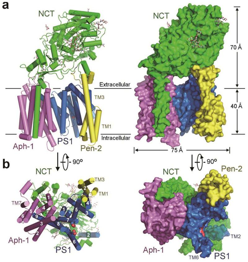
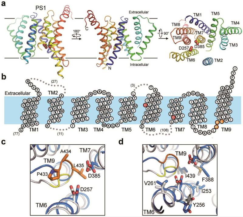
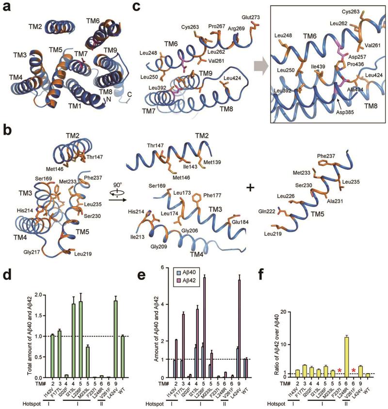
Dysfunction of the intramembrane protease γ-secretase is thought to cause Alzheimer's disease, with most mutations derived from Alzheimer's disease mapping to the catalytic subunit presenilin 1 (PS1). Here we report an atomic structure of human γ-secretase at 3.4 Å resolution, determined by single-particle cryo-electron microscopy. Mutations derived from Alzheimer's disease affect residues at two hotspots in PS1, each located at the centre of a distinct four transmembrane segment (TM) bundle. TM2 and, to a lesser extent, TM6 exhibit considerable flexibility, yielding a plastic active site and adaptable surrounding elements. The active site of PS1 is accessible from the convex side of the TM horseshoe, suggesting considerable conformational changes in nicastrin extracellular domain after substrate recruitment. Component protein APH-1 serves as a scaffold, anchoring the lone transmembrane helix from nicastrin and supporting the flexible conformation of PS1. Ordered phospholipids stabilize the complex inside the membrane. Our structure serves as a molecular basis for mechanistic understanding of γ-secretase function.


Figures

<b>Extended Data Figure 1</b>
Extended Data Figure 1. Cryo-EM, single-particle analysis of human γ-secretase
a, Representative raw particles from an original micrograph. b, Representative reference-free 2-D class averages of the γ-secretase particles. Two classes identified by a red rectangle box (lower right corner) may contain some density for the extended cytosolic loop sequences between TM6 and TM7 of PS1, which are disordered in the final maps. c, Resolution estimation of the EM structure. The overall resolution is calculated to be 3.4 Å on the basis of gold-standard FSC curve. d, Color-coded resolution variations in the γ-secretase structure as estimated by ResMap. e, FSC curves of the final, Refmac-refined model versus the map it was refined against (in black); of a model refined in the first of the two independent maps used for the gold-standard FSC versus that same map (in red); and of a model refined in the first of the two independent maps versus the second independent map (in green). The small difference between the red and green curves indicates that the refinement of the atomic coordinates did not suffer from severe overfitting.
<b>Extended Data Figure 2</b>
Extended Data Figure 2. An atomic model of human γ-secretase
a, The γ-secretase structure is viewed parallel to the lipid membrane. Shown here is EM density for the entire γ-secretase complex. EM density is colored blue for PS1, yellow for Pen-2, magenta for Aph-1, and green for nicastrin. b, The density map for TM2 of PS1. Among the 20 TMs, TM2 of PS1 shows the highest degree of flexibility and only becomes visible at as rod-shaped density in a 7 Å low-pass filtered map. At this resolution, another rod-shaped density is visible next to TM2 and remains unaccounted for. c, EM density map and the atomic model are shown for all 7 TMs of Aph-1. 2-3 bulky residues are indicated for each TM. d, EM density map and the atomic model are shown for 7 TMs of hPS1. TM6 exhibits relatively poor EM density, likely due to its intrinsic flexibility. e, EM density map and the atomic model are shown for the 3 TMs of Pen-2. f, EM density map and the atomic model are shown for the only TM and select regions of nicastrin. g, EM density map and the atomic model for three representative glycans are shown. h, EM density map and putative assignment are shown for two lipid molecules.
<b>Extended Data Figure 3</b>
Extended Data Figure 3. Overall structure of human γ-secretase
a, Structure of human γ-secretase is shown in cartoon representation (top panels) and surface view (bottom panels) in four successively perpendicular views. The γ-secretase structure is viewed parallel to the lipid membrane. The coloring scheme is the same as in Fig. 1. Two lipid molecules are shown. Eleven glycosylated Asn residues and their glycans are displayed in stick. b, The γ-secretase structure is represented by electrostatic surface potential.
<b>Extended Data Figure 4</b>
Extended Data Figure 4. Electrostatic surface potential of PS1
PS1 exhibits a loosely folded structure, with a number of large cavities and empty spaces between adjacent TMs.
<b>Extended Data Figure 5</b>
Extended Data Figure 5. Structural comparison between human nicastrin and nicastrin from Dictyostelium purpureum (DpNCT)
Individual structures of human nicastrin and DpNCT are shown in the left and middle panels, respectively. The overlay is shown in the right panel, with an RMSD of 2.2 Å. Two perpendicular views for each structure are displayed here.
<b>Extended Data Figure 6</b>
Extended Data Figure 6. Pen-2 contains three small hydrophobic cores in its three TMs
Unlike previous prediction,, Pen-2 contains three, not two, TMs. Pen-2 contains a small hydrophobic core in the extracellular side and two in the transmembrane region. These three regions are boxed and shown in close-up views.
<b>Extended Data Figure 7</b>
Extended Data Figure 7. Results of crosslinking experiments corroborate the atomic model of γ-secretase
a, Crosslinking results for the interface between Aph-1 and nicastrin (NCT). Three mutant γ-secretase complexes were examined: Aph-1-V147C/NCT-I40C, Aph-1-V146C/NCT-A664C, and Aph-1-A4C/NCT-L673C. Shown in the upper panel is a SDS-PAGE gel blotted by a monoclonal antibody against the HA tag on Aph-1. Only in the absence of DTT, crosslinking led to high molecular weight complexes for the mutant γ-secretase, but not for the WT γ-secretase. The two bands likely represent Aph-1 crosslinked to mature NCT (mNCT) and immature NCT (iNCT). The structural basis is shown in the lower panel. The distances between the Cα atoms of the two residues targeted for cysteine mutation range 4.1-6.1 Å, which would facilitate convenient crosslinking reactions. b, Crosslinking results for the interface between PS1 and Pen-2. The mutant γ-secretase contains Pen-2-P97C and PS1-N190C. Shown in the upper panel is a SDS-PAGE gel blotted by a monoclonal antibody against the FLAG tag on Pen-2. c, Crosslinking results for the interface between Pen-2 and nicastrin. Two γ-secretase mutants were examined: Pen-2-T100C/NCT-V224C and Pen-2-L98C/NCT-H222C. d, Crosslinking results for the interface between Aph-1 and PS1. Two γ-secretase mutants were examined: Aph-1-T204C/PS1-F465C and Aph-1-A76C/PS1-I467C.
<b>Extended Data Figure 8</b>
Extended Data Figure 8. Implication on substrate access to γ-secretase
Structure of γ-secretase is displayed in three relevant views. The middle panel shows the overall structure, with key features labeled. The left panel displays electrostatic surface potential from the convex side of γ-secretase. The right panel suggests a putative path for substrate access to the active site of γ-secretase.
Figure 1
Figure 1. Atomic structure of human γ-secretase
a, The γ-secretase structure is shown in cartoon representation (left panel) and surface view (right panel). Eleven N-linked glycans are displayed in stick. b, The γ-secretase structure is viewed perpendicular to the lipid membrane from the intracellular side. TM2 of PS1 is most flexible and shown in a semi-transparent fashion. The catalytic residues Asp257 and Asp385 are located on the convex side of the TM horseshoe. All structural figures were prepared using UCSF Chimera or PyMol.
Figure 2
Figure 2. Atomic structure of PS1
a, PS1 has a loosely organized structure and exhibits considerable flexibility. The cartoon representation of PS1 is rainbow-colored. TM2 is visible only at low resolutions, and the density map contains no features for side chains. Nonetheless, an atomic model for TM2 was built based on sequence and structural homology between PS1 and PSH,. b, A membrane topology diagram of PS1. The two catalytic aspartate residues are colored red. c, The two catalytic aspartate residues of PS1 are in nearly perfect registry with those in PSH. The PAL sequence motif implicated in substrate recognition is shown. d, PS1 and PSH share similar features at their active sites.
Figure 3
Figure 3. AD-derived mutations map to two hotspots in PS1
a, An overall view of the PS1 residues targeted for mutations in AD patients. PS1 is viewed from the extracellular side. Mutated residues are colored orange. b, Close-up views of the mutation-targeted residues in TMs 2-5. The vast majority of these residues map to the center of this four-TM bundle. c, Close-up views of the mutation-targeted residues in TMs 6-9. The two catalytic residues Asp257 and Asp385 are shown. d, FAD-derived mutations in PS1 have varying degrees of effect on the combined Aβ40 and Aβ42 cleavage activity of γ-secretase. Shown here are results of 10 such γ-secretase mutants, each containing a specific mutation derived from FAD. The activity of WT γ-secretase is normalized to 1.0. e, FAD-derived mutations either suppressed the production of Aβ40 more than Aβ42 or increased the production of Aβ40 less than Aβ42. f, All but two FAD-derived mutations led to increased Aβ42/Aβ40 ratios. Two mutations F237I and V261F in PS1 abrogated Aβ40 cleavage altogether, disallowing calculation of the Aβ42/Aβ40 ratio. Each experiment was independently repeated three times. All error bars represent standard deviations.
Figure 4
Figure 4. Structural features of nicastrin
a, Two perpendicular views of nicastrin. The Lid from the small lobe is highlighted in red. Surface glycans are shown. b, The Lid hovers above a hydrophilic pocket in the large lobe. Two large glycans on Asn55 and Asn435 sandwich the Lid and interact with surrounding residues. c, Glu333 and Tyr337 are surrounded by a number of charged and polar residues in the pocket. These structural features are consistent with the pocket being a binding site for substrate protein. d, Trp164 from the Lid makes van der Waals contacts to Pro424, Phe448, and the aliphatic side chain of Gln420. e, Phe287 from the large lobe may serve as the hydrophobic pivot. Phe287 interacts with four hydrophobic residues from the small lobe.
Figure 5
Figure 5. Assembly interfaces among the four components of γ-secretase in the transmembrane region
a, An overall view of the packing interfaces in the transmembrane region. The three boxed interfaces are detailed in panels b-d. b, The C-terminal three residues Phe465-Tyr466-Ile467 of PS1 insert into a cavity formed by TMs in Aph-1. c, Nicastrin interacts with Pen-2 through van der Waals contacts on a flat interface. d, Pen-2 binds to PS1 through mostly van der Waals contacts. In particular, Phe94 of Pen-2 is nestled in the greasy pocket of PS1, formed by Phe179, Leu182, Phe186, Val193, Tyr195, and Va198. e, Two phospholipids appear to stabilize the inter-component interfaces in γ-secretase. One lipid is bound at the interface between PS1 and Aph-1, whereas the other is intercalated between the lone TM of nicastrin and TMs 1/4/5/7 of Aph-1 (left panel). The aliphatic tails of the latter phospholipid may interact with a number of hydrophobic residues whereas its phosphate group likely hydrogen bonds to Arg115 and Gln116 (right panel).

References

    1. Alzheimer A. About a peculiar disease of the cerebral cortex. Centralblatt für Nervenheilkunde Psychiatrie. 1907;30:177–179.
    1. Hardy JA, Higgins GA. Alzheimer’s disease: the amyloid cascade hypothesis. Science. 1992;256:184–5. - PubMed
    1. De Strooper B, Iwatsubo T, Wolfe MS. Presenilins and gamma-Secretase: Structure, Function, and Role in Alzheimer Disease. Cold Spring Harbor perspectives in medicine. 2012;2:a006304. - PMC - PubMed
    1. Goate A, Hardy J. Twenty years of Alzheimer’s disease-causing mutations. J Neurochem. 2012;120(Suppl 1):3–8. - PubMed
    1. Tanzi RE, Bertram L. Twenty years of the Alzheimer’s disease amyloid hypothesis: a genetic perspective. Cell. 2005;120:545–55. - PubMed

Publication types

MeSH terms

Associated data