RAP1-mediated MEK/ERK pathway defects in Kabuki syndrome

Nina Bögershausen et al. J Clin Invest. 2015 Sep.

Abstract

The genetic disorder Kabuki syndrome (KS) is characterized by developmental delay and congenital anomalies. Dominant mutations in the chromatin regulators lysine (K)-specific methyltransferase 2D (KMT2D) (also known as MLL2) and lysine (K)-specific demethylase 6A (KDM6A) underlie the majority of cases. Although the functions of these chromatin-modifying proteins have been studied extensively, the physiological systems regulated by them are largely unknown. Using whole-exome sequencing, we identified a mutation in RAP1A that was converted to homozygosity as the result of uniparental isodisomy (UPD) in a patient with KS and a de novo, dominant mutation in RAP1B in a second individual with a KS-like phenotype. We elucidated a genetic and functional interaction between the respective KS-associated genes and their products in zebrafish models and patient cell lines. Specifically, we determined that dysfunction of known KS genes and the genes identified in this study results in aberrant MEK/ERK signaling as well as disruption of F-actin polymerization and cell intercalation. Moreover, these phenotypes could be rescued in zebrafish models by rebalancing MEK/ERK signaling via administration of small molecule inhibitors of MEK. Taken together, our studies suggest that the KS pathophysiology overlaps with the RASopathies and provide a potential direction for treatment design.


Figures
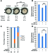
Figure 11
Figure 11. MEK/ERK signaling alterations can be rebalanced by pharmacological treatment in zebrafish models of KS.
(A) Coinjection of MEK inhibitor PD184161 ameliorates the CE defects in rap1 morphants. ***P < 0.001, χ2 test (n > 80). (B) Coinjection of MEK inhibitor PD184161 ameliorates the jaw defects in rap1 morphants. ***P < 0.001, 2-tailed Student’s t test (n = 10). (C) Coinjection of PD184161 ameliorates the CE defects in kmt2d morphants. ***P < 0.001, χ2 test. (D) Coinjection of PD184161 ameliorates the jaw defects in kmt2d morphants. ***P < 0.001, 2-tailed Student’s t test (n = 10). Error bars show SEM.
Figure 10
Figure 10. Rap1 signals via RAF1 in early zebrafish development.
(A) Western blot analysis for the lysates from 10 somites stage (midsomitic stage) zebrafish embryos. Depletion of rap1 and kmt2d elevates the abundance of pMEK1/2 in comparison with control embryos. Expression of WT but not RAP1AR163T mRNA reduces the activation of MEK1/2. Relative level of pMEK abundance is summarized in the lower panel. *P < 0.05, n = 3. (B) rap1aMO (1 ng) and rap1b (1 ng) were injected into embryos. Coinjection of raf1MO ameliorates the CE defects in rap1 morphants. ***P < 0.001, χ2 test. Error bars show SEM. (C) Coinjection of raf1MO ameliorates the jaw defects in rap1 morphants. ***P < 0.001, 2-tailed Student’s t test (n = 10). (D) rap1aMO (0.5 ng) and rap1b (0.5 ng) were injected into embryos. MAPK activator PAF-C16 induces CE defects in WT embryos and enhances the CE defects of rap1 morphants. Statistical analysis using the χ2 test. Error bars show SEM. (E) MAPK activator PAF-C16 enhances the jaw defects in rap1 morphants. Statistical analysis using a 2-tailed Student’s t test (n > 20). Error bars show SEM.
Figure 9
Figure 9. RAP1 signals via BRAF in human adult fibroblasts.
(A) Activation of BRAF is reduced in RAP1AR163T fibroblasts (n = 4). (B) Reduced activation of BRAF in patient fibroblasts can be rescued by transfection with WT RAP1A (n = 3). (C) Reduced activation of MEK1/2 and ERK1/2 in RAP1AR163T fibroblasts can be partially rescued by transfection with WT RAP1A (n = 2). (D) Western blot analysis of siRNA KD of BRAF reveals enhanced MAPK signaling defects in KMT2D defective human fibroblasts indicated by further reduction of pMEK (left), whereas the reduced activation of MEK can be rescued by BRAF overexpression (right). RAF1 activation reveals no difference upon BRAF KD or overexpression in KMT2DR5027* fibroblasts (n = 3). (E) RT-PCR analysis of BRAF (left) and RAF1 expression (right) upon siRNA KD of BRAF after 24 and 72 hours. After 24 hours, BRAF expression was about 40% reduced (left). RAF1 expression levels are independent of BRAF expression levels in KMT2DR5027* fibroblasts after BRAF KD in comparison with mock-treated KMT2DR5027* fibroblasts (right). Error bars depict SD (n = 3). (F) Coimmunoprecipitation studies revealed no alterations in the interaction of RAP1AWT-GFP or RAP1AR163T-GFP with BRAF after EGF stimulation (n = 3).
Figure 8
Figure 8. MEK/ERK signaling is perturbed in RAP1A and KMT2D defective cells.
(A) Schematic representation of MAPK regulation by RAP1. (B) Western blot analysis shows reduced phosphorylation of MEK1/2 in RAP1AR163T patient fibroblasts after stimulation with PDGF (minutes are indicated; n = 4). (C) Western blot analysis shows reduced phosphorylation of MEK1/2 and ERK1/2 in patient fibroblasts carrying the KMT2D p.R5027* mutation after stimulation with PDGF (minutes are indicated; n = 3). (D) Phosphorylation of MEK1/2 and ERK1/2 is reduced in LCLs from 3 different patients with KS carrying confirmed KMT2D mutations (c.15640C>T; p.R5214C, c.14946G>A; p.W4982*, and c.13895delC; p.P4632Hfs*8, respectively; n = 2). (E) Reduced phosphorylation of MEK1/2 and ERK1/2 in primary MEFs from homozygous and heterozygous Kmt2d-KO mice after stimulation with PDGF (minutes are indicated; n = 3).
Figure 7
Figure 7. KMT2D regulates the transcription of RAP1B in zebrafish and human.
(A) Coinjection of rap1MO with kmt2dMO enhances the CE defects in rap1 morphants. ***P < 0.001. (B) Expression of WT but not RAP1AR163T mRNA rescues CE defects in kmt2d morphants. **P < 0.01, χ2 test. Error bars show SEM. (C) Real-time RT-PCR from the RNA of 10- to 12-somite stage control and kmt2d morphant embryos. MO KD of kmt2d reduces the expression of rap1b. ***P < 0.001, 2-tailed Student’s t test (n = 3). (D) ChIP experiments show a marked reduction in H3K4 trimethylation (H3K4me3) of the RAP1B promoter in KMT2DR5027* patient fibroblasts. Results are given as mean ± SD of all 4 primer pairs used for quantitative PCR for patient and control fibroblast in 1 representative ChIP experiment (n = 2). (E) Real-time RT-PCR of RNA from the same patient fibroblasts revealed a reduction in RAP1B expression. Results are given as mean ± SD of 2 independent experiments for patient and control fibroblasts.
Figure 6
Figure 6. Depletion of rap1 and kmt2d changes the layout of jaw development in 5-dpf zebrafish embryos.
(A) Alcian blue staining of jaw cartilage. Rap1 and kmt2d morphants have a lower CH arch (lateral view) and shorter distance between MK and CH arches (ventral view; double arrow) in comparison with control embryos. The phenotype is rescued by human WT RAP1A mRNA in rap1 morphants. (B) Quantitative measurement of the distance between MK and CH arches. Expression of WT but not RAP1AR163T mRNA rescues the jaw defects in rap1 morphants. Error bars show SEM. ***P < 0.001, 2-tailed Student’s t test (n = 10). (C) Flat-mount of Alcian blue–stained CH arch. Red dashed line represents the boundary of CH arch. (D) Cell number of CH arch is not significantly different between control, rap1, and kmt2d morphant embryos. Statistical analysis using 2-tailed Student’s t test (n = 4). (E) The number of cells between CH arch boundaries is higher in rap1 and kmt2d morphants compared with control embryos (P < 0.0001). Statistical analysis using the χ2 test (n = 4). Error bars show SD.
Figure 5
Figure 5. Depletion of kmt2d and kdm6a affects CE movements in zebrafish embryos.
(A) Two independent MOs targeting kmt2d as well as kdm6aMO all result in CE movement defects. Human KDM6A mRNA rescues CE defects in kdm6a morphants, showing the specificity of kdm6aMO. ***P < 0.001, χ2 test. Error bars show SEM. (B) Depletion of kmt2d and kdm6a significantly increases the W/L ratio of somites in midsomatic embryos. Error bars show SEM. **P < 0.01, 2-tailed Student’s t test (n = 10). (C) RT-PCR for kmt2d expression showed efficient KD of kmt2d by kmt2dMO1 (n = 15). C, control.
Figure 4
Figure 4. The de novo mutation c.451A>G (p.Lys151Glu) in RAP1B identified in a patient with Kabuki-like syndrome.
(A) Facial features of the patient with the RAP1B mutation. (B) Sanger sequencing confirmation of the de novo RAP1B mutation c.451A>G. (C) The affected amino acid residue lysine 151 is highly conserved among a wide range of species. (D) In vivo CE complementation assay indicates that RAP1B c.451A>G (p.Lys151Glu = K151E) is a loss-of-function mutation. Statistical analysis using the χ2 test. Error bars show SEM. (E) Expression of human RAP1B WT mRNA,but not RAP1BK151E mRNA ameliorates the jaw defects in rap1 zebrafish mutants. Statistical analysis using a 2-tailed Student’s t test (n > 20). *P < 0.05; ***P < 0.001.
Figure 3
Figure 3. Rap1Cas9/gRNA embryos exhibit phenotypes similar to rap1 morphant.
(A) 45% and 20% CE defects were observed in embryos injected with gRNA targeting rap1a or rap1b. Combinatorial injection of guide RNAs for both genes (rap1Cas9/gRNA) induced CE defects in approximately 70% of embryos. Statistical analysis used the χ2 test. (B) Genotyping for genome editing of rap1a. Targeted clones (denoted with asterisks) are determined by appearance of size-changed band, extra band, smear, or inefficient PCR. Cont, control; Mr., marker (Invitrogen 1 kb Plus). (C) Genotyping for genome editing of rap1b. Targeted clones (denoted with asterisks) are determined by the same criteria as for rap1a genotyping.
Figure 2
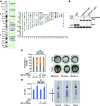
Figure 2. Uniparental disomy detection by whole-exome sequencing.
(A) Right: variants identified by exome sequencing are plotted against chromosomes; colored dots indicate Mendelian inconsistencies (green: paternal UPD; blue: maternal UPD; male sample); left: pUPD of chromosome 1 was confirmed by microsatellite marker analysis (green: pUPD, black: homozygous). (B) Active RAP1 pull-down assay shows a markedly reduced activation of RAP1AR163T-GFP after EGF stimulation compared with RAP1AWT-GFP (n = 4). (C) MO KD of rap1 causes CE defects in zebrafish that are partially rescued by WT, but not RAP1AR163T mRNA. ***P < 0.001, χ2 test. Error bars show SEM. Arrowheads show the body gap angle. Bars show the width of somites. (D) MO KD of rap1 causes a shift in the width-length ratio of somites in zebrafish embryos that is rescued by WT but not RAP1AR163T mRNA. Class I and class II embryos were merged for the statistical analysis. **P < 0.01, 2-tailed Student’s t test (n = 10). Error bars show SEM.
Figure 1
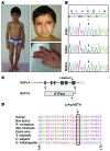
Figure 1. The de novo mutation c.488G>C (p.Arg163Thr) in RAP1A identified in a patient with KS.
(A) Clinical presentation. Left: note the proportionate short stature, short neck, and shoulder asymmetry as a result of Sprengel’s deformity. Upper right: facial features indicative of KS: wide palpebral fissures, long eyelashes, arched, flared eyebrows, large ears, flat midface, and full cheeks. Lower right: hand of the patient with persistent fingertip pads. (B) Sanger sequencing confirmation of the RAP1A c.488G>C mutation showing homozygosity in the patient due to UPD. (C) Schematic representation of the RAP1A gene and the RAP1A protein; black arrows indicate the localization of the identified homozygous missense mutation. (D) The affected amino acid residue is highly conserved among a wide range of species.

References

    1. Bögershausen N, Bruford E, Wollnik B. Skirting the pitfalls: a clear-cut nomenclature for H3K4 methyltransferases. Clin Genet. 2013;83(3):212–214. doi: 10.1111/cge.12050. - DOI - PMC - PubMed
    1. Ng SB, et al. Exome sequencing identifies MLL2 mutations as a cause of Kabuki syndrome. Nat Genet. 2010;42(9):790–793. doi: 10.1038/ng.646. - DOI - PMC - PubMed
    1. Lederer D, et al. Deletion of KDM6A, a histone demethylase interacting with MLL2, in three patients with Kabuki syndrome. Am J Hum Genet. 2012;90(1):119–124. doi: 10.1016/j.ajhg.2011.11.021. - DOI - PMC - PubMed
    1. Miyake N, et al. KDM6A point mutations cause Kabuki syndrome. Hum Mutat. 2013;34(1):108–110. doi: 10.1002/humu.22229. - DOI - PubMed
    1. Bögershausen N, Wollnik B. Unmasking Kabuki syndrome. Clin Genet. 2013;83(3):201–211. doi: 10.1111/cge.12051. - DOI - PubMed

Publication types

MeSH terms

Supplementary concepts