. 2015 Aug 17;4(8):e163.
doi: 10.1038/oncsis.2015.21.

Urothelial cells undergo epithelial-to-mesenchymal transition after exposure to muscle invasive bladder cancer exosomes

Affiliations

Urothelial cells undergo epithelial-to-mesenchymal transition after exposure to muscle invasive bladder cancer exosomes

C A Franzen et al. Oncogenesis. .

Abstract

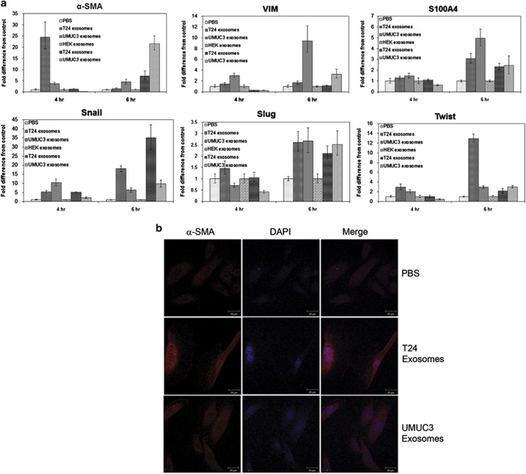
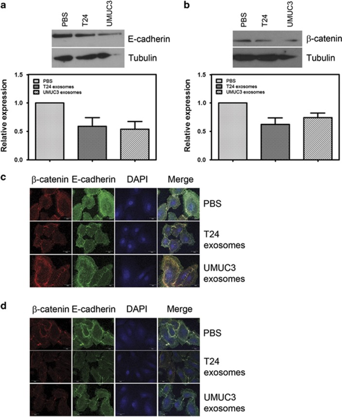
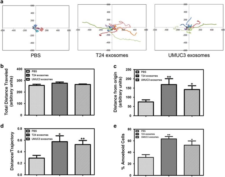
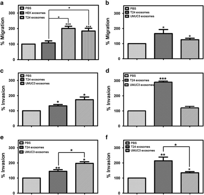
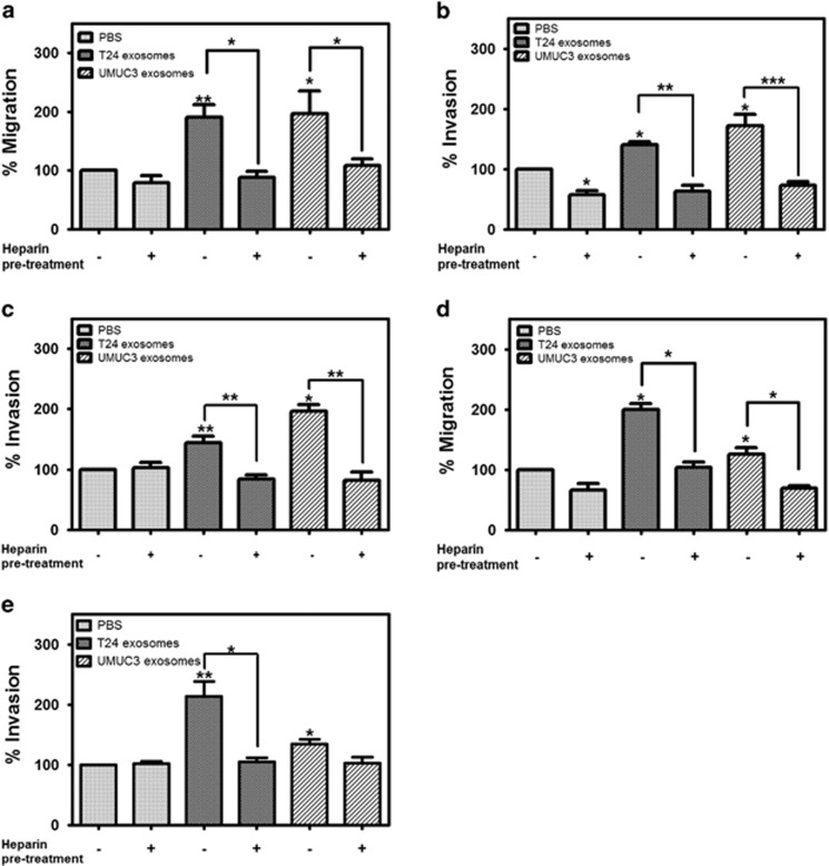
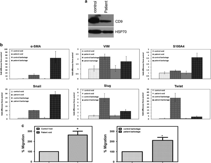
Bladder cancer, the fourth most common noncutaneous malignancy in the United States, is characterized by high recurrence rate, with a subset of these cancers progressing to a deadly muscle invasive form of disease. Exosomes are small secreted vesicles that contain proteins, mRNA and miRNA, thus potentially modulating signaling pathways in recipient cells. Epithelial-to-mesenchymal transition (EMT) is a process by which epithelial cells lose their cell polarity and cell-cell adhesion and gain migratory and invasive properties to become mesenchymal stem cells. EMT has been implicated in the initiation of metastasis for cancer progression. We investigated the ability of bladder cancer-shed exosomes to induce EMT in urothelial cells. Exosomes were isolated by ultracentrifugation from T24 or UMUC3 invasive bladder cancer cell conditioned media or from patient urine or bladder barbotage samples. Exosomes were then added to the urothelial cells and EMT was assessed. Urothelial cells treated with bladder cancer exosomes showed an increased expression in several mesenchymal markers, including α-smooth muscle actin, S100A4 and snail, as compared with phosphate-buffered saline (PBS)-treated cells. Moreover, treatment of urothelial cells with bladder cancer exosomes resulted in decreased expression of epithelial markers E-cadherin and β-catenin, as compared with the control, PBS-treated cells. Bladder cancer exosomes also increased the migration and invasion of urothelial cells, and this was blocked by heparin pretreatment. We further showed that exosomes isolated from patient urine and bladder barbotage samples were able to induce the expression of several mesenchymal markers in recipient urothelial cells. In conclusion, the research presented here represents both a new insight into the role of exosomes in transition of bladder cancer into invasive disease, as well as an introduction to a new platform for exosome research in urothelial cells.


Figures

Figure 1
Figure 1
MIBC exosomes increase expression of mesenchymal markers in urothelial cells. (a) qRT–PCR for mesenchymal genes expressed in urothelial cells treated with PBS, human embryonic kidney (HEK) exosomes or MIBC exosomes for 4 or 6 h. qRT–PCR was repeated at least three times for each gene. (b) Confocal microscopy images of α-SMA expression in urothelial cells treated with PBS or MIBC exosomes for 24 h. Confocal microscopy was performed at least three times. A representative image is shown. Scale bar=20 μm. DAPI, 4,6-diamidino-2-phenylindole.
Figure 2
Figure 2
MIBC exosomes decrease expression and alter localization of E-cadherin and β-catenin in urothelial cells. (a) Representative western blotting of E-cadherin expression in urothelial cells after 48 h of treatment with PBS or MIBC exosomes. The bar graph shows the quantitation of E-cadherin expression from four experiments. (b) Representative western blotting of β-catenin expression in urothelial cells after 48 h of treatment with PBS or MIBC exosomes. The bar graph shows the quantitation of β-catenin expression from four experiments. (c, d). Confocal microscopy images of β-catenin and E-cadherin expression and localization in urothelial cells treated with PBS or MIBC exosomes for (c) 24 or (d) 48 h. Confocal microscopy was performed at least three times. A representative image is shown. Scale bar=20 μm. DAPI, 4,6-diamidino-2-phenylindole.
Figure 3
Figure 3
MIBC exosomes alter motility in primary urothelial cells. Urothelial cells were plated on Col IV-coated cover glass with PBS or MIBC exosomes and allowed to attach for 1 h before being placed in the ASMDW for live cell imaging. (a) Wind-rose plots of cell tracks from time-lapse microscopy. Each wind-rose plot shows centroid tracks from 10 representative tracks, with the initial position of each track superimposed at 0,0 for clarity. (b) Total distance traveled over 6 h was calculated using the ImageJ imaging software. (c) The average distance from the origin over 6 h for cells treated with MIBC exosomes versus control (40 cells/condition were analyzed). (d) The distance/trajectory was calculated as a ratio of the distance from the origin traveled over the total distance traveled. (e) The percentage of cells undergoing amoeboid cell motility was calculated as the number of cells displaying amoeboid-type movement divided by the total number of cells for each treatment. Data are represented as average±s.e.m. *Difference between control and MIBC exosome-treated cells (P<0.05) by a paired Student's t-test. **Difference between control and MIBC exosome-treated cells (P<0.01) by a paired Student's t-test.
Figure 4
Figure 4
MIBC exosomes enhance the migration and invasion of primary urothelial cells. (a) Urothelial cells were plated on uncoated transwell inserts in 500 μl serum-free media with MIBC exosomes, human embryonic kidney (HEK) exosomes or PBS for a migration assay. (b) Urothelial cells were plated on uncoated transwell inserts in 500 μl serum-free media, and MIBC exosomes (30 μg/ml) or PBS were plated in the bottom chamber. (c) Urothelial cells were plated on Col IV-coated transwell inserts in 500 μl serum-free media with MIBC exosomes (30 μg/ml) or PBS for an invasion assay. (d) Urothelial cells were plated on Col IV-coated transwell inserts in 500 μl serum-free media, and MIBC exosomes (30 μg/ml) or PBS were plated in the bottom chamber. (e) Urothelial cells were plated on matrigel-coated transwell inserts in 500 μl serum-free media with MIBC exosomes (30 μg/ml) or PBS for an invasion assay. (f) Urothelial cells were plated on matrigel-coated transwell inserts in 500 μl serum-free media, and MIBC exosomes (30 μg/ml) or PBS were plated in the bottom chamber. Migration and assays were carried out in triplicate. Graphs represent averages (normalized to the PBS control-treated cells)±s.e.m. *P<0.05, **P<0.01, ***P<0.001, by a paired Student's t-test.
Figure 5
Figure 5
Heparin blocks the effect of MIBC exosomes on cell migration and invasion. (ac) Urothelial cells were pretreated with heparin (10 μg/ml) for 30 min and then plated for migration on uncoated transwell inserts (a), Col IV-coated inserts (b) or matrigel-coated inserts (c) in 500 μl serum-free media with MIBC exosomes or PBS for migration or invasion assays. (d, e) MIBC exosomes were pretreated with heparin for 30 min. Urothelial cells were plated on uncoated transwell inserts (d) or matrigel-coated inserts (e) in 500 μl serum-free media, and MIBC exosomes (30 μg/ml) or PBS were plated in the bottom chamber. Migration and invasion assays were carried out in triplicate. Graphs represent averages (normalized to the PBS control-treated cells)±s.e.m. *P<0.05, **P<0.01, ***P<0.001, by a paired Student's t-test.
Figure 6
Figure 6
Exosomes isolated from bladder cancer patient urine and barbotage samples increase expression of mesenchymal markers in and enhance the migration of primary urothelial cells. (a) Western blotting demonstrating that exosomal markers are expressed in the exosomes isolated from control and bladder cancer urinary exosomes. (b) qRT–PCR for mesenchymal genes expressed in urothelial cells treated with control or patient urinary or barbotage exosomes for 4 h. qRT–PCR was repeated in triplicate three times for each gene. (c) Urothelial cells were plated on uncoated transwell inserts in 500 μl serum-free media with control urinary exosomes or bladder cancer patient urinary exosomes for a migration assay. (d) Urothelial cells were plated on uncoated transwell inserts in 500 μl serum free media with control barbotage exosomes or bladder cancer patient barbotage exosomes for a migration assay. Migration assays were carried out in triplicate. Graphs represent averages (normalized to the control-treated cells)±s.e.m. *P<0.05, by a paired Student's t-test.

Similar articles

Cited by

References

    1. 1Resnick MJ, Chang SS. Optimizing outcomes for octogenarians with invasive bladder cancer: one size does not fit all. Urol Oncol 2013; 31: 1–4. - PubMed
    1. 2Sylvester RJ. Natural history, recurrence, and progression in superficial bladder cancer. ScientificWorldJournal 2006; 6: 2617–2625. - PMC - PubMed
    1. 3Messing EM. Urothelial tumors of the urinary tract. In: Walsh PC, Retik AB, Vaughan ED Jr, Wein AJ (eds). Campbell's Urology, 8th edn. Saunders: Philadelphia, PA, USA, 2002, pp 2750–2751.
    1. 4Messing EM. Urothelial tumors of the bladder. In: Wein AJ, Kavoussi LR, Novick AC, Partin AW, Peters CA (eds). Campbell-Walsh's Urology. Saunders: Philadelphia, PA, USA, 2007, pp 17–2430.
    1. 5Denzer K, Kleijmeer MJ, Heijnen HF, Stoorvogel W, Geuze HJ. Exosome: from internal vesicle of the multivesicular body to intercellular signaling device. J Cell Sci 2000; 113(Pt 19): 3365–3374. - PubMed