. 2016 Aug;68(4):1489-98.
doi: 10.1007/s10616-015-9909-2. Epub 2015 Aug 18.

A comparative study of colorimetric cell proliferation assays in immune cells

Affiliations

A comparative study of colorimetric cell proliferation assays in immune cells

Madoka Koyanagi et al. Cytotechnology. 2016 Aug.

Abstract

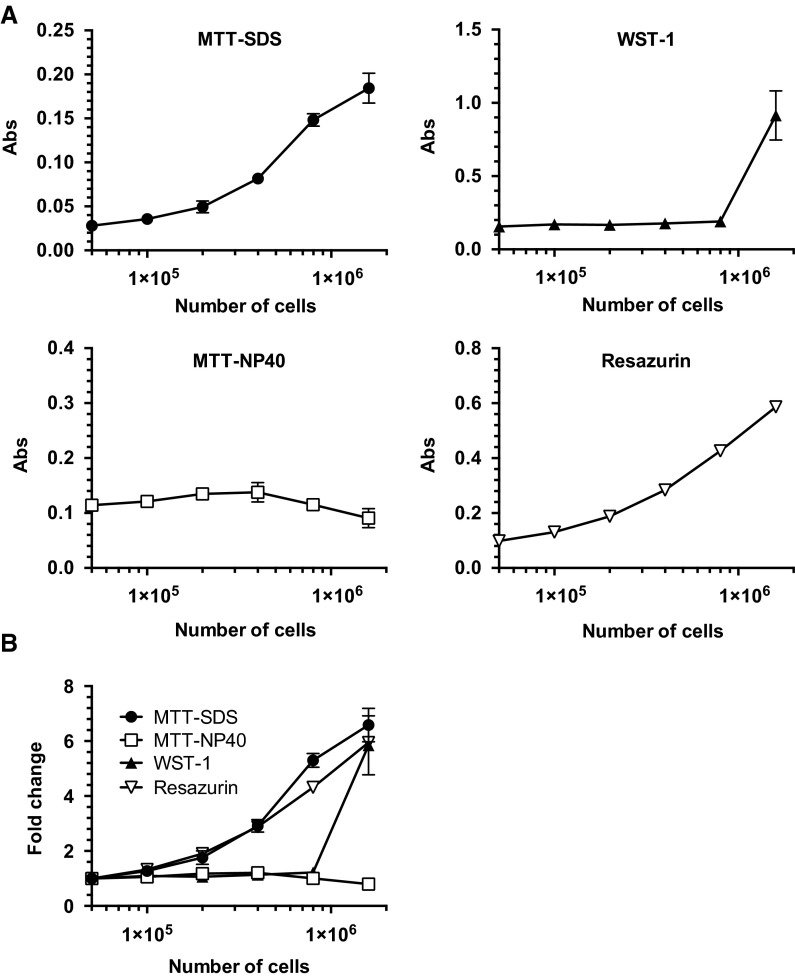
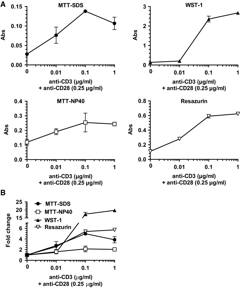
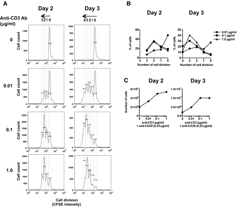
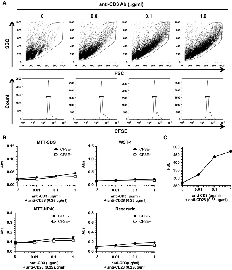
Cell proliferation assays are basic and essential techniques for assessing cellular function. Various colorimetric assays, such as MTT-, WST-1-, and resazurin-based assays, are available; however, studies directly comparing the suitability of each method for immune cell proliferation are scarce. Thus, we aimed to determine the best reagent and its optimal conditions based on variables such as cell number range, stimulation dose, kinetics, and compatibility with the cell division assay using CFSE fluorescence dye which is able to directly monitor divided cells by flow cytometry. In the absence of stimulation, MTT solubilized with SDS (MTT-SDS) and resazurin appeared to accurately reflect the cell numbers in a linear fashion. On the other hand, WST-1 exhibited a higher stimulation index following strong stimulation, whereas MTT-SDS and resazurin exhibited a better sensitivity to weak stimulation. A longer duration for stimulation did not necessarily increase sensitivity. CFSE staining revealed incremental cell division in response to anti-CD3 antibody stimulation in a dose-dependent manner. The cell numbers indirectly estimated from cell division profiles were consistent with the dose-response curve in the absorbance of MTT-SDS and resazurin. The absorbance does not increase before cell division, irrespective of T cell activation status, suggesting that these reagents reflect the cell number but not the cellular volume. Collectively, resazurin and MTT-SDS seem to be more reliable than others, and thus appear applicable in various conditions for the immune cell experiments.

Keywords: MTT; Resazurin; T cell; WST-1.


Figures

Fig. 1
Fig. 1
Cell number range for MTT, WST-1, and resazurin. a A serial number of mouse whole spleen cells were seeded into a 96-well plate in duplicates or triplicates, and they were incubated without stimulation in the absence of any reagent for the first 1 h at 37 °C. Three different reagents were then individually added, followed by incubation for the last 4 h at 37 °C. Each absorbance value was assessed by a plate reader. b The figures in a were normalized to the respective minimal values and combined into the same graph to compare their fold change. These are representative of at least three experiments with similar results
Fig. 2
Fig. 2
Dose-response curve of MTT, WST-1, and resazurin. a Mouse spleen cells were examined for the dose–response curve of MTT, WST-1, and resazurin against a serially diluted amount of anti-CD3 Ab (0.01, 0.1, and 1.0 μg/mL) in the constant presence of anti-CD28 Ab costimulation (0.25 μg/mL) for 2 days. b The figures in a were normalized to the respective minimal values and combined into the same graph to compare their fold change. These are representative of six experiments with similar results
Fig. 3
Fig. 3
Kinetics of responses to stimulation. Mouse whole spleen cells were examined for the kinetic response of MTT, WST-1, and resazurin to stimulation using anti-CD3 Ab (0.5 μg/mL) in the presence of anti-CD28 Ab (0.25 μg/mL) for 1–4 days. These are representative of two experiments with similar results
Fig. 4
Fig. 4
Monitoring of cell division with CFSE fluorescence intensity dilution assay. a Mouse whole spleen cells were stained with CFSE fluorescent dye and stimulated with a serially diluted amount of anti-CD3 (0.01, 0.1, and 1.0 μg/mL) and -CD28 Abs (0.25 μg/mL) for 2–3 days. The cell division was directly monitored by CFSE fluorescence intensity dilution using a flow cytometer. Numerals above the histogram indicate cell division number. b The percentages in each cell division number were expressed as a graph. c The increased cell numbers by anti-CD3 Ab stimulation were estimated from percentages in each cell division in b and expressed as a graph. These are representative of five experiments with similar results
Fig. 5
Fig. 5
TCR signal does not substantially raise MTT activity before cell division. Murine whole spleen cells were stained with or without CFSE fluorescent dye and stimulated with a serially diluted amount of anti-CD3 and -CD28 Abs for 16 h. a The cells were subjected to flow cytometry for FSC/SSC analysis and CFSE fluorescence intensity dilution assay. FSC and SSC reflect cell size and intracellular granular structures, respectively. b The same cells were given MTT, WST-1, or resazurin for the last 4 h of 16 h and subjected to the absorbance assays by a plate reader. These are representative of five experiments with similar results. c The FSC result shown in a is expressed as a graph

Similar articles

  • Basic Colorimetric Proliferation Assays: MTT, WST, and Resazurin.
    Präbst K, Engelhardt H, Ringgeler S, Hübner H. Präbst K, et al. Methods Mol Biol. 2017;1601:1-17. doi: 10.1007/978-1-4939-6960-9_1. Methods Mol Biol. 2017. PMID: 28470513
  • Small Molecule Interferences in Resazurin and MTT-Based Metabolic Assays in the Absence of Cells.
    Neufeld BH, Tapia JB, Lutzke A, Reynolds MM. Neufeld BH, et al. Anal Chem. 2018 Jun 5;90(11):6867-6876. doi: 10.1021/acs.analchem.8b01043. Epub 2018 May 17. Anal Chem. 2018. PMID: 29746096
  • Use of the viability reagent PrestoBlue in comparison with alamarBlue and MTT to assess the viability of human corneal epithelial cells.
    Xu M, McCanna DJ, Sivak JG. Xu M, et al. J Pharmacol Toxicol Methods. 2015 Jan-Feb;71:1-7. doi: 10.1016/j.vascn.2014.11.003. Epub 2014 Nov 15. J Pharmacol Toxicol Methods. 2015. PMID: 25464019
  • Lymphocyte proliferation response during Eimeria tenella infection assessed by a new, reliable, nonradioactive colorimetric assay.
    Miyamoto T, Min W, Lillehoj HS. Miyamoto T, et al. Avian Dis. 2002 Jan-Mar;46(1):10-6. doi: 10.1637/0005-2086(2002)046[0010:LPRDET]2.0.CO;2. Avian Dis. 2002. PMID: 11922320
  • Cell Viability Assays.
    Riss TL, Moravec RA, Niles AL, Duellman S, Benink HA, Worzella TJ, Minor L. Riss TL, et al. 2013 May 1 [updated 2016 Jul 1]. In: Markossian S, Grossman A, Baskir H, Arkin M, Auld D, Austin C, Baell J, Brimacombe K, Chung TDY, Coussens NP, Dahlin JL, Devanarayan V, Foley TL, Glicksman M, Gorshkov K, Grotegut S, Hall MD, Hoare S, Inglese J, Iversen PW, Lal-Nag M, Li Z, Manro JR, McGee J, Norvil A, Pearson M, Riss T, Saradjian P, Sittampalam GS, Tarselli MA, Trask OJ Jr, Weidner JR, Wildey MJ, Wilson K, Xia M, Xu X, editors. Assay Guidance Manual [Internet]. Bethesda (MD): Eli Lilly & Company and the National Center for Advancing Translational Sciences; 2004–. 2013 May 1 [updated 2016 Jul 1]. In: Markossian S, Grossman A, Baskir H, Arkin M, Auld D, Austin C, Baell J, Brimacombe K, Chung TDY, Coussens NP, Dahlin JL, Devanarayan V, Foley TL, Glicksman M, Gorshkov K, Grotegut S, Hall MD, Hoare S, Inglese J, Iversen PW, Lal-Nag M, Li Z, Manro JR, McGee J, Norvil A, Pearson M, Riss T, Saradjian P, Sittampalam GS, Tarselli MA, Trask OJ Jr, Weidner JR, Wildey MJ, Wilson K, Xia M, Xu X, editors. Assay Guidance Manual [Internet]. Bethesda (MD): Eli Lilly & Company and the National Center for Advancing Translational Sciences; 2004–. PMID: 23805433 Free Books & Documents. Review.

Cited by

References

    1. Arimura Y, Kato H, Dianzani U, Okamoto T, Kamekura S, Buonfiglio D, Miyoshi-Akiyama T, Uchiyama T, Yagi J. A co-stimulatory molecule on activated T cells, H4/ICOS, delivers specific signals in Th cells and regulate their responses. Int Immunol. 2002;14:555–566. doi: 10.1093/intimm/dxf022. - DOI - PubMed
    1. Arimura Y, Shiroki F, Kuwahara S, Kato H, Dianzani U, Uchiyama T, Yagi J. Akt is a neutral amplifier for Th cell differentiation. J Biol Chem. 2004;279:11408–11416. doi: 10.1074/jbc.M309063200. - DOI - PubMed
    1. Arimura Y, Ezaki T, Koyanagi M, Uchiyama T, Koyasu S, Yagi J. Reduced T cell expansion by a superantigen as a result of impaired B cell development in mice deficient for the p85α regulatory subunit of PI3K. J Leukoc Biol. 2010;87:493–500. doi: 10.1189/jlb.0708440. - DOI - PubMed
    1. Berridge MV, Herst PM, Tan AS. Tetrazolium dyes as tools in cell biology: new insight into their cellular reduction. Biotechnol Annu Rev. 2005;11:11004–11007. - PubMed
    1. Chen L, Koyanagi M, Fukada K, Imanishi K, Yagi J, Kato H, Miyoshi-Akiyama T, Zhang R, Miwa K, Uchiyama T. Continuous exposure of mice to superantigenic toxins induces a high-level protracted expansion and an immunological memory in the toxin-reactive CD4+ T cells. J Immunol. 2002;168:3817–3824. doi: 10.4049/jimmunol.168.8.3817. - DOI - PubMed

LinkOut - more resources