. 2015 Aug 18:5:13291.
doi: 10.1038/srep13291.

Systemic modeling myeloma-osteoclast interactions under normoxic/hypoxic condition using a novel computational approach

Affiliations

Systemic modeling myeloma-osteoclast interactions under normoxic/hypoxic condition using a novel computational approach

Zhiwei Ji et al. Sci Rep. .

Abstract

Interaction of myeloma cells with osteoclasts (OC) can enhance tumor cell expansion through activation of complex signaling transduction networks. Both cells reside in the bone marrow, a hypoxic niche. How OC-myeloma interaction in a hypoxic environment affects myeloma cell growth and their response to drug treatment is poorly understood. In this study, we i) cultured myeloma cells in the presence/absence of OCs under normoxia and hypoxia conditions and did protein profiling analysis using reverse phase protein array; ii) computationally developed an Integer Linear Programming approach to infer OC-mediated myeloma cell-specific signaling pathways under normoxic and hypoxic conditions. Our modeling analysis indicated that in the presence OCs, (1) cell growth-associated signaling pathways, PI3K/AKT and MEK/ERK, were activated and apoptotic regulatory proteins, BAX and BIM, down-regulated under normoxic condition; (2) β1 Integrin/FAK signaling pathway was activated in myeloma cells under hypoxic condition. Simulation of drug treatment effects by perturbing the inferred cell-specific pathways showed that targeting myeloma cells with the combination of PI3K and integrin inhibitors potentially (1) inhibited cell proliferation by reducing the expression/activation of NF-κB, S6, c-Myc, and c-Jun under normoxic condition; (2) blocked myeloma cell migration and invasion by reducing the expression of FAK and PKC under hypoxic condition.


Figures
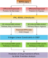
Figure 1
Figure 1. Flowchart of the proposed DILP approach.
Figure 2
Figure 2. Effects of osteoclasts on myeloma cell. RPMI 8226 myeloma cells were cultured in the presence or absence of OCs under normoxia (21% O2, 5% CO2) and hypoxia (5% O2, 5% CO2) conditions.
Myeloma cells were harvested at 72 h centrifugation. Myeloma cell growth was measured using dsDNA assay. Panel A shows myeloma growth in the presence or absence OCs under normoxic condition. Panel B shows myeloma cell growth in the presence or absence of OCs under hypoxic condition. **means p < 0.01.
Figure 3
Figure 3. Generic pathway maps of myeloma cells in the presence of osteoclasts in normoxia and hypoxia.
(A) OC-mediated myeloma cell-related generic pathway map in normoxia; (B) OC-mediated myeloma cell-related generic pathway map in hypoxia. The nodes with a dark color were predicted using our model.
Figure 4
Figure 4. Inferred specific pathways of myeloma cells in the presence/absence of OCs under normoxic condition.
Our model predicted that the functional module with red color was up-regulated, which potentially increases of cell proliferation and decrease of cell apoptosis. Similarity, the module with green color was down-regulated, which indicated the decrease of apoptosis. The predicted states of key proteins in this network were represented in detail in Supplementary Fig. S4.
Figure 5
Figure 5. Inferred specific pathways of myeloma cells in the presence/absence of OCs under hypoxic condition.
Several functional modules were highlighted to indicate the changes of phenotypes under different cellular contexts. (A) The specific pathways of myeloma cells in the absence of OCs under hypoxic condition. (B) The specific pathways of myeloma cells in the presence of OCs under hypoxic condition. The predicted states of key proteins in both networks were displayed in Supplementary Fig. S5 (A,B).
Figure 6
Figure 6. Experimental validation of key proteins involved in the inferred signaling pathways.
(A) RPMI 8226 myeloma cells were cultured in the presence or absence of OCs under normoxic condition for 24 h. Cell lysates were collected and subjected to western blot analysis with p-Akt, p-MEK, p-ERK and c-MYC antibodies. (B) Myeloma cells were cultured in the presence OCs under normoxic and hypoxic conditions for 24 h. Myeloma cell lysates were subjected to western blot analysis with the FAK, c-MYC, p53 and fibronectin antibodies.
Figure 7
Figure 7. Simulation of treatment effects by perturbing the inferred cell-specific pathways with combination of PI3K and integrin inhibitors.
The changes of downstream modules were highlighted. (A) Predicted treatment effects of PI3K and integrin inhibitors on OC-mediated myeloma cells-specific pathways in normoxia; (B) Predicted treatment effects of PI3K and integrin inhibitors on OC-mediated myeloma cells-specific pathways in hypoxia.
Figure 8
Figure 8. Five cases of linking patterns of signaling proteins in pathway network topology.
(A) single activation; (B) single inhibition; (C) multiple activations; (D) multiple inhibition; (E) mixed reactions. (A,B) show that, the state of downstream protein is determined by its unique parental node through single reaction of activation/inhibition, respectively; In (C,D) the state of a downstream protein can be changed if at least one of the upstream proteins is up-regulated/down-regulated and others are un-changed. In Fig. 8E, the state of downstream protein might be un-changed (0), up-regulated (1) or down-regulated (−1) if it has the potential of being both up- and down-regulated by its parental nodes, simultaneously.

References

    1. Hu J., Van Valckenborgh E., Menu E., De Bruyne E. & Vanderkerken K. Understanding the hypoxic niche of multiple myeloma: therapeutic implications and contributions of mouse models. Dis. Model Mech. 5, 763–771 (2012). - PMC - PubMed
    1. Manier S., Sacco A., Leleu X., Ghobrial I. M. & Roccaro A. M. Bone marrow microenvironment in multiple myeloma progression. J. Biomed Biotechnol. 2012, 157496 (2012). - PMC - PubMed
    1. Hu J. et al. Targeting the multiple myeloma hypoxic niche with TH-302, a hypoxia-activated prodrug. Blood 116, 1524–1527 (2010). - PubMed
    1. Azab A. K. et al. Hypoxia promotes dissemination of multiple myeloma through acquisition of epithelial to mesenchymal transition-like features. Blood 119, 5782–5794 (2012). - PMC - PubMed
    1. Ribatti D., Moschetta M. & Vacca A. Microenvironment and multiple myeloma spread. Thrombosis Research 133, S102–S106 (2014). - PubMed

Publication types