. 2015;15(18):3661-9.
doi: 10.1039/c5lc00666j.

A cost-effective fluorescence mini-microscope for biomedical applications

Affiliations

A cost-effective fluorescence mini-microscope for biomedical applications

Yu Shrike Zhang et al. Lab Chip. 2015.

Abstract

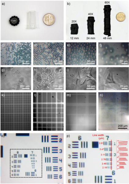
We have designed and fabricated a miniature microscope from off-the-shelf components and a webcam, with built-in fluorescence capability for biomedical applications. The mini-microscope was able to detect both biochemical parameters, such as cell/tissue viability (e.g. live/dead assay), and biophysical properties of the microenvironment such as oxygen levels in microfabricated tissues based on an oxygen-sensitive fluorescent dye. This mini-microscope has adjustable magnifications from 8-60×, achieves a resolution as high as <2 μm, and possesses a long working distance of 4.5 mm (at a magnification of 8×). The mini-microscope was able to chronologically monitor cell migration and analyze beating of microfluidic liver and cardiac bioreactors in real time, respectively. The mini-microscope system is cheap, and its modularity allows convenient integration with a wide variety of pre-existing platforms including, but not limited to, cell culture plates, microfluidic devices, and organs-on-a-chip systems. Therefore, we envision its widespread application in cell biology, tissue engineering, biosensing, microfluidics, and organs-on-chips, which can potentially replace conventional bench-top microscopy where long-term in situ and large-scale imaging/analysis is required.


Figures

Fig. 1
Fig. 1
a) Schematic showing design of the mini-microscope integrated with a bioreactor. b) Photograph showing the actual device; the inset shows the device under LED illumination. c, d) Fabrication of the imaging unit of the mini-microscope: a webcam (Logitech C-160) is first disassembled to obtain the CMOS chip, after which the lens is detached, flipped, and then re-attached to the base to achieve magnification.
Fig. 2
Fig. 2
a, b) Photographs showing the assembly of the objective with different magnifications by varying the height of the tubing. c-f and g-j) Mini-microscopic images of 16-μm polystyrene particles and NIH/3T3 fibroblasts at four different magnifications of 8X, 20X, 40X, and 60X. k-n) Full frame images of a hemocytometer captured from the mini-microscope at different magnifications where the field-of-views were calculated to be 1060 μm × 850 μm = 0.901 mm2, 500 μm × 400 μm = 0.200 mm2, 235 μm × 190 μm = 0.045 mm2, and 130 μm × 105 μm = 0.014 mm2, respectively. o, p) Mini-microscopic images of a resolution target showing Groups 6 and 7 targets where the microscope could easily resolve lines/spacing as small as 2.19 μm; the inset shows the intensity profile across the red line in the Group 7 targets.
Fig. 3
Fig. 3
a) Schematic showing RGB configuration of the CMOS sensor of the mini-microscope. b-d) RGB and R/G/B images showing the filter-free separation of R, G, B colors by taking advantage of the digital channel unmixing using a custom-coded MATLAB program. e-g) Quantification data showing the accuracy and sensitivity of the digital channel separation approach. h) Bright-field image showing HepG2 cells grown in a liver-on-a-chip device. i) The raw image obtained from the mini-microscope showing the same HepG2 cells stained with calcein and illuminated with a monochromatic LED at 490 nm. j-l) Images from R/G/B channels after separation, respectively. m) Pseudo-colored and contrast enhanced image of the G channel showing the fluorescence from the stained cells with minimal interference from the illuminating B channel.
Fig. 4
Fig. 4
a-c) Dual-channel fluorescence imaging with the mini-microscope and the bench-top microscope (Nikon Eclipse) for (a) green fluorescent and (b) red fluorescent beads; superimposed images of the two channels are indicated in (c). d, e) Mini-microscopic images of live/dead staining results showing HepG2 cells non-treated or treated with 10 mM acetaminophen for 24 h in the liver bioreactors. f) Bench-top microscope (Zeiss Axio Observer D1) image showing the same cell colony as in (e), indicating the accuracy in the detection of the live/dead cells using the mini-microscope.
Fig. 5
Fig. 5
a-d) Time lapse mini-microscopic images showing the migration of HepG2 cells at the bottom of the bioreactor over a course of 72 h. e) Schematic and photograph showing the preparation of a glass substrate coated with GelMA-CNTs for cardiomyocytes culture. f) Mini-microscopic image showing rat neonatal cardiomyocytes seeded onto the substrate and embedded in the chamber of the bioreactor at Day 5. g) Real-time measurement of cardiomyocytes beating in situ, using custom-coded MATLAB program with the bioreactor-integrated mini-microscope. h) Measurement of cardiomyocytes beating using a bench-top microscope after the cardiac bioreactor was removed from the incubator for 30 min.
Fig. 6
Fig. 6
a) Microfluidic device to fabricate uniform PDMS microspheres. b) PDMS microbeads fabricated by the microfluidic device and infiltration of ruthenium dye to produce oxygen-sensitive beads. c) Calibration curve showing fluorescence intensity versus oxygen concentration. d-i) Fluorescence images obtained from the mini-microscope showing beads at different oxygen concentrations. Ruthenium channel was used for imaging. j) Schematic diagram showing the setup for real-time sensing of oxygen levels in a liver bioreactor. k, l) Bright-field and fluorescence images showing the cells and the oxygen sensing beads at 24 h post seeding.

References

    1. Zernike F. Physica. 1942;9:686–698.
    1. Pluta M. Nomarski's DIC microscopy: a review. 1994
    1. Lichtman JW, Conchello J-A. Nat. Methods. 2005;2:910–919. - PubMed
    1. Hell SW, Wichmann J. Opt. Lett. 1994;19:780–782. - PubMed
    1. Klar TA, Hell SW. Opt. Lett. 1999;24:954–956. - PubMed

Publication types