

High-Resolution Rapid Diagnostic Imaging of Whole Prostate Biopsies Using Video-Rate Fluorescence Structured Illumination Microscopy
- PMID: 26282168
- PMCID: PMC4592466
- DOI: 10.1158/0008-5472.CAN-14-3806
High-Resolution Rapid Diagnostic Imaging of Whole Prostate Biopsies Using Video-Rate Fluorescence Structured Illumination Microscopy
Abstract
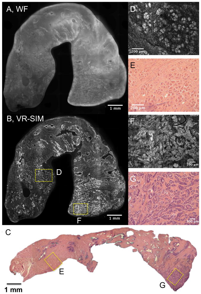
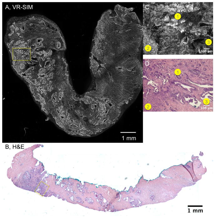
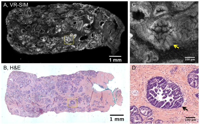
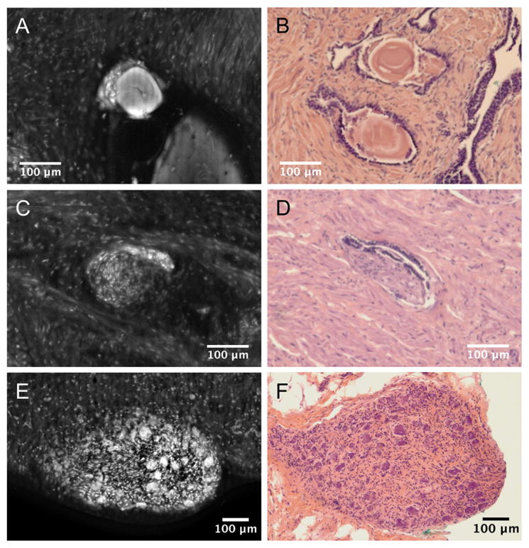
Rapid assessment of prostate core biopsy pathology at the point-of-procedure could provide benefit in a variety of clinical situations. Even with advanced transrectal ultrasound guidance and saturation biopsy protocols, prostate cancer can be missed in up to half of all initial biopsy procedures. In addition, collection of tumor specimens for downstream histologic, molecular, and genetic analysis is hindered by low tumor yield due to inability to identify prostate cancer grossly. However, current point-of-procedure pathology protocols, such as frozen section analysis (FSA), are destructive and too time- and labor-intensive to be practical or economical. Ex vivo microscopy of the excised specimens, stained with fast-acting fluorescent histology dyes, could be an attractive nondestructive alternative to FSA. In this work, we report the first demonstration of video-rate structured illumination microscopy (VR-SIM) for rapid high-resolution diagnostic imaging of prostate biopsies in realistic point-of-procedure timeframes. Large mosaic images of prostate biopsies stained with acridine orange are rendered in seconds and contain excellent contrast and detail, exhibiting close correlation with corresponding hematoxylin and eosin histology. A clinically relevant review of VR-SIM images of 34 unfixed and uncut prostate core biopsies by two independent pathologists resulted in an area under the receiver operative curve (AUC) of 0.82-0.88, with a sensitivity ranging from 63% to 88% and a specificity ranging from 78% to 89%. When biopsies contained more than 5% tumor content, the sensitivity improved to 75% to 92%. The image quality, speed, minimal complexity, and ease of use of VR-SIM could prove to be features in favor of adoption as an alternative to destructive pathology at the point-of-procedure.
©2015 American Association for Cancer Research.
Conflict of interest statement
Figures
References
-
- Epstein JI, Netto GJ, Epstein JI. Biopsy interpretation of the prostate. 4. Philadelphia: Wolters Kluwer Health/Lippincott Williams & Wilkins; 2008. p. x.p. 358.
-
- Wein AJ, Kavoussi LR, Campbell MF. In: Campbell-Walsh urology. 10. Wein Alan J, Kavoussi Louis R, et al., editors. Philadelphia, PA: Elsevier Saunders; 2012.
-
- Levy DA, Jones JS. Management of rising prostate-specific antigen after a negative biopsy. Current urology reports. 2011;12(3):197–202. - PubMed
-
- Ukimura O, Coleman JA, de la Taille A, Emberton M, Epstein JI, Freedland SJ, et al. Contemporary role of systematic prostate biopsies: indications, techniques, and implications for patient care. Eur Urol. 2013;63(2):214–30. - PubMed
-
- Campos-Fernandes JL, Bastien L, Nicolaiew N, Robert G, Terry S, Vacherot F, et al. Prostate cancer detection rate in patients with repeated extended 21-sample needle biopsy. Eur Urol. 2009;55(3):600–6. - PubMed
Publication types
MeSH terms
Substances
Grants and funding
LinkOut - more resources
Full Text Sources
Other Literature Sources
Medical
