. 2015 Aug 19:10:126.
doi: 10.1186/s13018-015-0265-x.

The effects of a dynamic patellar realignment brace on disease determinants for patellofemoral instability in the upright weight-bearing condition

Affiliations

The effects of a dynamic patellar realignment brace on disease determinants for patellofemoral instability in the upright weight-bearing condition

Christoph Becher et al. J Orthop Surg Res. .

Abstract

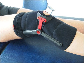
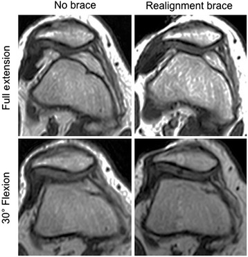
Background: Patellar stabilizing braces are used to alleviate pain and prevent subluxation/dislocation by having biomechanical effects in terms of improved patellar tracking. The purpose of this study is to analyze the effects of the dynamic patellar realignment brace, Patella Pro (Otto Bock GmbH, Duderstadt, Germany), on disease determinants in subjects with patellofemoral instability using upright weight-bearing magnetic resonance imaging (MRI).

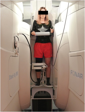
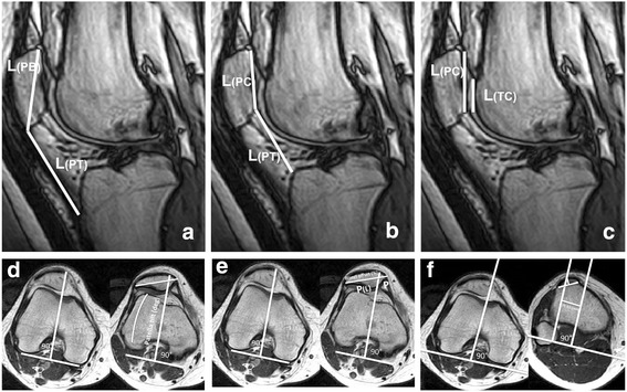
Methods: Twenty subjects (8 males and 12 females) with lateral patellofemoral instability were studied in an open-configuration magnetic resonance imaging scanner in an upright weight-bearing position at full extension (0° flexion) and 15° and 30° flexion with and without the realignment brace. Disease determinants were defined by common patellofemoral indices that included the Insall-Salvati Index, Caton-Deschamps Index, and the Patellotrochlear Index to determine patella height and patella tilt angle, bisect offset, and tuberositas tibiae-trochlear groove (TT-TG) distance to determine patellar rotation and translation with respect to the femur and the alignment of the extensor mechanism.

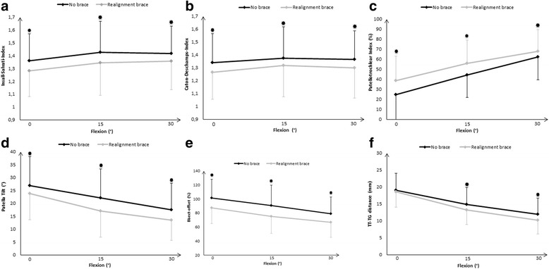
Results: Analyses of variance revealed a significant effect of the brace with reduction of the three patellar height ratios, patella tilt angle, and bisect offset as well as TT-TG distance. Post hoc pairwise comparisons of the corresponding conditions with and without the realignment brace revealed significantly reduced patella height ratios, patella tilt angles, and bisect offsets at full extension and 15° and 30° flexion. No significant differences between the TT-TG distances at full extension but significant reductions at 15° and 30° flexion were observed when using the realignment brace compared to no brace.

Conclusions: This study suggests that the dynamic patellar realignment brace is capable of improving disease determinants in the upright weight-bearing condition in the range of 0° to 30° flexion in patients with patellofemoral instability.


Figures

Fig. 1
Fig. 1
Patellar realignment brace (Patella Pro)
Fig. 2
Fig. 2
The upright open-MRI scanner with a subject examined at full extension weight-bearing condition
Fig. 3
Fig. 3
Descriptions of the measurement of the patellofemoral indices. a Insall–Salvati Index, L(PT) / L(PB). b Caton–Deschamps Index, L(PT) / L (PC). c Patellotrochlear Index (%), (L(TC) / L(PC)) × 100. d Patella tilt angle (°), the angle formed by lines joining the posterior femoral condyles and the maximum width of the patella. e Bisect offset (%), (P(L)/P) × 100. f TT–TG distance, distance between the midpoint of the patellar tendon at the insertion of the tibial tuberosity and a reference line through the first craniocaudal transverse slice that depicts complete cartilaginous trochlea coverage
Fig. 4
Fig. 4
af Comparison of the patellofemoral indices with the realignment brace (gray lines) and without the brace (black lines) at full extension (0° flexion) and 15° and 30° flexion with upright weight-bearing condition. Data presented as mean ± SD. Asterisks indicate significant group differences based on post hoc pairwise comparisons (p ≤ 0.05); according to Bonferroni correction (0.05 / 36), a p < 0.0014 was considered as significant
Fig. 5
Fig. 5
Axial imaging of a subject with patellofemoral instability with and without the realignment brace at 0° and 30°. The joint congruence appears considerably improved at the condition with the brace

References

    1. Amis AA, Firer P, Mountney J, Senavongse W, Thomas NP. Anatomy and biomechanics of the medial patellofemoral ligament. Knee. 2003;10:215–20. doi: 10.1016/S0968-0160(03)00006-1. - DOI - PubMed
    1. Dejour H, Walch G, Nove-Josserand L, Guier C. Factors of patellar instability: an anatomic radiographic study. Knee Surg Sports Traumatol Arthrosc. 1994;2:19–26. doi: 10.1007/BF01552649. - DOI - PubMed
    1. Smith TO, Davies L, Toms AP, Hing CB, Donell ST. The reliability and validity of radiological assessment for patellar instability. A systematic review and meta-analysis. Skeletal Radiol. 2011;40:399–414. doi: 10.1007/s00256-010-0961-x. - DOI - PubMed
    1. Pandit S, Frampton C, Stoddart J, Lynskey T. Magnetic resonance imaging assessment of tibial tuberosity-trochlear groove distance: normal values for males and females. Int Orthop. 2011;35:1799–803. doi: 10.1007/s00264-011-1240-8. - DOI - PMC - PubMed
    1. Schoettle PB, Zanetti M, Seifert B, Pfirrmann CW, Fucentese SF, Romero J. The tibial tuberosity-trochlear groove distance; a comparative study between CT and MRI scanning. Knee. 2006;13:26–31. doi: 10.1016/j.knee.2005.06.003. - DOI - PubMed

LinkOut - more resources