. 2015 Nov;11(8):2013-23.
doi: 10.1016/j.nano.2015.07.013. Epub 2015 Aug 15.

3D tumor tissue analogs and their orthotopic implants for understanding tumor-targeting of microenvironment-responsive nanosized chemotherapy and radiation

Affiliations

3D tumor tissue analogs and their orthotopic implants for understanding tumor-targeting of microenvironment-responsive nanosized chemotherapy and radiation

Pallavi Sethi et al. Nanomedicine. 2015 Nov.

Abstract

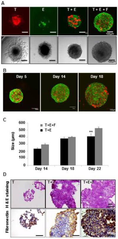
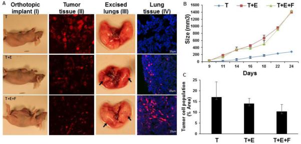
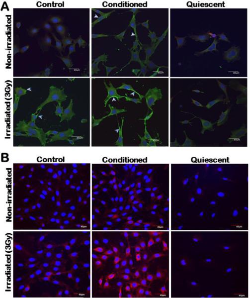
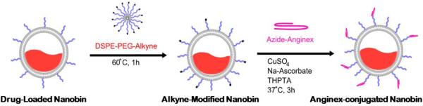
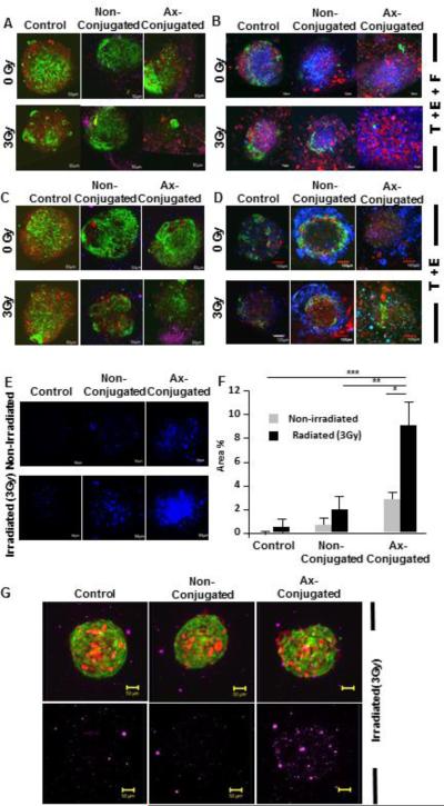
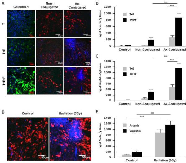
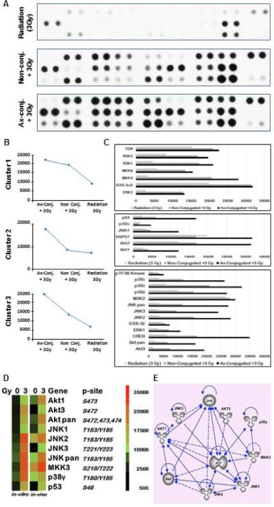
An appropriate representation of the tumor microenvironment in tumor models can have a pronounced impact on directing combinatorial treatment strategies and cancer nanotherapeutics. The present study develops a novel 3D co-culture spheroid model (3D TNBC) incorporating tumor cells, endothelial cells and fibroblasts as color-coded murine tumor tissue analogs (TTA) to better represent the tumor milieu of triple negative breast cancer in vitro. Implantation of TTA orthotopically in nude mice, resulted in enhanced growth and aggressive metastasis to ectopic sites. Subsequently, the utility of the model is demonstrated for preferential targeting of irradiated tumor endothelial cells via radiation-induced stromal enrichment of galectin-1 using anginex conjugated nanoparticles (nanobins) carrying arsenic trioxide and cisplatin. Demonstration of a multimodal nanotherapeutic system and inclusion of the biological response to radiation using an in vitro/in vivo tumor model incorporating characteristics of tumor microenvironment presents an advance in preclinical evaluation of existing and novel cancer nanotherapies.

From the clinical editor: Existing in-vivo tumor models are established by implanting tumor cells into nude mice. Here, the authors described their approach 3D spheres containing tumor cells, enodothelial cells and fibroblasts. This would mimic tumor micro-environment more realistically. This interesting 3D model should reflect more accurately tumor response to various drugs and would enable the design of new treatment modalities.

Keywords: 3 dimensional triple negative breast cancer (3D TNBC) model; 3D co-cultures; Galectin-1; Targeted nanoparticle; Tumor cell spheroids; Tumor microenvironment; Tumor tissue analogs (TTA).


Figures

Figure 1
Figure 1. Murine TTA from multicell 3D co-cultures in vitro demonstrate robust growth and tissue-like morphology
Confocal (Upper panel) and DIC (Lower panel) images of 3D cultures at day 14 in hanging drops of medium of (A) mCherry (Red) 4T1 tumor cells only [T], GFP-C166 endothelial cell only [E], co-culture of tumor cells and endothelial cells [T+E], and co-culture of murine Tumor cells, endothelial cells and mouse embryonic fibroblasts (MEF) [T+E+F]. (B) TTA [T+E+F] at day 5, 14 and 18. Confocal images are an overlay of green (C166 endothelial cells) and Red (4T1 tumor cells) fluorescence. (C) Growth comparison of [T+E] and [T+E+F] TTA; (D) H&E (Upper panel) and immunostaining for extracellular matrix protein (fibronectin) (Lower panel) in tumor cell aggregates [T] and TTA [T+E, T+E+F] at 20X magnification.
Figure 2
Figure 2. Enhanced growth and metastasis of tumors originating from orthotopic xenografts of 3D TTA
(A) Athymic nude mice bearing mammary fat pad orthotopic tumors originating from 3D cultures (TTA) of 4T1 tumor cells [T], 4T1 tumor and C166 endothelial cells [T+E] and 4T1 tumor, C166 endothelial and MEF [T+E+F] (I); 4T1 mCherry expressing tumor cells in tissue sections from tumors originating from orthotopic tumors as described earlier (II); Metastasis in diseased lung excised 24 days post-implantation (III), and presence of mCherry 4T1 tumor cells (Red) cryosections of lung tissue (IV) was found to be more aggressive in tumors originating from [T+E] or [T+E+F] TTA. (B) Tumor growth curve in orthotopic xenografts (n=8) of 3D cultures. Error bars represent mean ± SEM. (C) Reduced tumor cell population in Tumor tissue sections of orthotopic xenografts of 3D TTA. The mCherry (red) fluorescent protein expressing tumor cell population in tumor tissues originating from orthotopic xenografts of [T], [T+E] and [T+E+F] was quantified by densitomentry using the ImageJ software and graphically represented. Error bars represent mean ± SEM.
Figure 3
Figure 3. Radiation exposure augments expression of galectin-1 including the cell surface in murine endothelial cells when incubated in conditioned medium from tumor cells
(A) 2H11 and (B) C166 endothelial cells grown in regular (control), serum free (quiescent) and conditioned medium from 4T1 murine breast carcinoma cells respectively (upper panel) were subjected to 3Gy of radiation exposure (lower panel). Cells were incubated with goat anti-galectin-1 primary antibody (Invitrogen) followed by incubation with secondary alexafluor 488 (Invitrogen) [Green] in 2H11 cells and secondary alexafluor 633 [Red] in C166 cells. Nuclei were stained with DAPI [Blue]. Scale bar, 40 μm. While there is profuse overexpression of galectin-1 in both endothelial cell types maintained in conditioned medium, the white arrows in 2H11 cells indicate higher accumulation of galectin-1 on endothelial cell surface. Galectin-1 expression in cells grown in serum free condition representative of quiescent/normal endothelial cell phenotype with and without radiation exposure was not noticeable.
Figure 4
Figure 4. Anginex labeling strategy and for arsenic-cisplatin dual drug loaded nanoparticles
The figure is a schematic of the anginex labeling strategy.
Figure 5
Figure 5. Elimination of endothelial cells (green), increased cell damage and enhanced nanoparticle uptake in tumor tissue analogs (T+E+F) resulting from radiation induced targeting of anginex conjugated arsenic-cisplatin (as-cs) loaded nanobins
Confocal images (10X) of TTAs of [T+E+F] (A) and [T+E] (C) respectively 48 hr post-incubation with Axconjugated and non-conjugated as-cs loaded nanobins (15 μM) following radiation exposure (3Gy). The nanobins were labeled with a far red fluorescent lipophilic carbocyanine dye, DID. The images are an overlay of Red (tumor cells), Green (Endothelial cells) and Purple (DID labelled nanobins) fluorescence. An overlay of confocal images (10X) of tumor tissue analogs of [T+E+F] (B) and [T+E] (D) 8 days post-incubation with Ax-conjugated and non-conjugated ascs loaded nanobins (5 μM) with and without radiation exposure (3Gy) stained with Sytox blue (nuclear stain for cell damage). The images are an overlay of red (tumor cells), green (endothelial cells) and blue (Sytox blue) fluorescence. (E) Representative images of cell damage upon treatment as indicated by Sytox blue staining. (F) Toxicity of the radiation induced targeting of nanobins was evaluated by quantitation of the sytox blue staining using an ImageJ software. ***, P < 0.0001; **, P = 0.0006; *, P = 0.0004. (G) A comparison of an overlay of images from light sheet microscopy (LSM) excited with purple (DID labeled nanobins) fluorescence of irradiated TTA at 18 hr post treatment.
Figure 6
Figure 6. Elevated galectin-1 and Nanobin accumulation with significant drug uptake in orthotopic tumors originating from TTA (T+E+F)
Mice bearing orthotopic tumors originating from 3D tumor cell aggregates (T) and tumor tissue analogs of tumor-endothelial cells (T+E) and tumor-endothelial cells-Fibroblasts (T+E+F) were were exposed to radiation (3Gy) for 4 hr and then injected via tail vein with 4mg/kg of ax-conjugated or non-conjugated arsenic-cisplatin loaded nanobins (DID labelled). (A) Confocal images (10X) of tumor tissue sections after radiation exposure and immunostaining with galectin-1 (left panel); followed by incubation with non-conjugated (middle panel) and ax-conjugated (right panel) nanobins. The images are an overlay of red fluorescence (4T1-mCherry tumor cells) and nanobins labelled with DID fluorescing blue. ICP-MS analysis of arsenic (B) and cisplatin (C) concentration in tumor tissues of mice bearing T+E and T+E+F implants when treated with radiation and ax-conjugated or non-conjugated nanoparticles ***, P < 0.001. (D) Representative confocal images of tumor tissue sections and their ICP-MS analysis (E) for arsenic and cisplatin from T+E+F implants in mice incubated with anginex conjugated nanobins with and without pre-exposure to radiation (3 Gy). ***, P < 0.001.
Figure 7
Figure 7. Elevated expression of phospho proteins in the apoptotic and stress signaling pathway in response to targeted nanobins following radiation exposure (3Gy) in TTA and their orthotopic implants in nude mice
(A) A Phospho-MAPK antibody array was used to detect the changes in phosphorylated proteins in TTA (T+E+F) upon radiation exposure and incubation with Ax-conjugated or non-conjugated nanobins after radiation exposure (3Gy). (B) Based on the pixel densities of the signal in each spot of the array corresponding to the respective phosphorylated protein, they were grouped in one of the three clusters. (C) Quantification of pixel density as a measure of protein phosphorylation in the three clusters. (D) Heatmaps comparing the expression of the phosphorylated proteins in the TTA (in vitro) and upon treatment with anginex-conjugated nanobins with and without radiation exposure. Similar response is observed in tumor tissues at 24 hr post-treatment derived from orthotopic implants of TTAin nude mice (in vivo). (E) Ingenuity pathway analysis showing interaction and regulation of phospho proteins significantly activated in response to the nanotherapeutic treatment strategy by the phosphorylation of functional p53 (S46) that is expected to be activated by the galectin-1 targeting of endothelial cells.

References

    1. Neri D, Bicknell R. Tumour vascular targeting. Nature reviews. Cancer. 2005;5:436–446. doi:10.1038/nrc1627. - PubMed
    1. Upreti M, et al. Radiation-induced galectin-1 by endothelial cells: a promising molecular target for preferential drug delivery to the tumor vasculature. Journal of molecular medicine. 2013;91:497–506. doi:10.1007/s00109-012-0965-1. - PMC - PubMed
    1. Upreti M, et al. Tumor-Endothelial Cell Three-dimensional Spheroids: New Aspects to Enhance Radiation and Drug Therapeutics. Translational oncology. 2011;4:365–376. - PMC - PubMed
    1. Maeda H, Nakamura H, Fang J. The EPR effect for macromolecular drug delivery to solid tumors: Improvement of tumor uptake, lowering of systemic toxicity, and distinct tumor imaging in vivo. Adv Drug Deliv Rev. 2013;65:71–79. doi:10.1016/j.addr.2012.10.002. - PubMed
    1. Barua S, Mitragotri S. Challenges associated with Penetration of Nanoparticles across Cell and Tissue Barriers: A Review of Current Status and Future Prospects. Nano today. 2014;9:223–243. doi:10.1016/j.nantod.2014.04.008. - PMC - PubMed

MeSH terms