. 2015 Aug 18:6:8057.
doi: 10.1038/ncomms9057.

Stoichiometry for α-bungarotoxin block of α7 acetylcholine receptors

Affiliations

Stoichiometry for α-bungarotoxin block of α7 acetylcholine receptors

Corrie J B daCosta et al. Nat Commun. .

Abstract

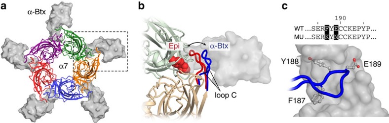
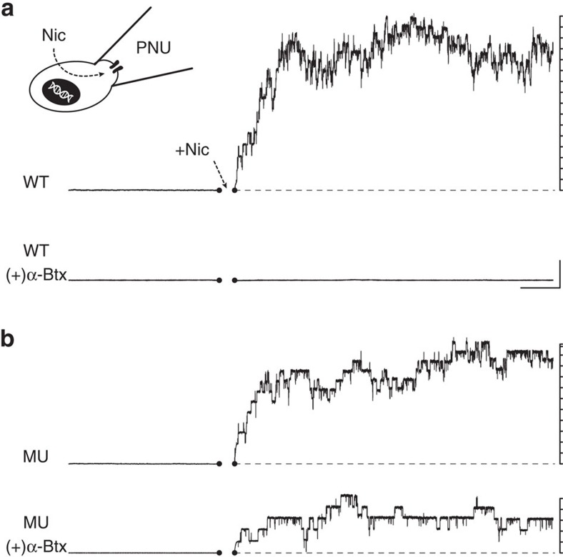
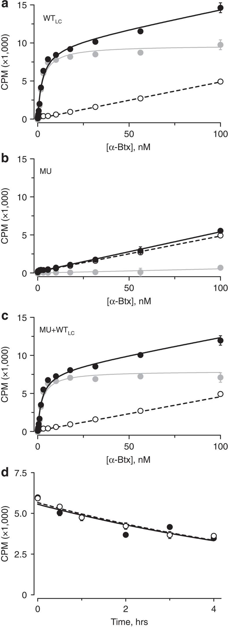
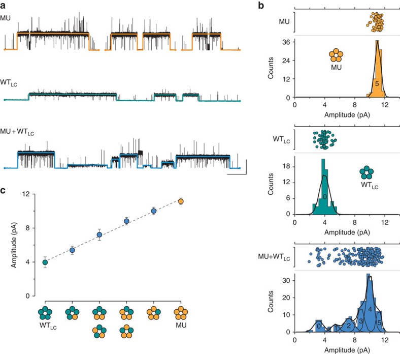
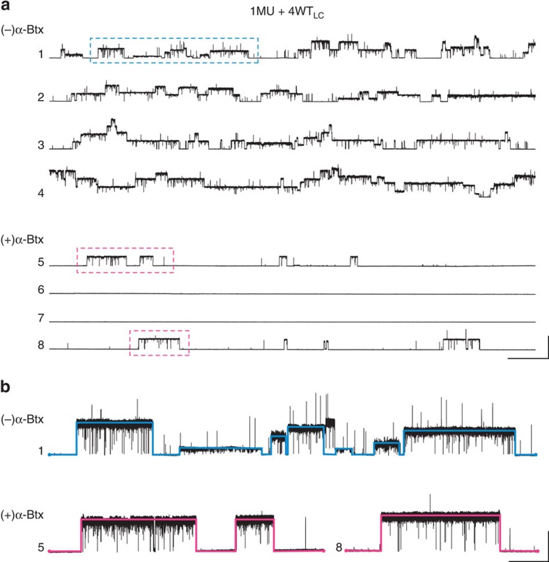
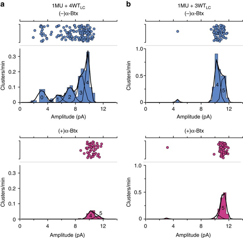
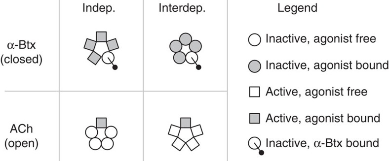
α-Bungarotoxin (α-Btx) binds to the five agonist binding sites on the homopentameric α7-acetylcholine receptor, yet the number of bound α-Btx molecules required to prevent agonist-induced channel opening remains unknown. To determine the stoichiometry for α-Btx blockade, we generate receptors comprised of wild-type and α-Btx-resistant subunits, tag one of the subunit types with conductance mutations to report subunit stoichiometry, and following incubation with α-Btx, monitor opening of individual receptor channels with defined subunit stoichiometry. We find that a single α-Btx-sensitive subunit confers nearly maximal suppression of channel opening, despite four binding sites remaining unoccupied by α-Btx and accessible to the agonist. Given structural evidence that α-Btx locks the agonist binding site in an inactive conformation, we conclude that the dominant mechanism of antagonism is non-competitive, originating from conformational arrest of the binding sites, and that the five α7 subunits are interdependent and maintain conformational symmetry in the open channel state.


Figures

Figure 1
Figure 1. Structural details of α-Btx binding to α7.
(a) Complex between the α7-acetylcholine receptor ligand binding domain chimera (α7; ribbons, where each subunit is a different colour) and α-Btx (grey surfaces; PDB: 4HQP). (b) Close up view of the boxed region in ‘a' showing α7 in complex with the agonist epibatidine (Epi, red spheres; PDB: 3SQ6) and α-Btx (grey surface). The conformations of ‘loop C' in both the Epi-α7 (red) and α-Btx-α7 (blue) complexes are overlaid to show how the toxin locks loop C in an extended conformation. With the exception of the epibatidine (red spheres) and the Epi-loop C (red), the structures depicted are from the α-Btx-α7 complex (PDB: 4HQP). (c) Sequence alignment of loop C residues in wild-type α7 (WT) and the toxin-resistant mutant (MU). Shown below is a close up of the interaction between WT-loop C (blue) and α-Btx (grey surface; PDB: 4HQP), where the wild-type side chains of the residues substituted in the mutant, which flank a canonical Tyrosine residue (Y188), are shown in a ball and stick representation.
Figure 2
Figure 2. A functional α-Btx-resistant α7 mutant.
Difference in toxin sensitivity for homopentameric receptors formed from (a) wild-type (WT, top two traces) and (b) mutant α-Btx-resistant subunits mutant ((MU), bottom two traces). After recording a stable baseline free of single channel openings (that is, no agonist present), nicotine (100 μM final concentration) was added to the bath solution surrounding each patch (arrow). For the lower traces, in both ‘a' and ‘b', cells were incubated with 50 nM α-Btx for 20 min before forming a cell-attached patch. A concentration of 10 μM PNU was included in all patch pipettes to prolong agonist-activated openings and facilitate their visualization (see Methods). Note that each recording was obtained from a different membrane patch, on a different cell, so the magnitude of the evoked currents is not comparable between recordings. Traces are filtered at 100 Hz, each increment of the vertical axes (right) indicates 10 pA, and scale bar, 5 s, 25 pA.
Figure 3
Figure 3. Steady state α-Btx binding measurements.
Radiolabeled [125I]-α-Btx binding to cells expressing (a) wild-type, low-conductance (WTLC) subunits, (b) mutant subunits (MU), or (c) mixtures of MU and WTLC subunits (1:4, MU:WTLC). In each case, total binding (closed circles, solid line), non-specific binding (open circles and dashed line), as well as specific binding (grey circles and line) are shown. Non-specific binding was determined from cells transfected with the cDNA encoding the human muscle AChR β1-subunit, which does not form α-Btx binding sites. The apparent Kds for cells expressing WTLC subunits, and mixtures of MU and WTLC subunits are 2.7 nM (2.26–3.14 nM) and 2.4 nM (1.90–2.84 nM), respectively, (95% confidence limits), where n=3 and the error bars represent one s.d. of the mean. (d) Time course of [125I]-α-Btx dissociation from cells expressing WTLC (closed circles and solid line), or mixtures of MU and WTLC subunits (1:4, MU:WTLC; open circles and dashed line). The dissociation half-life for cells expressing WTLC subunits and mixtures of MU and WTLC subunits are 322.6 min (239.4–494.3 min) and 316.7 min (258.0–410.0 min), respectively, (95% confidence limits), where n=2 and error bars are one s.d. of the mean.
Figure 4
Figure 4. Electrical fingerprinting to determine subunit composition from single channel amplitudes.
(a) Mutant α-Btx-resistant subunits produce channel openings with a uniform large current amplitude (MU, top), while wild-type, low conductance subunits (WTLC) produce openings with a uniform small current amplitude (middle trace). Co-expressing MU subunits with WTLC subunits (1:4; mutant:WTLC) gives rise to receptors with different subunit stoichiometry and channel openings with a distribution of amplitudes (bottom trace). For each trace, the detected amplitude for individual clusters of openings, corresponding to a single channel, is overlaid (see Methods for detection criteria; open is up; scale bar, 10 s, 10 pA). (b) Event-based amplitude histograms for pooled clusters of openings from mutant subunits (top; 7 recordings and 89 clusters), WTLC subunits (middle; 7 recordings and 62 clusters), and mixtures of mutant and WTLC subunits (bottom; 21 recordings and 225 clusters, pooled from 1:4 and 1:3 cDNA ratios). Amplitude scatter plots corresponding to the openings are also shown above each histogram. Histograms have been fit with a sum of Gaussian components, corresponding to the amplitude classes used to determine (c) the relationship between single-channel amplitude and the stoichiometry of MU and WTLC subunits. For each histogram in ‘b', the Gaussian components are labelled ‘0-5' according to the presumed number of incorporated mutant subunits. The mean amplitude and s.d. (σ) of each component was used to determine the amplitudes and associated error bars (1σ) used for the plot in ‘c'. The amplitudes for the MU and WTLC classes (yellow and green points) were determined from the corresponding MU and WTLC histograms, whereas for the channels with mixed stoichiometry (blue points) the amplitudes were from the corresponding components in the MU+WTLC histogram.
Figure 5
Figure 5. α-Btx inhibition of α7 receptors comprised of both α-Btx-resistant and wild-type subunits.
(a) Co-expression of mutant α-Btx-resistant subunits with α-Btx-sensitive wild-type, low-conductance subunits (1:4; MU:WTLC) gives rise to active patches with multiple levels of superimposed single channel openings (patches 1–4, top). In contrast, when cells that have been transfected by the same mixture are incubated with 50 nM α-Btx, patches have dramatically reduced single channel activity (patches 5–8, bottom). Each sweep represents six uninterrupted minutes of recording from a different patch. Traces are filtered at a bandwidth of 100 Hz; open is up; scale bar, 30 s, 25 pA. (b) Enlarged views of the boxed regions in ‘a', showing that when cells are not pre-incubated with α-Btx, single channel openings are of variable amplitudes and correspond to channels with different numbers of incorporated WTLC subunits (top trace). When cells are incubated with 50 nM α-Btx the infrequent single channel openings are always of large amplitude, corresponding to channels with no, or few, incorporated WTLC subunits. Traces in ‘b' are filtered at a bandwidth of 1 kHz; open is up; scale bar, 10 s, 10 pA.
Figure 6
Figure 6. Stoichiometry of α-Btx inhibition of α7.
Amplitude histograms, where the number of events in each bin has been scaled to their relative frequency (see Methods), for pooled clusters of openings from cells co-expressing mixtures of mutant (MU) and wild-type, low conductance (WTLC) subunits at cDNA ratios of (a) 1:4; MU:WTLC and (b) 1:3; mutant:WTLC. In each case, the top histogram represents the distribution and frequency of openings with a given amplitude when cells are not incubated with α-Btx. When cells transfected in parallel with the same cDNA mixture, are incubated with 50 nM α-Btx, the amplitude distributions change, reflecting relative inhibition by α-Btx (bottom histograms). Histograms have been fit with a sum of Gaussian components, which are labelled ‘0-5' corresponding to the number of incorporated MU subunits. For each histogram, the number of recordings, observed clusters and total recording time are as follows: A, top, 13 recordings, 153 clusters and 174.7 min; A, bottom, 21 recordings, 50 clusters and 396.6 min; B, top, 8 recordings, 72 clusters, 84.7 min; B, bottom, 7 recordings, 64 clusters and 115.7 min.
Figure 7
Figure 7. Scenarios for conformational coupling amongst subunits in α7.
In an independent scenario (left column), individual subunits can adopt different conformations irrespective of the other subunits within the pentamer, whereas in the interdependent scenario (right column), the conformations of individual subunits are coupled to each other, maintaining subunit conformational symmetry in the entire pentamer. In the situation where a single α-Btx is bound to α7, the channel is closed (top row), while in the absence of α-Btx, when a single acetylcholine (ACh) is bound, the channel is open (bottom row). The legend on the right explains the different shapes used to represent active (squares) and inactive (circles) conformations of each subunit, as well as their occupancy with either agonist (grey shading) or α-Btx (black ball and stick).

References

    1. Chang C. C. & Lee C. Y. Isolation of neurotoxins from the venom of bungarus multicinctus and their modes of neuromuscular blocking action. Arch. Int. Pharmacodyn. Ther. 144, 241–257 (1963). - PubMed
    1. Betzel C. et al.. The refined crystal structure of α-cobratoxin from Naja naja siamensis at 2.4-Å resolution. J. Biol. Chem. 266, 21530–21536 (1991). - PubMed
    1. Neubig R. R. & Cohen J. B. Equilibrium binding of [3H]tubocurarine and [3H]acetylcholine by Torpedo postsynaptic membranes: stoichiometry and ligand interactions. Biochemistry 18, 5464–5475 (1979). - PubMed
    1. Bourne Y., Talley T. T., Hansen S. B., Taylor P. & Marchot P. Crystal structure of a Cbtx-AChBP complex reveals essential interactions between snake α-neurotoxins and nicotinic receptors. EMBO J. 24, 1512–1522 (2005). - PMC - PubMed
    1. Hansen S. B. et al.. Structures of Aplysia AChBP complexes with nicotinic agonists and antagonists reveal distinctive binding interfaces and conformations. EMBO J. 24, 3635–3646 (2005). - PMC - PubMed

Publication types

Substances

LinkOut - more resources